array:25 [
  "pii" => "S0870255114000833"
  "issn" => "08702551"
  "doi" => "10.1016/j.repc.2013.11.009"
  "estado" => "S300"
  "fechaPublicacion" => "2014-04-01"
  "aid" => "457"
  "copyright" => "Sociedade Portuguesa de Cardiologia"
  "copyrightAnyo" => "2013"
  "documento" => "article"
  "crossmark" => 0
  "licencia" => "http://creativecommons.org/licenses/by-nc-nd/4.0/"
  "subdocumento" => "sco"
  "cita" => "Rev Port Cardiol. 2014;33:253-4"
  "abierto" => array:3 [
    "ES" => true
    "ES2" => true
    "LATM" => true
  ]
  "gratuito" => true
  "lecturas" => array:2 [
    "total" => 3827
    "formatos" => array:3 [
      "EPUB" => 173
      "HTML" => 2991
      "PDF" => 663
    ]
  ]
  "Traduccion" => array:1 [
    "en" => array:20 [
      "pii" => "S2174204914001081"
      "issn" => "21742049"
      "doi" => "10.1016/j.repce.2013.11.006"
      "estado" => "S300"
      "fechaPublicacion" => "2014-04-01"
      "aid" => "457"
      "copyright" => "Sociedade Portuguesa de Cardiologia"
      "documento" => "article"
      "crossmark" => 0
      "licencia" => "http://creativecommons.org/licenses/by-nc-nd/4.0/"
      "subdocumento" => "sco"
      "cita" => "Rev Port Cardiol. 2014;33:253-4"
      "abierto" => array:3 [
        "ES" => true
        "ES2" => true
        "LATM" => true
      ]
      "gratuito" => true
      "lecturas" => array:2 [
        "total" => 3169
        "formatos" => array:3 [
          "EPUB" => 160
          "HTML" => 2473
          "PDF" => 536
        ]
      ]
      "en" => array:10 [
        "idiomaDefecto" => true
        "titulo" => "Pulmonary embolism complicated by air embolism"
        "tienePdf" => "en"
        "tieneTextoCompleto" => "en"
        "paginas" => array:1 [
          0 => array:2 [
            "paginaInicial" => "253"
            "paginaFinal" => "254"
          ]
        ]
        "titulosAlternativos" => array:1 [
          "pt" => array:1 [
            "titulo" => "Trombo‐embolismo pulmonar complicado por embolia gasosa"
          ]
        ]
        "contieneTextoCompleto" => array:1 [
          "en" => true
        ]
        "contienePdf" => array:1 [
          "en" => true
        ]
        "resumenGrafico" => array:2 [
          "original" => 0
          "multimedia" => array:7 [
            "identificador" => "fig0020"
            "etiqueta" => "Figure 4"
            "tipo" => "MULTIMEDIAFIGURA"
            "mostrarFloat" => true
            "mostrarDisplay" => false
            "figura" => array:1 [
              0 => array:4 [
                "imagen" => "gr4.jpeg"
                "Alto" => 1675
                "Ancho" => 1500
                "Tamanyo" => 210485
              ]
            ]
            "descripcion" => array:1 [
              "en" => "<p id="spar0020" class="elsevierStyleSimplePara elsevierViewall">Topogram image suggesting the presence of air in the right ventricle before contrast injection &#40;red arrow&#41;&#46;</p>"
            ]
          ]
        ]
        "autores" => array:1 [
          0 => array:2 [
            "autoresLista" => "Catarina Mendon&#231;a, Alexandre Pinto"
            "autores" => array:2 [
              0 => array:2 [
                "nombre" => "Catarina"
                "apellidos" => "Mendon&#231;a"
              ]
              1 => array:2 [
                "nombre" => "Alexandre"
                "apellidos" => "Pinto"
              ]
            ]
          ]
        ]
      ]
      "idiomaDefecto" => "en"
      "Traduccion" => array:1 [
        "en" => array:9 [
          "pii" => "S0870255114000833"
          "doi" => "10.1016/j.repc.2013.11.009"
          "estado" => "S300"
          "subdocumento" => ""
          "abierto" => array:3 [
            "ES" => true
            "ES2" => true
            "LATM" => true
          ]
          "gratuito" => true
          "lecturas" => array:1 [
            "total" => 0
          ]
          "idiomaDefecto" => "en"
          "EPUB" => "https://multimedia.elsevier.es/PublicationsMultimediaV1/item/epub/S0870255114000833?idApp=UINPBA00004E"
        ]
      ]
      "EPUB" => "https://multimedia.elsevier.es/PublicationsMultimediaV1/item/epub/S2174204914001081?idApp=UINPBA00004E"
      "url" => "/21742049/0000003300000004/v1_201406010133/S2174204914001081/v1_201406010133/en/main.assets"
    ]
  ]
  "itemSiguiente" => array:20 [
    "pii" => "S0870255114000055"
    "issn" => "08702551"
    "doi" => "10.1016/j.repc.2013.10.007"
    "estado" => "S300"
    "fechaPublicacion" => "2014-04-01"
    "aid" => "410"
    "copyright" => "Sociedade Portuguesa de Cardiologia"
    "documento" => "article"
    "crossmark" => 0
    "licencia" => "http://creativecommons.org/licenses/by-nc-nd/4.0/"
    "subdocumento" => "sco"
    "cita" => "Rev Port Cardiol. 2014;33:255-7"
    "abierto" => array:3 [
      "ES" => true
      "ES2" => true
      "LATM" => true
    ]
    "gratuito" => true
    "lecturas" => array:2 [
      "total" => 3952
      "formatos" => array:3 [
        "EPUB" => 197
        "HTML" => 2869
        "PDF" => 886
      ]
    ]
    "pt" => array:11 [
      "idiomaDefecto" => true
      "cabecera" => "<span class="elsevierStyleTextfn">Imagem em cardiologia</span>"
      "titulo" => "Uma teia de aranha no cora&#231;&#227;o"
      "tienePdf" => "pt"
      "tieneTextoCompleto" => "pt"
      "paginas" => array:1 [
        0 => array:2 [
          "paginaInicial" => "255"
          "paginaFinal" => "257"
        ]
      ]
      "titulosAlternativos" => array:1 [
        "en" => array:1 [
          "titulo" => "A spiderweb in the heart"
        ]
      ]
      "contieneTextoCompleto" => array:1 [
        "pt" => true
      ]
      "contienePdf" => array:1 [
        "pt" => true
      ]
      "resumenGrafico" => array:2 [
        "original" => 0
        "multimedia" => array:7 [
          "identificador" => "fig0010"
          "etiqueta" => "Figura 2"
          "tipo" => "MULTIMEDIAFIGURA"
          "mostrarFloat" => true
          "mostrarDisplay" => false
          "figura" => array:1 [
            0 => array:4 [
              "imagen" => "gr2.jpeg"
              "Alto" => 1190
              "Ancho" => 1300
              "Tamanyo" => 290261
            ]
          ]
          "descripcion" => array:1 [
            "pt" => "<p id="spar0010" class="elsevierStyleSimplePara elsevierViewall">Amostra histol&#243;gica a&#41; pericardite fibrinosa &#40;hematoxilina&#8208;eosina x<span class="elsevierStyleHsp" style=""></span>100&#41;&#59; b&#41; L&#237;quido peric&#225;rdico hem&#225;tico com citologia negativa para malignidade &#40;hematoxilina&#8208;eosina x<span class="elsevierStyleHsp" style=""></span>100&#41;&#59; c&#41; peric&#225;rdio com c&#233;lulas mesoteliais com hiperplasia&#47;atipia &#40;calretinina x<span class="elsevierStyleHsp" style=""></span>100&#41;&#59; d&#41; infiltrado inflamat&#243;rio com numerosos macr&#243;fagos &#40;CD 68<span class="elsevierStyleHsp" style=""></span>x<span class="elsevierStyleHsp" style=""></span>100&#41;&#46;</p>"
          ]
        ]
      ]
      "autores" => array:1 [
        0 => array:2 [
          "autoresLista" => "Rita Salvado, Mafalda Guimar&#227;es, Miguel Ara&#250;jo, Diogo Costa, Jos&#233; Ara&#250;jo"
          "autores" => array:5 [
            0 => array:2 [
              "nombre" => "Rita"
              "apellidos" => "Salvado"
            ]
            1 => array:2 [
              "nombre" => "Mafalda"
              "apellidos" => "Guimar&#227;es"
            ]
            2 => array:2 [
              "nombre" => "Miguel"
              "apellidos" => "Ara&#250;jo"
            ]
            3 => array:2 [
              "nombre" => "Diogo"
              "apellidos" => "Costa"
            ]
            4 => array:2 [
              "nombre" => "Jos&#233;"
              "apellidos" => "Ara&#250;jo"
            ]
          ]
        ]
      ]
    ]
    "idiomaDefecto" => "pt"
    "Traduccion" => array:1 [
      "en" => array:9 [
        "pii" => "S2174204914000877"
        "doi" => "10.1016/j.repce.2013.10.039"
        "estado" => "S300"
        "subdocumento" => ""
        "abierto" => array:3 [
          "ES" => true
          "ES2" => true
          "LATM" => true
        ]
        "gratuito" => true
        "lecturas" => array:1 [
          "total" => 0
        ]
        "idiomaDefecto" => "en"
        "EPUB" => "https://multimedia.elsevier.es/PublicationsMultimediaV1/item/epub/S2174204914000877?idApp=UINPBA00004E"
      ]
    ]
    "EPUB" => "https://multimedia.elsevier.es/PublicationsMultimediaV1/item/epub/S0870255114000055?idApp=UINPBA00004E"
    "url" => "/08702551/0000003300000004/v1_201405230947/S0870255114000055/v1_201405230947/pt/main.assets"
  ]
  "itemAnterior" => array:20 [
    "pii" => "S0870255114000729"
    "issn" => "08702551"
    "doi" => "10.1016/j.repc.2013.11.007"
    "estado" => "S300"
    "fechaPublicacion" => "2014-04-01"
    "aid" => "446"
    "copyright" => "Sociedade Portuguesa de Cardiologia"
    "documento" => "article"
    "crossmark" => 0
    "licencia" => "http://creativecommons.org/licenses/by-nc-nd/4.0/"
    "subdocumento" => "sco"
    "cita" => "Rev Port Cardiol. 2014;33:251-2"
    "abierto" => array:3 [
      "ES" => true
      "ES2" => true
      "LATM" => true
    ]
    "gratuito" => true
    "lecturas" => array:2 [
      "total" => 6320
      "formatos" => array:3 [
        "EPUB" => 221
        "HTML" => 5167
        "PDF" => 932
      ]
    ]
    "pt" => array:11 [
      "idiomaDefecto" => true
      "cabecera" => "<span class="elsevierStyleTextfn">Imagem em cardiologia</span>"
      "titulo" => "Insufici&#234;ncia card&#237;aca aguda no adulto jovem &#8208; uma causa rara"
      "tienePdf" => "pt"
      "tieneTextoCompleto" => "pt"
      "paginas" => array:1 [
        0 => array:2 [
          "paginaInicial" => "251"
          "paginaFinal" => "252"
        ]
      ]
      "titulosAlternativos" => array:1 [
        "en" => array:1 [
          "titulo" => "Heart failure in a young adult&#58; A rare cause"
        ]
      ]
      "contieneTextoCompleto" => array:1 [
        "pt" => true
      ]
      "contienePdf" => array:1 [
        "pt" => true
      ]
      "resumenGrafico" => array:2 [
        "original" => 0
        "multimedia" => array:7 [
          "identificador" => "fig0015"
          "etiqueta" => "Figura 3"
          "tipo" => "MULTIMEDIAFIGURA"
          "mostrarFloat" => true
          "mostrarDisplay" => false
          "figura" => array:1 [
            0 => array:4 [
              "imagen" => "gr3.jpeg"
              "Alto" => 1006
              "Ancho" => 1584
              "Tamanyo" => 129184
            ]
          ]
          "descripcion" => array:1 [
            "pt" => "<p id="spar0015" class="elsevierStyleSimplePara elsevierViewall">Comunica&#231;&#227;o interventricular &#40;CIV&#41; subvalvular ao n&#237;vel do plano dos folhetos da v&#225;lvula mitral &#40;Val&#46; Mi&#46;&#41; e v&#225;lvula tric&#250;spide &#40;Val&#46; Tri&#46;&#41; &#8211; &#40;janela paraesternal eixo curto&#41;&#46;</p>"
          ]
        ]
      ]
      "autores" => array:1 [
        0 => array:2 [
          "autoresLista" => "Anne Delgado, Davide Moreira, Bruno Marmelo, Miguel Correia, Emanuel Correia, Oliveira Santos"
          "autores" => array:6 [
            0 => array:2 [
              "nombre" => "Anne"
              "apellidos" => "Delgado"
            ]
            1 => array:2 [
              "nombre" => "Davide"
              "apellidos" => "Moreira"
            ]
            2 => array:2 [
              "nombre" => "Bruno"
              "apellidos" => "Marmelo"
            ]
            3 => array:2 [
              "nombre" => "Miguel"
              "apellidos" => "Correia"
            ]
            4 => array:2 [
              "nombre" => "Emanuel"
              "apellidos" => "Correia"
            ]
            5 => array:2 [
              "nombre" => "Oliveira"
              "apellidos" => "Santos"
            ]
          ]
        ]
      ]
    ]
    "idiomaDefecto" => "pt"
    "Traduccion" => array:1 [
      "en" => array:9 [
        "pii" => "S2174204914000981"
        "doi" => "10.1016/j.repce.2013.11.004"
        "estado" => "S300"
        "subdocumento" => ""
        "abierto" => array:3 [
          "ES" => true
          "ES2" => true
          "LATM" => true
        ]
        "gratuito" => true
        "lecturas" => array:1 [
          "total" => 0
        ]
        "idiomaDefecto" => "en"
        "EPUB" => "https://multimedia.elsevier.es/PublicationsMultimediaV1/item/epub/S2174204914000981?idApp=UINPBA00004E"
      ]
    ]
    "EPUB" => "https://multimedia.elsevier.es/PublicationsMultimediaV1/item/epub/S0870255114000729?idApp=UINPBA00004E"
    "url" => "/08702551/0000003300000004/v1_201405230947/S0870255114000729/v1_201405230947/pt/main.assets"
  ]
  "en" => array:16 [
    "idiomaDefecto" => true
    "cabecera" => "<span class="elsevierStyleTextfn">Image in Cardiology</span>"
    "titulo" => "Pulmonary embolism complicated by air embolism"
    "tieneTextoCompleto" => true
    "paginas" => array:1 [
      0 => array:2 [
        "paginaInicial" => "253"
        "paginaFinal" => "254"
      ]
    ]
    "autores" => array:1 [
      0 => array:4 [
        "autoresLista" => "Catarina Mendon&#231;a, Alexandre Pinto"
        "autores" => array:2 [
          0 => array:4 [
            "nombre" => "Catarina"
            "apellidos" => "Mendon&#231;a"
            "email" => array:1 [
              0 => "catarinamdm&#64;gmail&#46;com"
            ]
            "referencia" => array:2 [
              0 => array:2 [
                "etiqueta" => "<span class="elsevierStyleSup">a</span>"
                "identificador" => "aff0005"
              ]
              1 => array:2 [
                "etiqueta" => "<span class="elsevierStyleSup">¿</span>"
                "identificador" => "cor0005"
              ]
            ]
          ]
          1 => array:3 [
            "nombre" => "Alexandre"
            "apellidos" => "Pinto"
            "referencia" => array:1 [
              0 => array:2 [
                "etiqueta" => "<span class="elsevierStyleSup">b</span>"
                "identificador" => "aff0010"
              ]
            ]
          ]
        ]
        "afiliaciones" => array:2 [
          0 => array:3 [
            "entidad" => "Servi&#231;o de Medicina&#44; Centro Hospitalar do Porto&#44; Porto&#44; Portugal"
            "etiqueta" => "a"
            "identificador" => "aff0005"
          ]
          1 => array:3 [
            "entidad" => "Unidade Interm&#233;dia M&#233;dico-Cir&#250;rgica&#44; Centro Hospitalar do Porto&#44; Porto&#44; Portugal"
            "etiqueta" => "b"
            "identificador" => "aff0010"
          ]
        ]
        "correspondencia" => array:1 [
          0 => array:3 [
            "identificador" => "cor0005"
            "etiqueta" => "&#8270;"
            "correspondencia" => "Corresponding author&#46;"
          ]
        ]
      ]
    ]
    "titulosAlternativos" => array:1 [
      "pt" => array:1 [
        "titulo" => "Trombo-embolismo pulmonar complicado por embolia gasosa"
      ]
    ]
    "resumenGrafico" => array:2 [
      "original" => 0
      "multimedia" => array:7 [
        "identificador" => "fig0010"
        "etiqueta" => "Figure 2"
        "tipo" => "MULTIMEDIAFIGURA"
        "mostrarFloat" => true
        "mostrarDisplay" => false
        "figura" => array:1 [
          0 => array:4 [
            "imagen" => "gr2.jpeg"
            "Alto" => 675
            "Ancho" => 900
            "Tamanyo" => 75415
          ]
        ]
        "descripcion" => array:1 [
          "en" => "<p id="spar0010" class="elsevierStyleSimplePara elsevierViewall">Air in the right ventricle&#44; coronal view &#40;green arrow&#41;&#46;</p>"
        ]
      ]
    ]
    "textoCompleto" => "<span class="elsevierStyleSections"><p id="par0005" class="elsevierStylePara elsevierViewall">Venous air embolism &#40;VAE&#41; is an entity with potential for severe morbidity and mortality&#46;<a class="elsevierStyleCrossRef" href="#bib0005"><span class="elsevierStyleSup">1</span></a> It is predominantly an iatrogenic complication of invasive diagnostic and therapeutic maneuvers&#46;<a class="elsevierStyleCrossRef" href="#bib0010"><span class="elsevierStyleSup">2</span></a></p><p id="par0010" class="elsevierStylePara elsevierViewall">A 84-year-old woman was referred to our emergency department from another hospital with acute dyspnea at rest and hypoxemia&#46; On admission a peripheral venous catheter had been inserted for fluid replacement and she had undergone computed tomography pulmonary angiography &#40;CTPA&#41; for suspected pulmonary embolism&#46; Soon after the exam the patient became confused and drowsy and her respiratory failure worsened significantly without hemodynamic instability&#46; CTPA confirmed the presence of bilateral central and peripheral thrombus and a large amount of air in the brachiocephalic veins&#44; pulmonary artery &#40;<a class="elsevierStyleCrossRef" href="#fig0005">Figure 1</a>&#41; and right ventricle &#40;<a class="elsevierStyleCrossRefs" href="#fig0010">Figures 2 and 3</a>&#41;&#46; She was considered to have VAE unrelated to the administration of contrast&#44; given a topogram image that suggested the presence of air in the right ventricle before contrast injection &#40;<a class="elsevierStyleCrossRef" href="#fig0020">Figure 4</a>&#41;&#46; It was therefore assumed that the VAE resulted from the placement or manipulation of the peripheral venous catheter previously inserted in another institution&#46; The patient was immediately given normal saline&#44; a Hudson mask was applied and she was placed in left lateral decubitus position &#40;Durant&#39;s maneuver&#41; and in the Trendelenburg position&#44; to keep the right ventricular outflow tract lower than the right ventricular cavity causing the air to migrate upwards&#44; minimizing the likelihood of embolism&#46; Her clinical status improved rapidly and she recovered without sequelae&#46; Bedside transthoracic echocardiography showed no acute ventricular dilation or dysfunction&#44; and the patient was transferred to another hospital for hyperbaric oxygen therapy&#46;</p><elsevierMultimedia ident="fig0005"></elsevierMultimedia><elsevierMultimedia ident="fig0010"></elsevierMultimedia><elsevierMultimedia ident="fig0015"></elsevierMultimedia><elsevierMultimedia ident="fig0020"></elsevierMultimedia><p id="par0015" class="elsevierStylePara elsevierViewall">The patient returned to our hospital after two days&#44; and was discharged home three days later&#46;</p><span id="sec0005" class="elsevierStyleSection elsevierViewall"><span class="elsevierStyleSectionTitle" id="sect0005">Ethical disclosures</span><span id="sec0010" class="elsevierStyleSection elsevierViewall"><span class="elsevierStyleSectionTitle" id="sect0010">Protection of human and animal subjects</span><p id="par0020" class="elsevierStylePara elsevierViewall">The authors declare that no experiments were performed on humans or animals for this study&#46;</p></span><span id="sec0015" class="elsevierStyleSection elsevierViewall"><span class="elsevierStyleSectionTitle" id="sect0015">Confidentiality of data</span><p id="par0025" class="elsevierStylePara elsevierViewall">The authors declare that they have followed the protocols of their work center on the publication of patient data and that all the patients included in the study received sufficient information and gave their written informed consent to participate in the study&#46;</p></span><span id="sec0020" class="elsevierStyleSection elsevierViewall"><span class="elsevierStyleSectionTitle" id="sect0020">Right to privacy and informed consent</span><p id="par0030" class="elsevierStylePara elsevierViewall">The authors have obtained the written informed consent of the patients or subjects mentioned in the article&#46; The corresponding author is in possession of this document&#46;</p></span></span><span id="sec0025" class="elsevierStyleSection elsevierViewall"><span class="elsevierStyleSectionTitle" id="sect0025">Conflicts of interest</span><p id="par0035" class="elsevierStylePara elsevierViewall">The authors have no conflicts of interest to declare&#46;</p></span></span>"
    "textoCompletoSecciones" => array:1 [
      "secciones" => array:3 [
        0 => array:3 [
          "identificador" => "sec0005"
          "titulo" => "Ethical disclosures"
          "secciones" => array:3 [
            0 => array:2 [
              "identificador" => "sec0010"
              "titulo" => "Protection of human and animal subjects"
            ]
            1 => array:2 [
              "identificador" => "sec0015"
              "titulo" => "Confidentiality of data"
            ]
            2 => array:2 [
              "identificador" => "sec0020"
              "titulo" => "Right to privacy and informed consent"
            ]
          ]
        ]
        1 => array:2 [
          "identificador" => "sec0025"
          "titulo" => "Conflicts of interest"
        ]
        2 => array:1 [
          "titulo" => "References"
        ]
      ]
    ]
    "pdfFichero" => "main.pdf"
    "tienePdf" => true
    "fechaRecibido" => "2013-09-04"
    "fechaAceptado" => "2013-11-12"
    "multimedia" => array:4 [
      0 => array:7 [
        "identificador" => "fig0005"
        "etiqueta" => "Figure 1"
        "tipo" => "MULTIMEDIAFIGURA"
        "mostrarFloat" => true
        "mostrarDisplay" => false
        "figura" => array:1 [
          0 => array:4 [
            "imagen" => "gr1.jpeg"
            "Alto" => 595
            "Ancho" => 900
            "Tamanyo" => 79648
          ]
        ]
        "descripcion" => array:1 [
          "en" => "<p id="spar0005" class="elsevierStyleSimplePara elsevierViewall">Air in the brachiocephalic veins and pulmonary artery &#40;blue arrow&#41;&#46;</p>"
        ]
      ]
      1 => array:7 [
        "identificador" => "fig0010"
        "etiqueta" => "Figure 2"
        "tipo" => "MULTIMEDIAFIGURA"
        "mostrarFloat" => true
        "mostrarDisplay" => false
        "figura" => array:1 [
          0 => array:4 [
            "imagen" => "gr2.jpeg"
            "Alto" => 675
            "Ancho" => 900
            "Tamanyo" => 75415
          ]
        ]
        "descripcion" => array:1 [
          "en" => "<p id="spar0010" class="elsevierStyleSimplePara elsevierViewall">Air in the right ventricle&#44; coronal view &#40;green arrow&#41;&#46;</p>"
        ]
      ]
      2 => array:7 [
        "identificador" => "fig0015"
        "etiqueta" => "Figure 3"
        "tipo" => "MULTIMEDIAFIGURA"
        "mostrarFloat" => true
        "mostrarDisplay" => false
        "figura" => array:1 [
          0 => array:4 [
            "imagen" => "gr3.jpeg"
            "Alto" => 778
            "Ancho" => 900
            "Tamanyo" => 88759
          ]
        ]
        "descripcion" => array:1 [
          "en" => "<p id="spar0015" class="elsevierStyleSimplePara elsevierViewall">Air in the right ventricle&#44; sagittal view &#40;green arrow&#41;&#46;</p>"
        ]
      ]
      3 => array:7 [
        "identificador" => "fig0020"
        "etiqueta" => "Figure 4"
        "tipo" => "MULTIMEDIAFIGURA"
        "mostrarFloat" => true
        "mostrarDisplay" => false
        "figura" => array:1 [
          0 => array:4 [
            "imagen" => "gr4.jpeg"
            "Alto" => 1005
            "Ancho" => 900
            "Tamanyo" => 122954
          ]
        ]
        "descripcion" => array:1 [
          "en" => "<p id="spar0020" class="elsevierStyleSimplePara elsevierViewall">Topogram image suggesting the presence of air in the right ventricle before contrast injection &#40;red arrow&#41;&#46;</p>"
        ]
      ]
    ]
    "bibliografia" => array:2 [
      "titulo" => "References"
      "seccion" => array:1 [
        0 => array:2 [
          "identificador" => "bibs0005"
          "bibliografiaReferencia" => array:2 [
            0 => array:3 [
              "identificador" => "bib0005"
              "etiqueta" => "1"
              "referencia" => array:1 [
                0 => array:2 [
                  "contribucion" => array:1 [
                    0 => array:2 [
                      "titulo" => "Nonthrombotic pulmonary embolism"
                      "autores" => array:1 [
                        0 => array:2 [
                          "etal" => true
                          "autores" => array:3 [
                            0 => "P&#46;G&#46; Jorens"
                            1 => "E&#46; Van Marck"
                            2 => "A&#46; Snoeckx"
                          ]
                        ]
                      ]
                    ]
                  ]
                  "host" => array:1 [
                    0 => array:2 [
                      "doi" => "10.1183/09031936.00141708"
                      "Revista" => array:6 [
                        "tituloSerie" => "Eur Respir J"
                        "fecha" => "2009"
                        "volumen" => "34"
                        "paginaInicial" => "452"
                        "paginaFinal" => "474"
                        "link" => array:1 [
                          0 => array:2 [
                            "url" => "https://www.ncbi.nlm.nih.gov/pubmed/19648522"
                            "web" => "Medline"
                          ]
                        ]
                      ]
                    ]
                  ]
                ]
              ]
            ]
            1 => array:3 [
              "identificador" => "bib0010"
              "etiqueta" => "2"
              "referencia" => array:1 [
                0 => array:2 [
                  "contribucion" => array:1 [
                    0 => array:2 [
                      "titulo" => "Venous air embolism&#58; clinical and experimental considerations"
                      "autores" => array:1 [
                        0 => array:2 [
                          "etal" => false
                          "autores" => array:1 [
                            0 => "S&#46;L&#46; Orebaugh"
                          ]
                        ]
                      ]
                    ]
                  ]
                  "host" => array:1 [
                    0 => array:1 [
                      "Revista" => array:6 [
                        "tituloSerie" => "Crit Care Med"
                        "fecha" => "1992"
                        "volumen" => "20"
                        "paginaInicial" => "1169"
                        "paginaFinal" => "1177"
                        "link" => array:1 [
                          0 => array:2 [
                            "url" => "https://www.ncbi.nlm.nih.gov/pubmed/1643897"
                            "web" => "Medline"
                          ]
                        ]
                      ]
                    ]
                  ]
                ]
              ]
            ]
          ]
        ]
      ]
    ]
  ]
  "idiomaDefecto" => "en"
  "url" => "/08702551/0000003300000004/v1_201405230947/S0870255114000833/v1_201405230947/en/main.assets"
  "Apartado" => array:4 [
    "identificador" => "357"
    "tipo" => "SECCION"
    "pt" => array:2 [
      "titulo" => "Imagens em cardiologia"
      "idiomaDefecto" => true
    ]
    "idiomaDefecto" => "pt"
  ]
  "PDF" => "https://static.elsevier.es/multimedia/08702551/0000003300000004/v1_201405230947/S0870255114000833/v1_201405230947/en/main.pdf?idApp=UINPBA00004E&text.app=https://revportcardiol.org/"
  "EPUB" => "https://multimedia.elsevier.es/PublicationsMultimediaV1/item/epub/S0870255114000833?idApp=UINPBA00004E"
]
Partilhar
Informação da revista
Vol. 33. Núm. 4.
Páginas 253-254 (abril 2014)
Partilhar
Partilhar
Baixar PDF
Mais opções do artigo
Vol. 33. Núm. 4.
Páginas 253-254 (abril 2014)
Image in Cardiology
Open Access
Pulmonary embolism complicated by air embolism
Trombo-embolismo pulmonar complicado por embolia gasosa
Visitas
6825
Catarina Mendonçaa,
Autor para correspondência
catarinamdm@gmail.com

Corresponding author.
, Alexandre Pintob
a Serviço de Medicina, Centro Hospitalar do Porto, Porto, Portugal
b Unidade Intermédia Médico-Cirúrgica, Centro Hospitalar do Porto, Porto, Portugal
Este item recebeu

Under a Creative Commons license
Informação do artigo
Texto Completo
Bibliografia
Baixar PDF
Estatísticas
Figuras (4)
Mostrar maisMostrar menos
Texto Completo

Venous air embolism (VAE) is an entity with potential for severe morbidity and mortality.1 It is predominantly an iatrogenic complication of invasive diagnostic and therapeutic maneuvers.2

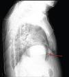
A 84-year-old woman was referred to our emergency department from another hospital with acute dyspnea at rest and hypoxemia. On admission a peripheral venous catheter had been inserted for fluid replacement and she had undergone computed tomography pulmonary angiography (CTPA) for suspected pulmonary embolism. Soon after the exam the patient became confused and drowsy and her respiratory failure worsened significantly without hemodynamic instability. CTPA confirmed the presence of bilateral central and peripheral thrombus and a large amount of air in the brachiocephalic veins, pulmonary artery (Figure 1) and right ventricle (Figures 2 and 3). She was considered to have VAE unrelated to the administration of contrast, given a topogram image that suggested the presence of air in the right ventricle before contrast injection (Figure 4). It was therefore assumed that the VAE resulted from the placement or manipulation of the peripheral venous catheter previously inserted in another institution. The patient was immediately given normal saline, a Hudson mask was applied and she was placed in left lateral decubitus position (Durant's maneuver) and in the Trendelenburg position, to keep the right ventricular outflow tract lower than the right ventricular cavity causing the air to migrate upwards, minimizing the likelihood of embolism. Her clinical status improved rapidly and she recovered without sequelae. Bedside transthoracic echocardiography showed no acute ventricular dilation or dysfunction, and the patient was transferred to another hospital for hyperbaric oxygen therapy.

Figure 1.

Air in the brachiocephalic veins and pulmonary artery (blue arrow).

(0.08MB).
Figure 2.

Air in the right ventricle, coronal view (green arrow).

(0.07MB).
Figure 3.

Air in the right ventricle, sagittal view (green arrow).

(0.08MB).
Figure 4.

Topogram image suggesting the presence of air in the right ventricle before contrast injection (red arrow).

(0.12MB).

The patient returned to our hospital after two days, and was discharged home three days later.

Ethical disclosuresProtection of human and animal subjects

The authors declare that no experiments were performed on humans or animals for this study.

Confidentiality of data

The authors declare that they have followed the protocols of their work center on the publication of patient data and that all the patients included in the study received sufficient information and gave their written informed consent to participate in the study.

Right to privacy and informed consent

The authors have obtained the written informed consent of the patients or subjects mentioned in the article. The corresponding author is in possession of this document.

Conflicts of interest

The authors have no conflicts of interest to declare.

References
[1]
P.G. Jorens, E. Van Marck, A. Snoeckx, et al.
Nonthrombotic pulmonary embolism.
Eur Respir J, 34 (2009), pp. 452-474
[2]
S.L. Orebaugh.
Venous air embolism: clinical and experimental considerations.
Crit Care Med, 20 (1992), pp. 1169-1177
Copyright © 2013. Sociedade Portuguesa de Cardiologia
Baixar PDF
Idiomas
Revista Portuguesa de Cardiologia
Opções de artigo
Ferramentas
en pt

Are you a health professional able to prescribe or dispense drugs?

Você é um profissional de saúde habilitado a prescrever ou dispensar medicamentos

Ao assinalar que é «Profissional de Saúde», declara conhecer e aceitar que a responsável pelo tratamento dos dados pessoais dos utilizadores da página de internet da Revista Portuguesa de Cardiologia (RPC), é esta entidade, com sede no Campo Grande, n.º 28, 13.º, 1700-093 Lisboa, com os telefones 217 970 685 e 217 817 630, fax 217 931 095 e com o endereço de correio eletrónico revista@spc.pt. Declaro para todos os fins, que assumo inteira responsabilidade pela veracidade e exatidão da afirmação aqui fornecida.