Cardiac metastases are more common than primary tumors. Several types of malignant tumors have been reported to metastasize to the heart, mainly lung cancer, but in the setting of esophageal cancer, myocardial metastasis is comparatively rare. We report a case of a cardiac metastasis from esophageal squamous cell carcinoma detected 9 months after surgically curative esophagectomy, which presented mimicking acute myocardial infarction. The use of different imaging modalities was fundamental to a correct diagnosis considering the challenging presentation.
As metástases cardíacas são mais frequentes que os tumores primário. Tem sido descrito que diversos tipos de neoplasias apresentam capacidade de envolvimento do coração, particularmente os carcinomas do pulmão. No caso do carcinoma do esófago, a metastização miocárdica é comparativamente mais rara. Os autores descrevem o caso de um doente com uma metástase cardíaca proveniente de um carcinoma epidermóide do esófago diagnosticada 9 meses após esofagectomia com intuito curativo e que mimetizou um enfarte agudo do miocárdio. Face a esta forma de apresentação tão invulgar, o contributo de diferentes modalidades de imagem foi fundamental para o diagnóstico final.
Metastatic cardiac involvement, much more frequent than primary tumors of the heart,1 occurs in up to 18% of all patients with malignancies.2 Direct extension or regional lymphatic invasion is the most frequent route for invading the heart, while metastasis to the myocardium is less common.2 The clinical presentation of these cardiac lesions can vary, including heart failure symptoms or pericardial effusion. Nonetheless, they can evolve silently and be discovered only at autopsy. Since most cardiac metastases appear in patients in advanced stages of the disease, prognosis is poor and therapeutic options limited. The authors report a case of a cardiac metastasis from esophageal squamous cell carcinoma which presented mimicking acute myocardial infarction. The use of different imaging modalities was fundamental to a correct diagnosis considering the challenging presentation.
Case reportA 81-year-old Caucasian man, with a history of esophageal squamous cell carcinoma diagnosed 9 months earlier treated by total esophagectomy (stage pT3, N0, Mx, R0), was admitted to our institution due to acute stroke. He had no known previous cardiovascular risk factors. The day after admission, he complained of chest discomfort and an electrocardiogram (ECG) was performed, revealing ST-segment elevation in leads V2–V4. Due to the suspicion of acute myocardial infarction (AMI), a coronary angiogram was carried out. No significant coronary artery lesions were found (Figure 1).
Transthoracic echocardiography revealed a large echogenic mass (66mm×60mm), involving the ventricular septum and protruding anteriorly and to the right (Fig. 2). Biventricular systolic function and inflow pattern were normal.
Two-dimensional transthoracic echocardiogram (top: parasternal long-axis view; middle: apical four-chamber view; bottom: subcostal view) showing involvement of the ventricular septum by the mass, which also protrudes into the right ventricular chamber. Maximum dimensions of 60mm×66mm assessed in subcostal view.
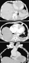
For a better characterization of the cardiac mass, a computed tomography (CT) scan was performed, further confirming its large extension. The inferior and apical halves of the right ventricle were involved, invasion of the ventricular septum and pericardium was apparent, and it also surrounded the left anterior descending artery (LAD) (Fig. 3, arrow).
Since the mass was in close contact with the anterior thoracic wall, a CT-guided needle aspiration biopsy was carried out (Fig. 4), which showed large epithelial cells, arranged in nests, with irregular, pleomorphic, central nuclei with dense cytoplasm, and keratin pearls, secondary to cardiac metastasis from the former epidermoid esophageal cancer.
During hospitalization, the patient's clinical status gradually worsened and he ultimately died of pneumonia.
DiscussionMetastases to the heart from tumors in other organs are much more common than primary cardiac tumors.1 A high proportion of malignant tumors have been described as metastasizing to the heart, with melanoma and bronchogenic and mammary carcinoma being reported as having the highest risk. When specifically sought, the incidence of cardiac metastases ranges from 2.3% to 18.3%, according to the imaging technique used and the type of primary tumor.2 Myocardial metastasis from esophageal cancer is comparatively rare.3 A study analyzing autopsy findings for 111 cases of esophageal cancer4 reported that tumor spread to the pericardium was observed in 13% of cases but myocardial metastasis was uncommon. Direct invasion and hematogenous and lymphatic spread are the most frequent pathways for tumors to invade the heart.2 Direct invasion is usual with esophageal cancer, and so solitary hematopoietic cardiac metastasis is rare.3 In our patient, hematogenous spread through vessel invasion was a possible route for cardiac involvement, since the tumor was located on the ventricular septum and protruded into the right ventricular cavity. Pericardium invasion may be explained by direct extension of the mass but, because of CT evidence of enlarged cervical and mediastinal lymph nodes, lymphatic spread cannot be completely ruled out.
The most frequent signs and symptoms of cardiac metastasis are congestive heart failure, arrhythmias, electrocardiographic abnormalities and pericardial effusion, but lesions can also be silent and death can occur suddenly.5 This case report highlights an uncommon presentation, with chest pain and ECG changes being misdiagnosed as AMI. The proximity of the cardiac mass to the LAD coronary artery might explain the ST-segment elevation in the anterior leads. Given the non-specific and diverse nature of clinical manifestations, a high level of diagnostic suspicion is needed. Early recognition of cardiac involvement could have important therapeutic and prognostic implications. Different imaging modalities are currently used for the detection and anatomic characterization of cardiac masses. Echocardiography has been the most commonly used and readily available noninvasive imaging technique, enabling clarification of the location, size, shape, number and mobility of masses. Transesophageal echocardiography may be more informative than the transthoracic approach, overcoming the drawback of poor acoustic window quality due to unfavorable patient body habitus. Since some masses, such as myxomas, show specific echogenic properties, they can be identified with a high degree of accuracy. However, echocardiography is usually limited in tissue characterization, and therefore other imaging methods are needed for further assessment. Magnetic resonance imaging (MRI) is presently considered the modality of choice to evaluate cardiac tumors, offering improved resolution, a larger field of view and superior soft-tissue contrast compared with echocardiography. In addition, it is noninvasive and does not require ionizing radiation or exposure to nephrotoxic contrast agents. T1-weighted, T2-weighted, and gadolinium-enhanced sequences are used for anatomic definition and tissue characterization, while cine gradient-echo imaging is used to assess functional behavior.6,7 Hence, it may noninvasively help in determining the etiology of the cardiac mass. However, its inability to evaluate calcification constitutes an important limitation of MRI. Computed tomography (CT), more specifically multidetector CT (MDCT), has been increasingly used for cardiac imaging. MDCT is particularly useful for the evaluation of calcification and fat content of a cardiac mass.8
Since cardiac involvement is usually detected in the context of widespread metastases in the terminal stage of cancer, outcome is poor in the majority of cases. The main aims of surgical resection, chemotherapy and radiotherapy are to relieve life-threatening symptoms and prevent sudden death; they have no curative purpose.3
ConclusionsIn conclusion, this case represents a rare presentation of a cardiac metastasis from esophageal squamous cell carcinoma surgically removed nine months before. Even though the frequency of myocardial metastasis is reportedly low, we should be aware that they may exist to enable early recognition and adopt the most suitable approach.
Conflicts of interestThe authors have no conflicts of interest to declare.


