Informação da revista
Vol. 31. Núm. 5.
Páginas 401-402 (maio 2012)
Partilhar
Partilhar
Baixar PDF
Mais opções do artigo
Vol. 31. Núm. 5.
Páginas 401-402 (maio 2012)
Image in cardiology
Open Access
Usefulness of cardiac MRI in the early diagnosis of endomyocardial fibrosis
Utilidade da ressonância magnética cardíaca no diagnóstico precoce da endomiocardiofibrose
Visitas
10004
Diego Leóna,
Autor para correspondência
diegoleon2000@gmail.com

Corresponding author.
, María Martína, Cecilia Corrosa, Elena Santamartab, Serafín Costillab, José L. Lamberta
a Cardiology Department, Hospital Universitario Central de Asturias, Oviedo, Spain
b Radiology Department, Hospital Universitario Central de Asturias, Oviedo, Spain
Este item recebeu

Under a Creative Commons license
Informação do artigo
Texto Completo
Baixar PDF
Estatísticas
Figuras (1)
Texto Completo

Endomyocardial fibrosis (EF) is characterized by marked endocardial thickening of the apex and subvalvular apparatus of one or both ventricles. It is considered a primary and chronic form of restrictive cardiomyopathy (the acute form is Löffler's disease) and is associated with inflammation and hypereosinophilia secondary to chronic processes such as parasite infection or autoimmune disease.

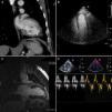
We describe the case of a 38-year-old Guinean man who was admitted to the Internal Medicine department for left upper quadrant pain. He had a previous history of malaria. Physical exam revealed a 4-cm hepatomegaly with tenderness in the epigastrium and hypochondrium without signs of heart failure. The hemogram showed marked eosinophilia (29500/mm3) and loa loa microfilariae were identified in the blood circulation. Thoraco-abdominal CT confirmed hepato- and splenomegaly and showed mild calcification in the left ventricular apex (Figure 1A, arrow). Given the suspicion of EF, the patient was referred for echocardiographic assessment (Figure 1B). Contrast echo revealed no suspicion of restrictive cardiomyopathy, the left ventricle was normal in size and systolic and diastolic function were both normal, with normal filling pressures (Figure 1C). Finally, on cardiac MRI subendocardial late gadolinium hyperenhancement was observed in the left ventricular apex (Figure 1D, arrow), reflecting myocardial fibrosis. The patient's clinical context supports the diagnosis of an initial stage of EF. Cardiac MRI is a very useful tool for the diagnosis of EF as it enables characterization and quantification of fibrous tissue, which has prognostic value.

Figure 1.

Imaging studies performed to establish diagnosis of endomyocardial fibrosis: Thoraco-abdominal CT (A), transthoratic contrast echo (B, C), and cardiac MRI (D).

(0.27MB).
Conflicts of interest

The authors have no conflicts of interest to declare.

Copyright © 2011. Sociedade Portuguesa de Cardiologia
Baixar PDF
Idiomas
Revista Portuguesa de Cardiologia
Opções de artigo
Ferramentas
en pt

Are you a health professional able to prescribe or dispense drugs?

Você é um profissional de saúde habilitado a prescrever ou dispensar medicamentos

Ao assinalar que é «Profissional de Saúde», declara conhecer e aceitar que a responsável pelo tratamento dos dados pessoais dos utilizadores da página de internet da Revista Portuguesa de Cardiologia (RPC), é esta entidade, com sede no Campo Grande, n.º 28, 13.º, 1700-093 Lisboa, com os telefones 217 970 685 e 217 817 630, fax 217 931 095 e com o endereço de correio eletrónico revista@spc.pt. Declaro para todos os fins, que assumo inteira responsabilidade pela veracidade e exatidão da afirmação aqui fornecida.