Informação da revista
Vol. 31. Núm. 6.
Páginas 465 (Junho 2012)
Partilhar
Partilhar
Baixar PDF
Mais opções do artigo
Vol. 31. Núm. 6.
Páginas 465 (Junho 2012)
Image in Cardiology
Open Access
Pulmonary dissection during diagnostic pulmonary angiography
Dissecção pulmonar durante uma angiografia pulmonar diagnóstica
Visitas
6098
Rui Baptistaa,b,
Autor para correspondência
ruibaptista@gmail.com

Corresponding author.
, Graça Castroa, António Marinho da Silvaa, Luís A. Providênciaa,b
a Serviço de Cardiologia, Hospitais da Universidade de Coimbra - Centro Hospitalar e Universitário de Coimbra, Coimbra, Portugal
b Faculdade de Medicina da Universidade de Coimbra, Coimbra, Portugal
Este item recebeu

Under a Creative Commons license
Informação do artigo
Texto Completo
Baixar PDF
Estatísticas
Figuras (1)
Material adicional (1)
Texto Completo

A 50-year-old woman with a history of deep venous thrombosis presented to our clinic for evaluation of pulmonary hypertension. On examination, she had a strong pulmonary component of the second heart sound and a grade 2/6 systolic murmur on the left sternal border. Echocardiography revealed severe right ventricular dilatation, with mildly depressed function and severe tricuspid regurgitation. Estimated pulmonary artery systolic pressure was 52mmHg. Contrast-enhanced chest computed tomography suggested the presence of thrombi in the pulmonary vasculature.

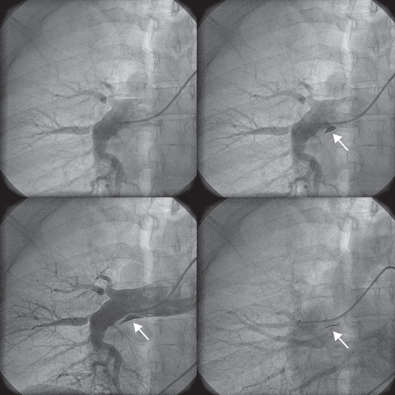
Right heart catheterization through the right femoral vein was then performed to confirm the clinical suspicion of chronic thromboembolic pulmonary hypertension and to assess operability. Access to the proximal segment of the right pulmonary artery was obtained using a 5-French NIH catheter. A 40-ml bolus of contrast material was then injected into the right pulmonary artery at a rate of 15ml/s (Supplementary Video 1). A small amount of subintimal opacification was seen after clearance of the contrast, a finding suggestive of iatrogenic pulmonary artery dissection (Fig. 1). This complication is thought to be due to fluctuations in the catheter tip due to respiration, causing high-pressure injection of contrast against a weakened pulmonary vessel wall. The patient remained completely asymptomatic and hemodynamically stable. No therapeutic measures were taken. Contrast-enhanced chest computed tomography performed the next day showed no evidence of pulmonary dissection. The patient is now on the waiting list for pulmonary endarterectomy.

Figure 1.

Subintimal opacification (arrow) after contrast clearance in the right pulmonary artery, indicating a small dissection. The pulmonary arteriography is suggestive of chronic thromboembolic disease.

(0,25MB).
Conflicts of interest

The authors have no conflicts of interest to declare.

Appendix A
Supplementary data

The following are the supplementary data to this article:

(4,75MB)

Copyright © 2011. Sociedade Portuguesa de Cardiologia
Idiomas
Revista Portuguesa de Cardiologia
Opções de artigo
Ferramentas
Material Suplementar
en pt

Are you a health professional able to prescribe or dispense drugs?

Você é um profissional de saúde habilitado a prescrever ou dispensar medicamentos

Ao assinalar que é «Profissional de Saúde», declara conhecer e aceitar que a responsável pelo tratamento dos dados pessoais dos utilizadores da página de internet da Revista Portuguesa de Cardiologia (RPC), é esta entidade, com sede no Campo Grande, n.º 28, 13.º, 1700-093 Lisboa, com os telefones 217 970 685 e 217 817 630, fax 217 931 095 e com o endereço de correio eletrónico revista@spc.pt. Declaro para todos os fins, que assumo inteira responsabilidade pela veracidade e exatidão da afirmação aqui fornecida.