Informação da revista
Vol. 31. Núm. 6.
Páginas 463-464 (Junho 2012)
Partilhar
Partilhar
Baixar PDF
Mais opções do artigo
Vol. 31. Núm. 6.
Páginas 463-464 (Junho 2012)
Images in cardiology
Open Access
Hemoptysis as the presenting symptom in cardiac sarcoma
Hemoptise como o sintoma de apresentação em sarcoma cardíaco
Visitas
7481
Itziar Solla-Ruiza,
Autor para correspondência
itziarsolla@yahoo.es

Corresponding author.
, Iñaki Villanueva-Benitoa, M. Begoña Iglesias-Rodriguezb, Vanesa Asorey-Veigac, Subhi R. Yasc, Miguel A. Pereira-Loureirod
a Cardiology Department, Meixoeiro Hospital, Vigo (Pontevedra), Spain
b Pathology Department, Meixoeiro Hospital, Vigo (Pontevedra), Spain
c Cardiac Surgery Department, Meixoeiro Hospital, Vigo (Pontevedra), Spain
d Anesthesiology and Intensive Care Department, Meixoeiro Hospital, Vigo (Pontevedra), Spain
Este item recebeu

Under a Creative Commons license
Informação do artigo
Texto Completo
Baixar PDF
Estatísticas
Figuras (4)
Mostrar maisMostrar menos
Material adicional (4)
Texto Completo

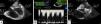
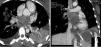
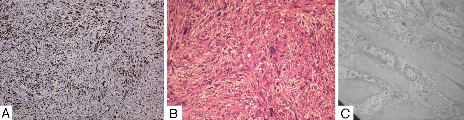
A healthy 25-year-old woman was admitted to the emergency department with a three-week history of gradual dyspnea on exertion, asthenia and weight loss. One week before admission, her dyspnea had worsened and had also presented at rest, with cough and hemoptysis, so she decided to seek medical attention. Physical examination revealed a mid-diastolic cardiac murmur. The transthoracic echocardiogram showed a large mass with irregular echogenicity filling the whole of the left atrium. Transesophageal echocardiography was performed and revealed the mass in the left atrium prolapsing into the left ventricle during diastole and causing mitral valve obstruction (Fig. 1A; Online Videos A–C). Continuous-wave Doppler ultrasound indicated severe mitral valve obstruction (mean gradient of 16mmHg) (Fig. 1B). The study also revealed extension into all the pulmonary veins (Fig. 1C). A computed tomogram of the thorax was performed and confirmed the diagnosis (Fig. 2; Online Video D). Cardiac surgery was performed with complete tumor resection (Fig. 3). The histopathological examination with immunohistochemical stain (Fig. 4A), hematoxylin–eosin stain (Fig. 4B) and electron microscopy (Fig. 4C) showed an undifferentiated pleomorphic sarcoma (pleomorphic malignant fibrous histiocytoma). The patient started chemotherapy and was discharged 17 days after surgery. Unfortunately, twenty days after discharge, she was readmitted to the hospital because of headache. Cranial computed tomography revealed multiple intracranial lesions suggestive of metastases. She underwent 10 sessions of holocranial radiotherapy. Six months after surgery she is alive but multiorgan metastatic disease has been demonstrated.

Figure 1.

(A) Transesophageal echocardiogram 4-chamber view: a large tumor with irregular echogenicity filling the whole of the left atrium. (B) Transesophageal echocardiogram: mitral valve obstruction. (C) Transesophageal echocardiogram focusing on the left atrium (53°): mass in the left atrium extending into the pulmonary veins.

(0,13MB).
Figure 2.

Cardiac computed tomography (CT): CT images showing the tumor occupying almost the entire left atrial chamber and infiltrating the pulmonary veins.

(0,14MB).
Figure 3.

Cardiac surgery and surgical specimen.

(0,16MB).
Figure 4.

(A) Immunohistochemical stain. (B) Hematoxylin–eosin stain. (C) Electron microscopy.

(0,24MB).
Conflicts of interest

The authors have no conflicts of interest to declare.

Appendix A
Supplementary data

The following are the supplementary data to this article:

(10,88MB)

Video A–C Transesophageal echocardiogram: multiple views of the left atrial mass prolapsing into the left ventricular chamber through the mitral valve and causing severe mitral obstruction.

(11,79MB)
(11,78MB)
(3,74MB)

Video D Cardiac computed tomography (CT): CT axial view showing the tumor occupying almost the entire left atrial chamber and extending into the pulmonary veins.

Copyright © 2012. Sociedade Portuguesa de Cardiologia
Idiomas
Revista Portuguesa de Cardiologia
Opções de artigo
Ferramentas
Material Suplementar
en pt

Are you a health professional able to prescribe or dispense drugs?

Você é um profissional de saúde habilitado a prescrever ou dispensar medicamentos

Ao assinalar que é «Profissional de Saúde», declara conhecer e aceitar que a responsável pelo tratamento dos dados pessoais dos utilizadores da página de internet da Revista Portuguesa de Cardiologia (RPC), é esta entidade, com sede no Campo Grande, n.º 28, 13.º, 1700-093 Lisboa, com os telefones 217 970 685 e 217 817 630, fax 217 931 095 e com o endereço de correio eletrónico revista@spc.pt. Declaro para todos os fins, que assumo inteira responsabilidade pela veracidade e exatidão da afirmação aqui fornecida.