


An 82-year-old woman, with a previous mechanical mitral valve (MMV) replacement for rheumatic mitral stenosis, presented with symptomatic severe aortic stenosis. The transthoracic echocardiogram (TTE) showed an aortic valve mean gradient of 57 mmHg, aortic valve area of 0.5 cm2 and left ventricular ejection fraction (LVEF) of 48%. The patient had a high operative risk (Euroscore II: 24%), suitable femoral access, an aortic valve calcium score of 2700 Agatston units, and a mean annulus diameter of 21.5 mm. The heart team opted for replacement of the damaged valve. The new valve was then placed inside the diseased valve, and transcatheter aortic valve replacement (TAVI) was performed using a self-expanding, recapturable Corevalve Evolut Pro 26 mm.
On the first attempt, the TAVI was placed in high position to avoid interference with MMV, but this attempt was aborted due to several “pop out” events. The TAVI was then repositioned lower and in this final implantation position there was underexpansion of the proximal strut, but no interference with MMV or with the TAVI leaflets. Post-dilatation was not performed due to potential risk of disruption of the MMV.
The post-procedure TTE showed a moderate paravalvular leak, with normal transvalvular gradients across the aortic and mitral valves (mean gradients of 9 mmHg and 4 mmHg, respectively).
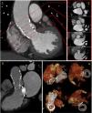
A post-procedural cardiac computed tomography to assess prosthesis morphology and the residual regurgitation mechanism was performed1 (Panel 1).
At one-year follow-up, the patient was stable, with no congestion, no hemolysis, improved LVEF (>55%), a mild paravalvular leak, and normal transvalvular gradients across both the aortic and mitral valves (mean gradients of 7 mmHg and 3 mmHg, respectively).
Transcatheter aortic valve replacement in MMV patients requires careful device selection and precise positioning due to the balance between avoiding MMV interference with low implantation and minimizing embolization risk from high positioning in mildly calcified valves.2 For this patient we considered this position the best possible result (Figure 1).

Post-procedural cardiac CT to access prosthesis morphology: modified coronal plane (1), short-axis (2), long-axis 3-chamber view (3) and volume rendering depicting the relation of TAVI prosthesis with the MMV with the restriction in expansion (elliptical shape) in the distal struts (4). This deformation is below the plane of the valve (CoraValve has a supraannular design).
We obtained informed consent from the patient to publish this case report, as well as any images associated with the case. The patient verbally consented to the publication of her medical case in a peer-reviewed medical journal. The authors of this article (J.C.P. and A.R.B.) obtained written informed consent from the patient, in accordance with COPE guidelines. The authors declare that the figures in the article do not enable patient identification. Dates were omitted to comply with confidentiality. This case report was exempt from ethics board approval.
FundingNone declared.
Conflict of interestNone declared.
