


A 63-year-old male smoker with a known history of hypertension was admitted to our hospital due to a two-month history of progressive dyspnea on exertion. He had initially presented two months previously with an acute episode of exertional chest pain. In that episode, the ECG was unremarkable and no ST elevation was detected; no further work-up was done at that time. Since that episode, the patient continued to have exertional chest pain with progressive dyspnea and weakness.
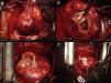
On admission to our hospital, ECG changes consistent with a previous posterolateral myocardial infarction were seen. Echocardiography showed a severely dysfunctional left ventricle with a large posterolateral wall aneurysm. Coronary angiography revealed a severely stenosed left anterior descending artery. The patient underwent emergent cardiac surgery which confirmed a large aneurysm in the posterolateral wall of the left ventricle (Figure 1A). During surgery, the endocardium and the papillary muscles of the mitral valve were seen through the neck of the aneurysm (Figure 1B). Surgical ventricular restoration and coronary revascularization were performed, and the left ventricular aneurysm was excluded using a pericardial bovine patch (Figure 1C). Subsequent reinforcement of ventricular tissues and sutures was carried out with Teflon bands (Figure 1D). A bypass of the left anterior descending coronary artery with the left internal mammary artery was performed for myocardial revascularization. The patient had an uneventful recovery and was discharged ten days after surgery.
(A) Surgical view of large aneurysm of the left ventricular posterolateral wall. (B) Neck of left ventricular aneurysm. The fibrous wall of the aneurysm, the endocardium and the papillary muscles of the mitral valve can be seen. (C) Exclusion of the aneurysm using a bovine pericardial patch sutured to the edges of the myocardium. (D) Occlusion and reinforcement of sutures with Teflon bands.
The authors declare that no experiments were performed on humans or animals for this study.
Confidentiality of dataThe authors declare that no patient data appear in this article.
Right to privacy and informed consentThe authors declare that no patient data appear in this article.
Conflicts of interestThe authors have no conflicts of interest to declare.
