


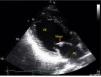
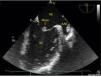

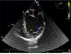
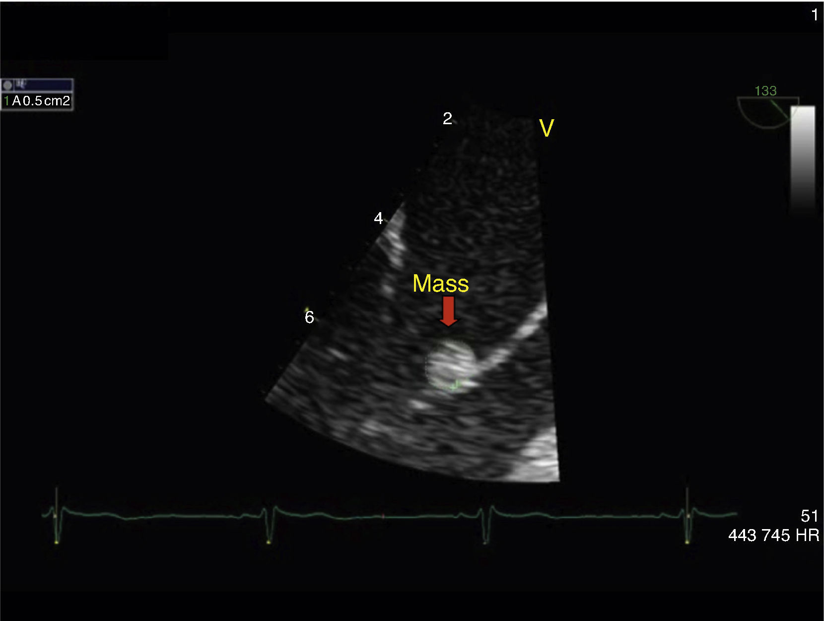
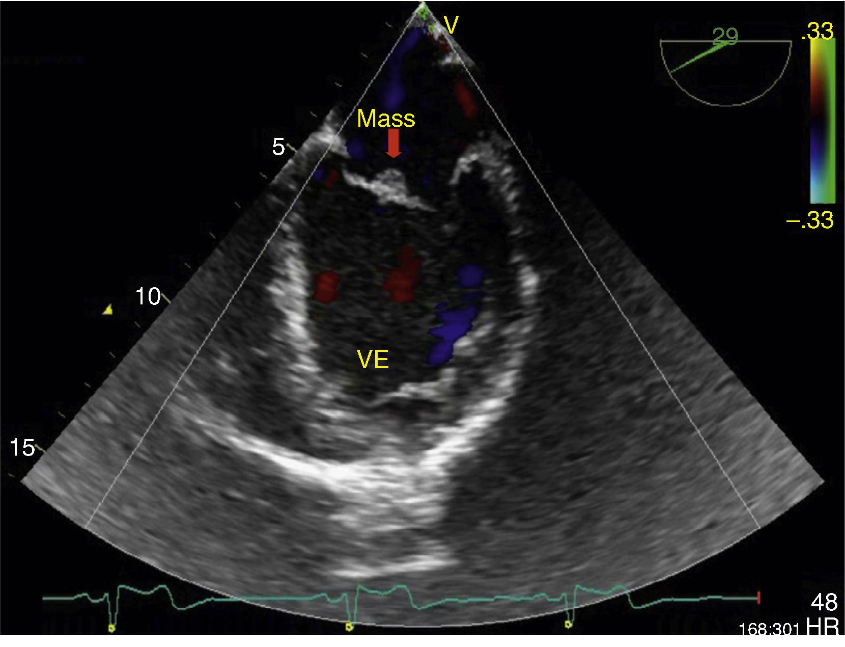
Primary cardiac tumors are rare in all age groups, with a reported prevalence of 0.001–0.03% in autopsy series. We report the case of a 37-year-old man, with no relevant medical history, who was admitted to the emergency department with sudden-onset right hemiparesis. Diagnostic studies revealed a small hypodense area in the left putamen and he was admitted to the stroke unit with a diagnosis of ischemic stroke. During investigation of the cause of stroke in such a young patient, he underwent transthoracic echocardiography, which showed a mass attached to the anterior leaflet of the mitral valve (Figure 1). Transesophageal echocardiography was then performed to clarify the picture, which confirmed a small echodense pedunculated mass, 0.5 cm2 in size, with well-defined borders, features typical of a fibroelastoma (Figures 2 and 3), attached to the atrial surface of the anterior mitral valve leaflet without causing mitral regurgitation (Figure 4). The rest of the echocardiographic examination revealed an estimated left ventricular ejection fraction of 65% and no other valvular abnormalities. The patient was subsequently referred to the cardiothoracic surgery center for surgical excision of the mass. Macroscopic examination showed a 0.6-cm whitish spiculated nodule with a soft elastic consistency. Histopathological study revealed cardiac myxoma. We present this case to highlight the unusual presentation, particularly its echocardiographic features.
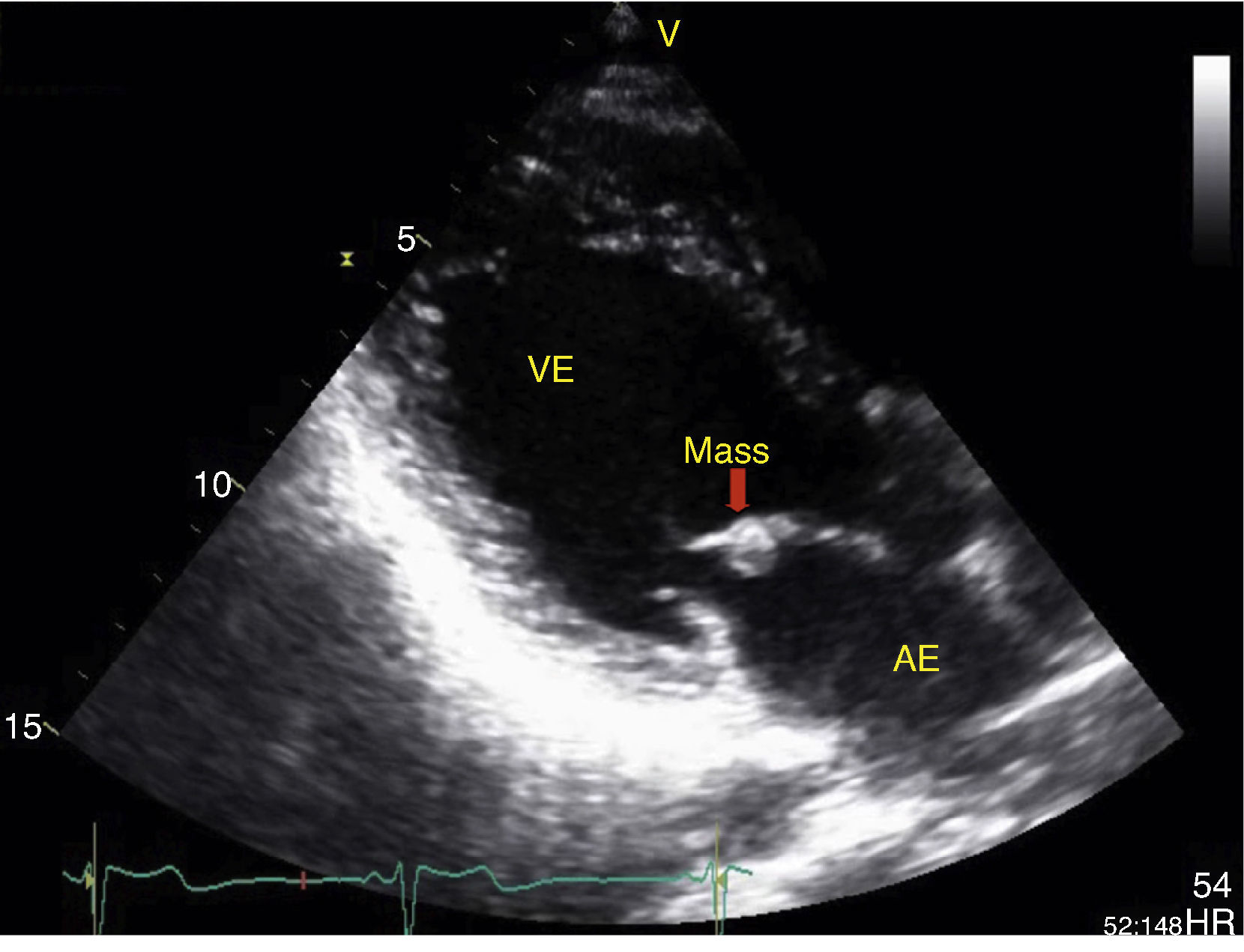
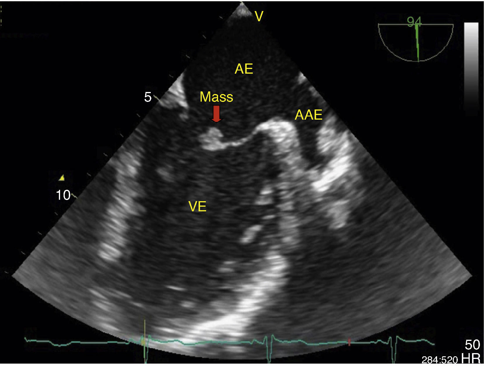
Transesophageal echocardiogram in long-axis view, showing an echodense pedunculated mass adhering to the atrial face of the anterior mitral valve leaflet. The left atrial appendage can also be seen, with no evidence of thrombus. AAE: left atrial appendage; AE: left atrium; VE: left ventricle.
The authors declare that the procedures followed were in accordance with the regulations of the relevant clinical research ethics committee and with those of the Code of Ethics of the World Medical Association (Declaration of Helsinki).
Confidentiality of dataThe authors declare that they have followed the protocols of their work center on the publication of patient data.
Right to privacy and informed consentThe authors have obtained the written informed consent of the patients or subjects mentioned in the article. The corresponding author is in possession of this document.
Conflicts of interestThe authors have no conflicts of interest to declare.
Please cite this article as: Ferreira R, Gonzaga A, Viana J, et al. Fibroma ou mixoma, eis a questão. Rev Port Cardiol. 2014;33:657–658.
