array:24 [
  "pii" => "S0870255112002624"
  "issn" => "08702551"
  "doi" => "10.1016/j.repc.2012.05.022"
  "estado" => "S300"
  "fechaPublicacion" => "2013-01-01"
  "aid" => "191"
  "copyright" => "Sociedade Portuguesa de Cardiologia"
  "copyrightAnyo" => "2012"
  "documento" => "article"
  "crossmark" => 0
  "licencia" => "http://creativecommons.org/licenses/by-nc-nd/4.0/"
  "subdocumento" => "sco"
  "cita" => "Rev Port Cardiol. 2013;32:69-70"
  "abierto" => array:3 [
    "ES" => true
    "ES2" => true
    "LATM" => true
  ]
  "gratuito" => true
  "lecturas" => array:2 [
    "total" => 4289
    "formatos" => array:3 [
      "EPUB" => 180
      "HTML" => 3202
      "PDF" => 907
    ]
  ]
  "itemSiguiente" => array:19 [
    "pii" => "S0870255112002600"
    "issn" => "08702551"
    "doi" => "10.1016/j.repc.2012.07.008"
    "estado" => "S300"
    "fechaPublicacion" => "2013-01-01"
    "aid" => "189"
    "copyright" => "Sociedade Portuguesa de Cardiologia"
    "documento" => "article"
    "crossmark" => 0
    "licencia" => "http://creativecommons.org/licenses/by-nc-nd/4.0/"
    "subdocumento" => "sco"
    "cita" => "Rev Port Cardiol. 2013;32:71-2"
    "abierto" => array:3 [
      "ES" => true
      "ES2" => true
      "LATM" => true
    ]
    "gratuito" => true
    "lecturas" => array:2 [
      "total" => 5754
      "formatos" => array:3 [
        "EPUB" => 155
        "HTML" => 4627
        "PDF" => 972
      ]
    ]
    "en" => array:11 [
      "idiomaDefecto" => true
      "cabecera" => "<span class="elsevierStyleTextfn">Images in cardiology</span>"
      "titulo" => "Pulmonary embolism with thromboembolus in transit"
      "tienePdf" => "en"
      "tieneTextoCompleto" => "en"
      "paginas" => array:1 [
        0 => array:2 [
          "paginaInicial" => "71"
          "paginaFinal" => "72"
        ]
      ]
      "titulosAlternativos" => array:1 [
        "pt" => array:1 [
          "titulo" => "Embolia pulmonar com trombo-&#234;mbolo em tr&#226;nsito"
        ]
      ]
      "contieneTextoCompleto" => array:1 [
        "en" => true
      ]
      "contienePdf" => array:1 [
        "en" => true
      ]
      "resumenGrafico" => array:2 [
        "original" => 0
        "multimedia" => array:7 [
          "identificador" => "fig0015"
          "etiqueta" => "Figure 3"
          "tipo" => "MULTIMEDIAFIGURA"
          "mostrarFloat" => true
          "mostrarDisplay" => false
          "figura" => array:1 [
            0 => array:4 [
              "imagen" => "gr3.jpeg"
              "Alto" => 424
              "Ancho" => 1265
              "Tamanyo" => 79180
            ]
          ]
          "descripcion" => array:1 [
            "en" => "<p id="spar0015" class="elsevierStyleSimplePara elsevierViewall">A central bilateral intraluminal filling defect &#40;arrows&#41; is seen&#44; consistent with pulmonary emboli &#40;A &#8211; transaxial view&#59; B and C &#8211; sagittal views&#41;&#46; A thromboembolus &#40;arrowhead&#41; is also seen in the right atrium &#40;C&#41;&#46; IVC&#58; inferior vena cava&#59; LV&#58; left ventricle&#59; RA&#58; right atrium&#59; RV&#58; right ventricle&#59; SVC&#58; superior vena cava&#46;</p>"
          ]
        ]
      ]
      "autores" => array:1 [
        0 => array:2 [
          "autoresLista" => "Miguel Silva Vieira, Diana Anjo, Nuno Antunes, Henrique Carvalho, Severo Torres"
          "autores" => array:5 [
            0 => array:2 [
              "nombre" => "Miguel"
              "apellidos" => "Silva Vieira"
            ]
            1 => array:2 [
              "nombre" => "Diana"
              "apellidos" => "Anjo"
            ]
            2 => array:2 [
              "nombre" => "Nuno"
              "apellidos" => "Antunes"
            ]
            3 => array:2 [
              "nombre" => "Henrique"
              "apellidos" => "Carvalho"
            ]
            4 => array:2 [
              "nombre" => "Severo"
              "apellidos" => "Torres"
            ]
          ]
        ]
      ]
    ]
    "idiomaDefecto" => "en"
    "EPUB" => "https://multimedia.elsevier.es/PublicationsMultimediaV1/item/epub/S0870255112002600?idApp=UINPBA00004E"
    "url" => "/08702551/0000003200000001/v1_201308021254/S0870255112002600/v1_201308021254/en/main.assets"
  ]
  "itemAnterior" => array:20 [
    "pii" => "S0870255112002533"
    "issn" => "08702551"
    "doi" => "10.1016/j.repc.2012.05.015"
    "estado" => "S300"
    "fechaPublicacion" => "2013-01-01"
    "aid" => "182"
    "copyright" => "Sociedade Portuguesa de Cardiologia"
    "documento" => "article"
    "crossmark" => 0
    "licencia" => "http://creativecommons.org/licenses/by-nc-nd/4.0/"
    "subdocumento" => "sco"
    "cita" => "Rev Port Cardiol. 2013;32:67-8"
    "abierto" => array:3 [
      "ES" => true
      "ES2" => true
      "LATM" => true
    ]
    "gratuito" => true
    "lecturas" => array:2 [
      "total" => 8076
      "formatos" => array:3 [
        "EPUB" => 179
        "HTML" => 6233
        "PDF" => 1664
      ]
    ]
    "pt" => array:11 [
      "idiomaDefecto" => true
      "cabecera" => "<span class="elsevierStyleTextfn">Imagem em Cardiologia</span>"
      "titulo" => "Retorno venoso pulmonar an&#243;malo parcial"
      "tienePdf" => "pt"
      "tieneTextoCompleto" => "pt"
      "paginas" => array:1 [
        0 => array:2 [
          "paginaInicial" => "67"
          "paginaFinal" => "68"
        ]
      ]
      "titulosAlternativos" => array:1 [
        "en" => array:1 [
          "titulo" => "Partial anomalous pulmonary venous return"
        ]
      ]
      "contieneTextoCompleto" => array:1 [
        "pt" => true
      ]
      "contienePdf" => array:1 [
        "pt" => true
      ]
      "resumenGrafico" => array:2 [
        "original" => 0
        "multimedia" => array:7 [
          "identificador" => "fig0005"
          "etiqueta" => "Figura 1"
          "tipo" => "MULTIMEDIAFIGURA"
          "mostrarFloat" => true
          "mostrarDisplay" => false
          "figura" => array:1 [
            0 => array:4 [
              "imagen" => "gr1.jpeg"
              "Alto" => 1078
              "Ancho" => 850
              "Tamanyo" => 103936
            ]
          ]
          "descripcion" => array:1 [
            "pt" => "<p id="spar0005" class="elsevierStyleSimplePara elsevierViewall">A&#46; Plano axial &#40;b-SSFP&#41;&#58; seta azul&#58; veia cava superior&#59; seta amarela&#58; veia inominada&#59; seta branca&#58; veia pulmonar an&#243;mala vertical&#46; B&#46; Plano axial &#40;b-SSFP&#41;&#59; seta azul&#58; veia cava superior&#59; seta vermelha&#58; arco a&#243;rtico&#59; seta branca&#58; veia pulmonar an&#243;mala vertical&#46;</p>"
          ]
        ]
      ]
      "autores" => array:1 [
        0 => array:2 [
          "autoresLista" => "Ana Rita Almeida, Lu&#237;s Lopes, Jo&#227;o Palas, Rui Lopes, Cec&#237;lia Bagulho, H&#233;lder Pereira"
          "autores" => array:6 [
            0 => array:2 [
              "nombre" => "Ana Rita"
              "apellidos" => "Almeida"
            ]
            1 => array:2 [
              "nombre" => "Lu&#237;s"
              "apellidos" => "Lopes"
            ]
            2 => array:2 [
              "nombre" => "Jo&#227;o"
              "apellidos" => "Palas"
            ]
            3 => array:2 [
              "nombre" => "Rui"
              "apellidos" => "Lopes"
            ]
            4 => array:2 [
              "nombre" => "Cec&#237;lia"
              "apellidos" => "Bagulho"
            ]
            5 => array:2 [
              "nombre" => "H&#233;lder"
              "apellidos" => "Pereira"
            ]
          ]
        ]
      ]
    ]
    "idiomaDefecto" => "pt"
    "Traduccion" => array:1 [
      "en" => array:9 [
        "pii" => "S2174204912002036"
        "doi" => "10.1016/j.repce.2012.11.004"
        "estado" => "S300"
        "subdocumento" => ""
        "abierto" => array:3 [
          "ES" => true
          "ES2" => true
          "LATM" => true
        ]
        "gratuito" => true
        "lecturas" => array:1 [
          "total" => 0
        ]
        "idiomaDefecto" => "en"
        "EPUB" => "https://multimedia.elsevier.es/PublicationsMultimediaV1/item/epub/S2174204912002036?idApp=UINPBA00004E"
      ]
    ]
    "EPUB" => "https://multimedia.elsevier.es/PublicationsMultimediaV1/item/epub/S0870255112002533?idApp=UINPBA00004E"
    "url" => "/08702551/0000003200000001/v1_201308021254/S0870255112002533/v1_201308021254/pt/main.assets"
  ]
  "en" => array:15 [
    "idiomaDefecto" => true
    "cabecera" => "<span class="elsevierStyleTextfn">Image in cardiology</span>"
    "titulo" => "Chest stab wound&#8212;A rare cause of late ventricular tachycardia"
    "tieneTextoCompleto" => true
    "paginas" => array:1 [
      0 => array:2 [
        "paginaInicial" => "69"
        "paginaFinal" => "70"
      ]
    ]
    "autores" => array:1 [
      0 => array:4 [
        "autoresLista" => "S&#237;lvia Ribeiro, Nuno Salom&#233;, Teresa Pinho, Helena Gon&#231;alves, Jo&#227;o Primo, Pedro Azevedo, Adelino Correia"
        "autores" => array:7 [
          0 => array:4 [
            "nombre" => "S&#237;lvia"
            "apellidos" => "Ribeiro"
            "email" => array:1 [
              0 => "silviamartinsribeiro&#64;gmail&#46;com"
            ]
            "referencia" => array:2 [
              0 => array:2 [
                "etiqueta" => "<span class="elsevierStyleSup">a</span>"
                "identificador" => "aff0005"
              ]
              1 => array:2 [
                "etiqueta" => "<span class="elsevierStyleSup">¿</span>"
                "identificador" => "cor0005"
              ]
            ]
          ]
          1 => array:3 [
            "nombre" => "Nuno"
            "apellidos" => "Salom&#233;"
            "referencia" => array:1 [
              0 => array:2 [
                "etiqueta" => "<span class="elsevierStyleSup">a</span>"
                "identificador" => "aff0005"
              ]
            ]
          ]
          2 => array:3 [
            "nombre" => "Teresa"
            "apellidos" => "Pinho"
            "referencia" => array:1 [
              0 => array:2 [
                "etiqueta" => "<span class="elsevierStyleSup">b</span>"
                "identificador" => "aff0010"
              ]
            ]
          ]
          3 => array:3 [
            "nombre" => "Helena"
            "apellidos" => "Gon&#231;alves"
            "referencia" => array:1 [
              0 => array:2 [
                "etiqueta" => "<span class="elsevierStyleSup">c</span>"
                "identificador" => "aff0015"
              ]
            ]
          ]
          4 => array:3 [
            "nombre" => "Jo&#227;o"
            "apellidos" => "Primo"
            "referencia" => array:1 [
              0 => array:2 [
                "etiqueta" => "<span class="elsevierStyleSup">c</span>"
                "identificador" => "aff0015"
              ]
            ]
          ]
          5 => array:3 [
            "nombre" => "Pedro"
            "apellidos" => "Azevedo"
            "referencia" => array:1 [
              0 => array:2 [
                "etiqueta" => "<span class="elsevierStyleSup">a</span>"
                "identificador" => "aff0005"
              ]
            ]
          ]
          6 => array:3 [
            "nombre" => "Adelino"
            "apellidos" => "Correia"
            "referencia" => array:1 [
              0 => array:2 [
                "etiqueta" => "<span class="elsevierStyleSup">a</span>"
                "identificador" => "aff0005"
              ]
            ]
          ]
        ]
        "afiliaciones" => array:3 [
          0 => array:3 [
            "entidad" => "Servi&#231;o de Cardiologia&#44; Hospital de Braga&#44; Braga&#44; Portugal"
            "etiqueta" => "<span class="elsevierStyleSup">a</span>"
            "identificador" => "aff0005"
          ]
          1 => array:3 [
            "entidad" => "Servi&#231;o de Cardiologia&#44; Hospital de S&#227;o Jo&#227;o&#44; Porto&#44; Portugal"
            "etiqueta" => "<span class="elsevierStyleSup">b</span>"
            "identificador" => "aff0010"
          ]
          2 => array:3 [
            "entidad" => "Servi&#231;o de Cardiologia&#44; Centro Hospitalar de Vila Nova de Gaia&#47;Espinho&#44; Vila Nova de Gaia&#44; Portugal"
            "etiqueta" => "<span class="elsevierStyleSup">c</span>"
            "identificador" => "aff0015"
          ]
        ]
        "correspondencia" => array:1 [
          0 => array:3 [
            "identificador" => "cor0005"
            "etiqueta" => "&#8270;"
            "correspondencia" => "Corresponding author&#46;"
          ]
        ]
      ]
    ]
    "titulosAlternativos" => array:1 [
      "pt" => array:1 [
        "titulo" => "Les&#227;o mioc&#225;rdica por ferimento de arma branca &#8211; uma causa rara de taquicardia ventricular tardia"
      ]
    ]
    "resumenGrafico" => array:2 [
      "original" => 0
      "multimedia" => array:7 [
        "identificador" => "fig0005"
        "etiqueta" => "Figure 1"
        "tipo" => "MULTIMEDIAFIGURA"
        "mostrarFloat" => true
        "mostrarDisplay" => false
        "figura" => array:1 [
          0 => array:4 [
            "imagen" => "gr1.jpeg"
            "Alto" => 2680
            "Ancho" => 2671
            "Tamanyo" => 659208
          ]
        ]
        "descripcion" => array:1 [
          "en" => "<p id="spar0005" class="elsevierStyleSimplePara elsevierViewall">A&#41; Admission ECG demonstrating a monomorphic ventricular tachycardia&#59; B&#41; CMR images in short axis&#44; long axis and four-chambers views obtained after gadolinium injection demonstrating transmural gadolinium enhancement in mid-apical anterior and lateral segments&#59; C&#41; morphology of ventricular tachycardia during the electrophysiological study and left ventricular endocardial mapping&#46;</p>"
        ]
      ]
    ]
    "textoCompleto" => "<span class="elsevierStyleSections"><p id="par0005" class="elsevierStylePara elsevierViewall">A 67-year-old man with a history of systemic arterial hypertension and a chest stab trauma with emergent cardiothoracic surgery 38 years previously was admitted to our cardiology department for sustained monomorphic ventricular tachycardia&#46;</p><p id="par0010" class="elsevierStylePara elsevierViewall">At admission to the emergency department the patient complained of palpitations&#44; sweating and general malaise&#46; He was hemodynamically stable&#46; The admission electrocardiogram showed regular wide complex tachycardia with right bundle branch block pattern consistent with ventricular tachycardia &#40;<a class="elsevierStyleCrossRef" href="#fig0005">Figure 1</a>A&#41;&#46; Chemical cardioversion with amiodarone was successfully performed&#46; The baseline ECG demonstrated sinus rhythm with Q waves in DI&#44; aVL and V3&#8211;V6&#46; Laboratory tests&#44; including troponin&#44; were unremarkable&#46; The transthoracic echocardiogram revealed normal global left ventricular systolic function with anterolateral hypokinesia&#46; Coronary angiography excluded epicardial coronary disease&#46; Cardiac magnetic resonance imaging revealed transmural gadolinium enhancement in the mid-apical anterior and lateral segments consistent with the chest stab wound &#40;<a class="elsevierStyleCrossRef" href="#fig0005">Figure 1</a>B&#41;&#46; The patient was referred for electrophysiological study&#44; during which ventricular tachycardia was induced with the same pattern as the admission ECG &#40;<a class="elsevierStyleCrossRef" href="#fig0005">Figure 1</a>C&#41; but it was not possible to perform ablation because of a probable epicardial circuit and an implantable cardioverter-defibrillator was implanted&#46; The patient is currently asymptomatic&#46;</p><elsevierMultimedia ident="fig0005"></elsevierMultimedia><p id="par0015" class="elsevierStylePara elsevierViewall">Ventricular tachycardia is a life-threatening arrhythmia&#46; In the majority of cases it is associated with structural heart disease&#44; particularly coronary disease&#46; We describe an unusual case of a 67-year-old man who survived a chest stab wound and presented 38 years later with ventricular tachycardia due to the anterolateral transmural scar&#46;</p><span id="sec0005" class="elsevierStyleSection elsevierViewall"><span class="elsevierStyleSectionTitle">Ethical disclosures</span><span id="sec0010" class="elsevierStyleSection elsevierViewall"><span class="elsevierStyleSectionTitle">Protection of human and animal subjects</span><p id="par0020" class="elsevierStylePara elsevierViewall">The authors declare that no experiments were performed on humans or animals for this study&#46;</p></span><span id="sec0015" class="elsevierStyleSection elsevierViewall"><span class="elsevierStyleSectionTitle">Confidentiality of data</span><p id="par0025" class="elsevierStylePara elsevierViewall">The authors declare that no patient data appear in this article&#46;</p></span><span id="sec0020" class="elsevierStyleSection elsevierViewall"><span class="elsevierStyleSectionTitle">Right to privacy and informed consent</span><p id="par0030" class="elsevierStylePara elsevierViewall">The authors declare that no patient data appear in this article&#46;</p></span></span><span id="sec0025" class="elsevierStyleSection elsevierViewall"><span class="elsevierStyleSectionTitle">Conflicts of interest</span><p id="par0035" class="elsevierStylePara elsevierViewall">The authors have no conflicts of interest to declare&#46;</p></span></span>"
    "textoCompletoSecciones" => array:1 [
      "secciones" => array:2 [
        0 => array:3 [
          "identificador" => "sec0005"
          "titulo" => "Ethical disclosures"
          "secciones" => array:3 [
            0 => array:2 [
              "identificador" => "sec0010"
              "titulo" => "Protection of human and animal subjects"
            ]
            1 => array:2 [
              "identificador" => "sec0015"
              "titulo" => "Confidentiality of data"
            ]
            2 => array:2 [
              "identificador" => "sec0020"
              "titulo" => "Right to privacy and informed consent"
            ]
          ]
        ]
        1 => array:2 [
          "identificador" => "sec0025"
          "titulo" => "Conflicts of interest"
        ]
      ]
    ]
    "pdfFichero" => "main.pdf"
    "tienePdf" => true
    "fechaRecibido" => "2012-05-10"
    "fechaAceptado" => "2012-05-27"
    "multimedia" => array:1 [
      0 => array:7 [
        "identificador" => "fig0005"
        "etiqueta" => "Figure 1"
        "tipo" => "MULTIMEDIAFIGURA"
        "mostrarFloat" => true
        "mostrarDisplay" => false
        "figura" => array:1 [
          0 => array:4 [
            "imagen" => "gr1.jpeg"
            "Alto" => 2680
            "Ancho" => 2671
            "Tamanyo" => 659208
          ]
        ]
        "descripcion" => array:1 [
          "en" => "<p id="spar0005" class="elsevierStyleSimplePara elsevierViewall">A&#41; Admission ECG demonstrating a monomorphic ventricular tachycardia&#59; B&#41; CMR images in short axis&#44; long axis and four-chambers views obtained after gadolinium injection demonstrating transmural gadolinium enhancement in mid-apical anterior and lateral segments&#59; C&#41; morphology of ventricular tachycardia during the electrophysiological study and left ventricular endocardial mapping&#46;</p>"
        ]
      ]
    ]
  ]
  "idiomaDefecto" => "en"
  "url" => "/08702551/0000003200000001/v1_201308021254/S0870255112002624/v1_201308021254/en/main.assets"
  "Apartado" => array:4 [
    "identificador" => "357"
    "tipo" => "SECCION"
    "pt" => array:2 [
      "titulo" => "Imagens em cardiologia"
      "idiomaDefecto" => true
    ]
    "idiomaDefecto" => "pt"
  ]
  "PDF" => "https://static.elsevier.es/multimedia/08702551/0000003200000001/v1_201308021254/S0870255112002624/v1_201308021254/en/main.pdf?idApp=UINPBA00004E&text.app=https://revportcardiol.org/"
  "EPUB" => "https://multimedia.elsevier.es/PublicationsMultimediaV1/item/epub/S0870255112002624?idApp=UINPBA00004E"
]
Partilhar
Informação da revista
Vol. 32. Núm. 1.
Páginas 69-70 (janeiro 2013)
Partilhar
Partilhar
Baixar PDF
Mais opções do artigo
Vol. 32. Núm. 1.
Páginas 69-70 (janeiro 2013)
Image in cardiology
Open Access
Chest stab wound—A rare cause of late ventricular tachycardia
Lesão miocárdica por ferimento de arma branca – uma causa rara de taquicardia ventricular tardia
Visitas
7628
Sílvia Ribeiroa,
Autor para correspondência
silviamartinsribeiro@gmail.com

Corresponding author.
, Nuno Saloméa, Teresa Pinhob, Helena Gonçalvesc, João Primoc, Pedro Azevedoa, Adelino Correiaa
a Serviço de Cardiologia, Hospital de Braga, Braga, Portugal
b Serviço de Cardiologia, Hospital de São João, Porto, Portugal
c Serviço de Cardiologia, Centro Hospitalar de Vila Nova de Gaia/Espinho, Vila Nova de Gaia, Portugal
Este item recebeu

Under a Creative Commons license
Informação do artigo
Texto Completo
Baixar PDF
Estatísticas
Figuras (1)
Texto Completo

A 67-year-old man with a history of systemic arterial hypertension and a chest stab trauma with emergent cardiothoracic surgery 38 years previously was admitted to our cardiology department for sustained monomorphic ventricular tachycardia.

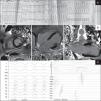
At admission to the emergency department the patient complained of palpitations, sweating and general malaise. He was hemodynamically stable. The admission electrocardiogram showed regular wide complex tachycardia with right bundle branch block pattern consistent with ventricular tachycardia (Figure 1A). Chemical cardioversion with amiodarone was successfully performed. The baseline ECG demonstrated sinus rhythm with Q waves in DI, aVL and V3–V6. Laboratory tests, including troponin, were unremarkable. The transthoracic echocardiogram revealed normal global left ventricular systolic function with anterolateral hypokinesia. Coronary angiography excluded epicardial coronary disease. Cardiac magnetic resonance imaging revealed transmural gadolinium enhancement in the mid-apical anterior and lateral segments consistent with the chest stab wound (Figure 1B). The patient was referred for electrophysiological study, during which ventricular tachycardia was induced with the same pattern as the admission ECG (Figure 1C) but it was not possible to perform ablation because of a probable epicardial circuit and an implantable cardioverter-defibrillator was implanted. The patient is currently asymptomatic.

Figure 1.

A) Admission ECG demonstrating a monomorphic ventricular tachycardia; B) CMR images in short axis, long axis and four-chambers views obtained after gadolinium injection demonstrating transmural gadolinium enhancement in mid-apical anterior and lateral segments; C) morphology of ventricular tachycardia during the electrophysiological study and left ventricular endocardial mapping.

(0.63MB).

Ventricular tachycardia is a life-threatening arrhythmia. In the majority of cases it is associated with structural heart disease, particularly coronary disease. We describe an unusual case of a 67-year-old man who survived a chest stab wound and presented 38 years later with ventricular tachycardia due to the anterolateral transmural scar.

Ethical disclosuresProtection of human and animal subjects

The authors declare that no experiments were performed on humans or animals for this study.

Confidentiality of data

The authors declare that no patient data appear in this article.

Right to privacy and informed consent

The authors declare that no patient data appear in this article.

Conflicts of interest

The authors have no conflicts of interest to declare.

Copyright © 2012. Sociedade Portuguesa de Cardiologia
Baixar PDF
Idiomas
Revista Portuguesa de Cardiologia
Opções de artigo
Ferramentas
en pt

Are you a health professional able to prescribe or dispense drugs?

Você é um profissional de saúde habilitado a prescrever ou dispensar medicamentos

Ao assinalar que é «Profissional de Saúde», declara conhecer e aceitar que a responsável pelo tratamento dos dados pessoais dos utilizadores da página de internet da Revista Portuguesa de Cardiologia (RPC), é esta entidade, com sede no Campo Grande, n.º 28, 13.º, 1700-093 Lisboa, com os telefones 217 970 685 e 217 817 630, fax 217 931 095 e com o endereço de correio eletrónico revista@spc.pt. Declaro para todos os fins, que assumo inteira responsabilidade pela veracidade e exatidão da afirmação aqui fornecida.