array:24 [
  "pii" => "S0870255117304985"
  "issn" => "08702551"
  "doi" => "10.1016/j.repc.2017.07.021"
  "estado" => "S300"
  "fechaPublicacion" => "2019-01-01"
  "aid" => "1255"
  "copyright" => "Sociedade Portuguesa de Cardiologia"
  "copyrightAnyo" => "2018"
  "documento" => "article"
  "crossmark" => 1
  "licencia" => "http://creativecommons.org/licenses/by-nc-nd/4.0/"
  "subdocumento" => "sco"
  "cita" => "Rev Port Cardiol. 2019;38:67-8"
  "abierto" => array:3 [
    "ES" => true
    "ES2" => true
    "LATM" => true
  ]
  "gratuito" => true
  "lecturas" => array:2 [
    "total" => 1348
    "formatos" => array:3 [
      "EPUB" => 105
      "HTML" => 896
      "PDF" => 347
    ]
  ]
  "itemSiguiente" => array:20 [
    "pii" => "S0870255118305171"
    "issn" => "08702551"
    "doi" => "10.1016/j.repc.2018.09.008"
    "estado" => "S300"
    "fechaPublicacion" => "2019-01-01"
    "aid" => "1316"
    "copyright" => "Sociedade Portuguesa de Cardiologia"
    "documento" => "simple-article"
    "crossmark" => 1
    "licencia" => "http://creativecommons.org/licenses/by-nc-nd/4.0/"
    "subdocumento" => "cor"
    "cita" => "Rev Port Cardiol. 2019;38:69"
    "abierto" => array:3 [
      "ES" => true
      "ES2" => true
      "LATM" => true
    ]
    "gratuito" => true
    "lecturas" => array:2 [
      "total" => 664
      "formatos" => array:3 [
        "EPUB" => 67
        "HTML" => 386
        "PDF" => 211
      ]
    ]
    "pt" => array:10 [
      "idiomaDefecto" => true
      "cabecera" => "<span class="elsevierStyleTextfn">Carta ao Editor</span>"
      "titulo" => "<span class="elsevierStyleItalic">Scores</span> de risco nas cirurgias card&#237;acas valvares"
      "tienePdf" => "pt"
      "tieneTextoCompleto" => "pt"
      "paginas" => array:1 [
        0 => array:1 [
          "paginaInicial" => "69"
        ]
      ]
      "titulosAlternativos" => array:1 [
        "en" => array:1 [
          "titulo" => "Risk scores in heart valve surgery"
        ]
      ]
      "contieneTextoCompleto" => array:1 [
        "pt" => true
      ]
      "contienePdf" => array:1 [
        "pt" => true
      ]
      "autores" => array:1 [
        0 => array:2 [
          "autoresLista" => "Filipa Waihrich de Oliveira, Adriane Marines dos Santos, Ana Cl&#225;udia Moraes Mena Barreto"
          "autores" => array:3 [
            0 => array:2 [
              "nombre" => "Filipa Waihrich"
              "apellidos" => "de Oliveira"
            ]
            1 => array:2 [
              "nombre" => "Adriane Marines"
              "apellidos" => "dos Santos"
            ]
            2 => array:2 [
              "nombre" => "Ana Cl&#225;udia Moraes Mena"
              "apellidos" => "Barreto"
            ]
          ]
        ]
      ]
    ]
    "idiomaDefecto" => "pt"
    "Traduccion" => array:1 [
      "en" => array:9 [
        "pii" => "S2174204919300030"
        "doi" => "10.1016/j.repce.2018.09.010"
        "estado" => "S300"
        "subdocumento" => ""
        "abierto" => array:3 [
          "ES" => true
          "ES2" => true
          "LATM" => true
        ]
        "gratuito" => true
        "lecturas" => array:1 [
          "total" => 0
        ]
        "idiomaDefecto" => "en"
        "EPUB" => "https://multimedia.elsevier.es/PublicationsMultimediaV1/item/epub/S2174204919300030?idApp=UINPBA00004E"
      ]
    ]
    "EPUB" => "https://multimedia.elsevier.es/PublicationsMultimediaV1/item/epub/S0870255118305171?idApp=UINPBA00004E"
    "url" => "/08702551/0000003800000001/v5_201911281009/S0870255118305171/v5_201911281009/pt/main.assets"
  ]
  "itemAnterior" => array:18 [
    "pii" => "S0870255116303511"
    "issn" => "08702551"
    "doi" => "10.1016/j.repc.2018.05.016"
    "estado" => "S300"
    "fechaPublicacion" => "2019-01-01"
    "aid" => "1335"
    "documento" => "simple-article"
    "crossmark" => 1
    "licencia" => "http://creativecommons.org/licenses/by-nc-nd/4.0/"
    "subdocumento" => "crp"
    "cita" => "Rev Port Cardiol. 2019;38:65&#46;e1-65&#46;e3"
    "abierto" => array:3 [
      "ES" => true
      "ES2" => true
      "LATM" => true
    ]
    "gratuito" => true
    "lecturas" => array:2 [
      "total" => 763
      "formatos" => array:3 [
        "EPUB" => 88
        "HTML" => 446
        "PDF" => 229
      ]
    ]
    "en" => array:13 [
      "idiomaDefecto" => true
      "cabecera" => "<span class="elsevierStyleTextfn">Case report</span>"
      "titulo" => "Superior vena cava syndrome early after pacemaker implantation in a patient with lung carcinoma&#58; A case report"
      "tienePdf" => "en"
      "tieneTextoCompleto" => "en"
      "tieneResumen" => array:2 [
        0 => "en"
        1 => "pt"
      ]
      "paginas" => array:1 [
        0 => array:2 [
          "paginaInicial" => "65&#46;e1"
          "paginaFinal" => "65&#46;e3"
        ]
      ]
      "titulosAlternativos" => array:1 [
        "pt" => array:1 [
          "titulo" => "S&#237;ndrome da veia cava superior logo ap&#243;s a implanta&#231;&#227;o de um <span class="elsevierStyleItalic">pacemaker</span> num doente com carcinoma no pulm&#227;o&#58; um relato de caso"
        ]
      ]
      "contieneResumen" => array:2 [
        "en" => true
        "pt" => true
      ]
      "contieneTextoCompleto" => array:1 [
        "en" => true
      ]
      "contienePdf" => array:1 [
        "en" => true
      ]
      "resumenGrafico" => array:2 [
        "original" => 0
        "multimedia" => array:7 [
          "identificador" => "fig0005"
          "etiqueta" => "Figure 1"
          "tipo" => "MULTIMEDIAFIGURA"
          "mostrarFloat" => true
          "mostrarDisplay" => false
          "figura" => array:1 [
            0 => array:4 [
              "imagen" => "gr1.jpeg"
              "Alto" => 553
              "Ancho" => 950
              "Tamanyo" => 79191
            ]
          ]
          "descripcion" => array:1 [
            "en" => "<p id="spar0015" class="elsevierStyleSimplePara elsevierViewall">The patient of superior vena cava syndrome&#46;</p>"
          ]
        ]
      ]
      "autores" => array:1 [
        0 => array:2 [
          "autoresLista" => "Liting Pan, Pengyong Yan, Daying Wang, Zongjun Liu"
          "autores" => array:4 [
            0 => array:2 [
              "nombre" => "Liting"
              "apellidos" => "Pan"
            ]
            1 => array:2 [
              "nombre" => "Pengyong"
              "apellidos" => "Yan"
            ]
            2 => array:2 [
              "nombre" => "Daying"
              "apellidos" => "Wang"
            ]
            3 => array:2 [
              "nombre" => "Zongjun"
              "apellidos" => "Liu"
            ]
          ]
        ]
      ]
    ]
    "idiomaDefecto" => "en"
    "EPUB" => "https://multimedia.elsevier.es/PublicationsMultimediaV1/item/epub/S0870255116303511?idApp=UINPBA00004E"
    "url" => "/08702551/0000003800000001/v5_201911281009/S0870255116303511/v5_201911281009/en/main.assets"
  ]
  "en" => array:15 [
    "idiomaDefecto" => true
    "cabecera" => "<span class="elsevierStyleTextfn">Image in Cardiology</span>"
    "titulo" => "Cardiogenic shock due to sexually transmitted disease &#8211; A rare cause of acute myocardial infarction"
    "tieneTextoCompleto" => true
    "paginas" => array:1 [
      0 => array:2 [
        "paginaInicial" => "67"
        "paginaFinal" => "68"
      ]
    ]
    "autores" => array:1 [
      0 => array:4 [
        "autoresLista" => "Marisa Passos Silva, Marta Ponte, Eul&#225;lia Pereira, Daniel Caeiro, Lino Santos, Vasco Ribeiro"
        "autores" => array:6 [
          0 => array:4 [
            "nombre" => "Marisa"
            "apellidos" => "Passos Silva"
            "email" => array:1 [
              0 => "marisa.passos.silva@gmail.com"
            ]
            "referencia" => array:1 [
              0 => array:2 [
                "etiqueta" => "<span class="elsevierStyleSup">&#42;</span>"
                "identificador" => "cor0005"
              ]
            ]
          ]
          1 => array:2 [
            "nombre" => "Marta"
            "apellidos" => "Ponte"
          ]
          2 => array:2 [
            "nombre" => "Eul&#225;lia"
            "apellidos" => "Pereira"
          ]
          3 => array:2 [
            "nombre" => "Daniel"
            "apellidos" => "Caeiro"
          ]
          4 => array:2 [
            "nombre" => "Lino"
            "apellidos" => "Santos"
          ]
          5 => array:2 [
            "nombre" => "Vasco"
            "apellidos" => "Ribeiro"
          ]
        ]
        "afiliaciones" => array:1 [
          0 => array:2 [
            "entidad" => "Cardiology Department&#44; Vila Nova de Gaia&#47;Espinho Hospital Center&#44; Vila Nova de Gaia&#44; Portugal"
            "identificador" => "aff0005"
          ]
        ]
        "correspondencia" => array:1 [
          0 => array:3 [
            "identificador" => "cor0005"
            "etiqueta" => "&#8270;"
            "correspondencia" => "Corresponding author&#46;"
          ]
        ]
      ]
    ]
    "titulosAlternativos" => array:1 [
      "pt" => array:1 [
        "titulo" => "Choque cardiog&#233;nico devido a doen&#231;a sexualmente transmiss&#237;vel &#8211; uma causa rara de enfarte agudo do mioc&#225;rdio"
      ]
    ]
    "resumenGrafico" => array:2 [
      "original" => 0
      "multimedia" => array:7 [
        "identificador" => "fig0005"
        "etiqueta" => "Figure 1"
        "tipo" => "MULTIMEDIAFIGURA"
        "mostrarFloat" => true
        "mostrarDisplay" => false
        "figura" => array:1 [
          0 => array:4 [
            "imagen" => "gr1.jpeg"
            "Alto" => 3333
            "Ancho" => 3333
            "Tamanyo" => 688851
          ]
        ]
        "descripcion" => array:1 [
          "en" => "<p id="spar0005" class="elsevierStyleSimplePara elsevierViewall">&#40;A&#41; Electrocardiogram on admission&#59; &#40;B&#41; left coronary artery with no atherosclerotic disease&#59; &#40;C&#41; aortography showing mild aortic regurgitation and no signs of ascending aortic dissection&#59; the origin of the left coronary artery can be visualized but the right coronary artery cannot&#59; &#40;D-G&#41; computed tomography angiography showing circumferential wall thickening of the thoracic aorta &#40;arrows&#41;&#44; causing occlusion of the right coronary ostium &#40;arrowheads&#41;&#59; &#40;H&#41; transesophageal echocardiography &#40;TOE&#41; showing circumferential thickening of the thoracic aorta&#59; &#40;I&#41; TOE showing dilated right ventricle&#46;</p>"
        ]
      ]
    ]
    "textoCompleto" => "<span class="elsevierStyleSections"><p id="par0045" class="elsevierStylePara elsevierViewall">A 55-year-old male patient&#44; who was a smoker and chronic alcoholic&#44; was admitted to the emergency department with epigastric pain and vomiting which had lasted 14 hours&#46; He had signs of cardiogenic shock&#46; The electrocardiogram showed atrial fibrillation with slow ventricular response and ST-segment elevation in leads II&#44; III&#44; aVF&#44; V1 and V2 &#40;A&#41;&#46; Cardiac angiography was performed immediately showing a normal left coronary artery &#40;<a class="elsevierStyleCrossRef" href="#fig0005">Figure 1</a>B and C&#41;&#59; the right coronary artery &#40;RCA&#41; could not be catheterized&#46; Computed tomography angiography was performed and showed aortic root &#40;AR&#41; and ascending aorta &#40;AA&#41; dilation with diffuse circumferential wall thickening occluding the ostium of the RCA &#40;<a class="elsevierStyleCrossRef" href="#fig0005">Figure 1</a>D-F&#41;&#46; The wall thickening also extended to the descending aorta &#40;<a class="elsevierStyleCrossRef" href="#fig0005">Figure 1</a>D and G&#41;&#46; On transesophageal echocardiography&#44; the circumferential thickening of the thoracic aorta had a coarse irregular luminal surface &#40;<a class="elsevierStyleCrossRef" href="#fig0005">Figure 1</a>H&#41;&#46; The right ventricle was dilated and with severely depressed systolic function &#40;<a class="elsevierStyleCrossRef" href="#fig0005">Figure 1</a>I&#41;&#46;</p><elsevierMultimedia ident="fig0005"></elsevierMultimedia><p id="par0050" class="elsevierStylePara elsevierViewall">The patient tested positive for syphilis serology&#44; with elevated titers of <span class="elsevierStyleItalic">Treponema pallidum</span> hemagglutination assay &#40;TPHA&#41; and venereal disease research laboratory &#40;VDRL&#41; testing&#46; The remaining microbiological&#44; serological and immunological tests were negative&#46; A diagnosis of syphilitic aortitis with involvement of the RCA ostium was then made&#46;</p><p id="par0055" class="elsevierStylePara elsevierViewall">Surgical intervention was ruled out and&#44; despite supportive measures&#44; the patient died 72 hours after admission&#46;</p><p id="par0060" class="elsevierStylePara elsevierViewall">Dilatation of the AR and the AA&#44; aortic thickness involving a coronary ostium and elevated VDRL and TPHA titers are highly suggestive of syphilitic aortitis&#46; Syphilis is an etiology that&#44; although rare&#44; should be taken into account in the diagnostic approach of patients with acute myocardial infarction and coronary ostial stenosis&#46;</p><span id="sec0025" class="elsevierStyleSection elsevierViewall"><span class="elsevierStyleSectionTitle" id="sect0005">Conflicts of interest</span><p id="par0040" class="elsevierStylePara elsevierViewall">The authors have no conflicts of interest to declare&#46;</p></span></span>"
    "textoCompletoSecciones" => array:1 [
      "secciones" => array:1 [
        0 => array:2 [
          "identificador" => "sec0025"
          "titulo" => "Conflicts of interest"
        ]
      ]
    ]
    "pdfFichero" => "main.pdf"
    "tienePdf" => true
    "fechaRecibido" => "2017-06-21"
    "fechaAceptado" => "2017-07-09"
    "multimedia" => array:1 [
      0 => array:7 [
        "identificador" => "fig0005"
        "etiqueta" => "Figure 1"
        "tipo" => "MULTIMEDIAFIGURA"
        "mostrarFloat" => true
        "mostrarDisplay" => false
        "figura" => array:1 [
          0 => array:4 [
            "imagen" => "gr1.jpeg"
            "Alto" => 3333
            "Ancho" => 3333
            "Tamanyo" => 688851
          ]
        ]
        "descripcion" => array:1 [
          "en" => "<p id="spar0005" class="elsevierStyleSimplePara elsevierViewall">&#40;A&#41; Electrocardiogram on admission&#59; &#40;B&#41; left coronary artery with no atherosclerotic disease&#59; &#40;C&#41; aortography showing mild aortic regurgitation and no signs of ascending aortic dissection&#59; the origin of the left coronary artery can be visualized but the right coronary artery cannot&#59; &#40;D-G&#41; computed tomography angiography showing circumferential wall thickening of the thoracic aorta &#40;arrows&#41;&#44; causing occlusion of the right coronary ostium &#40;arrowheads&#41;&#59; &#40;H&#41; transesophageal echocardiography &#40;TOE&#41; showing circumferential thickening of the thoracic aorta&#59; &#40;I&#41; TOE showing dilated right ventricle&#46;</p>"
        ]
      ]
    ]
  ]
  "idiomaDefecto" => "en"
  "url" => "/08702551/0000003800000001/v5_201911281009/S0870255117304985/v5_201911281009/en/main.assets"
  "Apartado" => array:4 [
    "identificador" => "75834"
    "tipo" => "SECCION"
    "en" => array:2 [
      "titulo" => "Imagem em Cardiologia"
      "idiomaDefecto" => true
    ]
    "idiomaDefecto" => "en"
  ]
  "PDF" => "https://static.elsevier.es/multimedia/08702551/0000003800000001/v5_201911281009/S0870255117304985/v5_201911281009/en/main.pdf?idApp=UINPBA00004E&text.app=https://revportcardiol.org/"
  "EPUB" => "https://multimedia.elsevier.es/PublicationsMultimediaV1/item/epub/S0870255117304985?idApp=UINPBA00004E"
]
Partilhar
Informação da revista
Vol. 38. Núm. 1.
Páginas 67-68 (janeiro 2019)
Partilhar
Partilhar
Baixar PDF
Mais opções do artigo
Vol. 38. Núm. 1.
Páginas 67-68 (janeiro 2019)
Image in Cardiology
Open Access
Cardiogenic shock due to sexually transmitted disease – A rare cause of acute myocardial infarction
Choque cardiogénico devido a doença sexualmente transmissível – uma causa rara de enfarte agudo do miocárdio
Visitas
4269
Marisa Passos Silva
Autor para correspondência
marisa.passos.silva@gmail.com

Corresponding author.
, Marta Ponte, Eulália Pereira, Daniel Caeiro, Lino Santos, Vasco Ribeiro
Cardiology Department, Vila Nova de Gaia/Espinho Hospital Center, Vila Nova de Gaia, Portugal
Este item recebeu

Under a Creative Commons license
Informação do artigo
Texto Completo
Baixar PDF
Estatísticas
Figuras (1)
Texto Completo

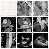
A 55-year-old male patient, who was a smoker and chronic alcoholic, was admitted to the emergency department with epigastric pain and vomiting which had lasted 14 hours. He had signs of cardiogenic shock. The electrocardiogram showed atrial fibrillation with slow ventricular response and ST-segment elevation in leads II, III, aVF, V1 and V2 (A). Cardiac angiography was performed immediately showing a normal left coronary artery (Figure 1B and C); the right coronary artery (RCA) could not be catheterized. Computed tomography angiography was performed and showed aortic root (AR) and ascending aorta (AA) dilation with diffuse circumferential wall thickening occluding the ostium of the RCA (Figure 1D-F). The wall thickening also extended to the descending aorta (Figure 1D and G). On transesophageal echocardiography, the circumferential thickening of the thoracic aorta had a coarse irregular luminal surface (Figure 1H). The right ventricle was dilated and with severely depressed systolic function (Figure 1I).

Figure 1.

(A) Electrocardiogram on admission; (B) left coronary artery with no atherosclerotic disease; (C) aortography showing mild aortic regurgitation and no signs of ascending aortic dissection; the origin of the left coronary artery can be visualized but the right coronary artery cannot; (D-G) computed tomography angiography showing circumferential wall thickening of the thoracic aorta (arrows), causing occlusion of the right coronary ostium (arrowheads); (H) transesophageal echocardiography (TOE) showing circumferential thickening of the thoracic aorta; (I) TOE showing dilated right ventricle.

(0.66MB).

The patient tested positive for syphilis serology, with elevated titers of Treponema pallidum hemagglutination assay (TPHA) and venereal disease research laboratory (VDRL) testing. The remaining microbiological, serological and immunological tests were negative. A diagnosis of syphilitic aortitis with involvement of the RCA ostium was then made.

Surgical intervention was ruled out and, despite supportive measures, the patient died 72 hours after admission.

Dilatation of the AR and the AA, aortic thickness involving a coronary ostium and elevated VDRL and TPHA titers are highly suggestive of syphilitic aortitis. Syphilis is an etiology that, although rare, should be taken into account in the diagnostic approach of patients with acute myocardial infarction and coronary ostial stenosis.

Conflicts of interest

The authors have no conflicts of interest to declare.

Copyright © 2018. Sociedade Portuguesa de Cardiologia
Baixar PDF
Idiomas
Revista Portuguesa de Cardiologia
Opções de artigo
Ferramentas
en pt

Are you a health professional able to prescribe or dispense drugs?

Você é um profissional de saúde habilitado a prescrever ou dispensar medicamentos

Ao assinalar que é «Profissional de Saúde», declara conhecer e aceitar que a responsável pelo tratamento dos dados pessoais dos utilizadores da página de internet da Revista Portuguesa de Cardiologia (RPC), é esta entidade, com sede no Campo Grande, n.º 28, 13.º, 1700-093 Lisboa, com os telefones 217 970 685 e 217 817 630, fax 217 931 095 e com o endereço de correio eletrónico revista@spc.pt. Declaro para todos os fins, que assumo inteira responsabilidade pela veracidade e exatidão da afirmação aqui fornecida.