array:22 [
  "pii" => "S0870255124002762"
  "issn" => "08702551"
  "doi" => "10.1016/j.repc.2024.07.007"
  "estado" => "S200"
  "fechaPublicacion" => "2024-10-18"
  "aid" => "2377"
  "copyright" => "Sociedade Portuguesa de Cardiologia"
  "copyrightAnyo" => "2024"
  "documento" => "article"
  "crossmark" => 0
  "subdocumento" => "sco"
  "abierto" => array:3 [
    "ES" => true
    "ES2" => true
    "LATM" => true
  ]
  "gratuito" => true
  "lecturas" => array:1 [
    "total" => 0
  ]
  "itemSiguiente" => array:17 [
    "pii" => "S0870255124001896"
    "issn" => "08702551"
    "doi" => "10.1016/j.repc.2024.04.007"
    "estado" => "S200"
    "fechaPublicacion" => "2024-10-22"
    "aid" => "2346"
    "copyright" => "Sociedade Portuguesa de Cardiologia"
    "documento" => "article"
    "crossmark" => 0
    "subdocumento" => "sco"
    "abierto" => array:3 [
      "ES" => true
      "ES2" => true
      "LATM" => true
    ]
    "gratuito" => true
    "lecturas" => array:1 [
      "total" => 0
    ]
    "en" => array:10 [
      "idiomaDefecto" => true
      "cabecera" => "<span class="elsevierStyleTextfn">Image in Cardiology</span>"
      "titulo" => "Rare images of a unileaflet mitral valve"
      "tienePdf" => "en"
      "tieneTextoCompleto" => "en"
      "titulosAlternativos" => array:1 [
        "pt" => array:1 [
          "titulo" => "Imagens raras de valva mitral com folheto &#250;nico"
        ]
      ]
      "contieneTextoCompleto" => array:1 [
        "en" => true
      ]
      "contienePdf" => array:1 [
        "en" => true
      ]
      "resumenGrafico" => array:2 [
        "original" => 0
        "multimedia" => array:7 [
          "identificador" => "fig0005"
          "etiqueta" => "Figure 1"
          "tipo" => "MULTIMEDIAFIGURA"
          "mostrarFloat" => true
          "mostrarDisplay" => false
          "figura" => array:1 [
            0 => array:4 [
              "imagen" => "gr1.jpeg"
              "Alto" => 895
              "Ancho" => 1922
              "Tamanyo" => 165506
            ]
          ]
          "descripcion" => array:1 [
            "en" => "<p id="spar0010" class="elsevierStyleSimplePara elsevierViewall">Transthoracic echocardiography&#46; Two-dimensional parasternal long-axis &#40;A&#41; and short-axis &#91;&#40;B&#41; mitral valve level&#44; &#40;C&#41; papillary muscle level&#93; views&#59; M-mode images at the level of the mitral leaflets of the present unileaflet mitral valve case &#40;D&#41; and of a normal bileaflet mitral valve &#40;E&#41;&#46; D point&#58; initial diastolic leaflet opening&#59; E point&#58; maximum leaflet opening during the phase of rapid ventricular filling ending at the F point&#59; A point&#58; maximum leaflet opening during atrial contraction&#59; C point&#58; leaflet coaptation point at the beginning of systole&#46; In the present case of a unileaflet mitral valve &#40;D&#41;&#44; there is loss of the F point &#40;asterisk&#41; with a flat E-A wave in constant contact with the anterior interventricular septum&#44; corresponding to the opening of the elongated anterior mitral leaflet throughout diastole&#44; while the coaptation point C is posteriorly displaced&#46;</p>"
          ]
        ]
      ]
      "autores" => array:1 [
        0 => array:2 [
          "autoresLista" => "Nikolaos Miaris, Dimitrios-Persefs Zampelis, Konstantina Ntalekou, Maria Karakosta, Alkistis-Eleni Kalesi, Nearchos Kasinos"
          "autores" => array:6 [
            0 => array:2 [
              "nombre" => "Nikolaos"
              "apellidos" => "Miaris"
            ]
            1 => array:2 [
              "nombre" => "Dimitrios-Persefs"
              "apellidos" => "Zampelis"
            ]
            2 => array:2 [
              "nombre" => "Konstantina"
              "apellidos" => "Ntalekou"
            ]
            3 => array:2 [
              "nombre" => "Maria"
              "apellidos" => "Karakosta"
            ]
            4 => array:2 [
              "nombre" => "Alkistis-Eleni"
              "apellidos" => "Kalesi"
            ]
            5 => array:2 [
              "nombre" => "Nearchos"
              "apellidos" => "Kasinos"
            ]
          ]
        ]
      ]
    ]
    "idiomaDefecto" => "en"
    "EPUB" => "https://multimedia.elsevier.es/PublicationsMultimediaV1/item/epub/S0870255124001896?idApp=UINPBA00004E"
    "url" => "/08702551/unassign/S0870255124001896/v1_202410220426/en/main.assets"
  ]
  "itemAnterior" => array:16 [
    "pii" => "S0870255122005224"
    "issn" => "08702551"
    "doi" => "10.1016/j.repc.2022.12.004"
    "estado" => "S100"
    "fechaPublicacion" => "2022-12-18"
    "aid" => "2078"
    "documento" => "simple-article"
    "crossmark" => 0
    "subdocumento" => "rem"
    "abierto" => array:3 [
      "ES" => true
      "ES2" => true
      "LATM" => true
    ]
    "gratuito" => true
    "lecturas" => array:1 [
      "total" => 0
    ]
    "en" => array:8 [
      "idiomaDefecto" => true
      "titulo" => "WITHDRAWN&#58; Cardioprotective role of GABA-B receptor activation on ventricular arrhythmia following myocardial infarction"
      "tienePdf" => "en"
      "tieneTextoCompleto" => 0
      "tieneResumen" => "en"
      "contieneResumen" => array:1 [
        "en" => true
      ]
      "contienePdf" => array:1 [
        "en" => true
      ]
      "autores" => array:1 [
        0 => array:2 [
          "autoresLista" => "T&#226;nia Martins-Marques"
          "autores" => array:1 [
            0 => array:2 [
              "nombre" => "T&#226;nia"
              "apellidos" => "Martins-Marques"
            ]
          ]
        ]
      ]
    ]
    "idiomaDefecto" => "en"
    "EPUB" => "https://multimedia.elsevier.es/PublicationsMultimediaV1/item/epub/S0870255122005224?idApp=UINPBA00004E"
    "url" => "/08702551/unassign/S0870255122005224/v2_202212230519/en/main.assets"
  ]
  "en" => array:15 [
    "idiomaDefecto" => true
    "cabecera" => "<span class="elsevierStyleTextfn">Image in Cardiology</span>"
    "titulo" => "Asymptomatic ruptured sinus of Valsalva aneurysm"
    "tieneTextoCompleto" => true
    "autores" => array:1 [
      0 => array:4 [
        "autoresLista" => "M&#243;nica Dias, Sofia Fernandes, Nuno Salom&#233;, Catarina Vieira, Juliana Martins"
        "autores" => array:5 [
          0 => array:4 [
            "nombre" => "M&#243;nica"
            "apellidos" => "Dias"
            "email" => array:1 [
              0 => "monica.dias_96@hotmail.com"
            ]
            "referencia" => array:1 [
              0 => array:2 [
                "etiqueta" => "<span class="elsevierStyleSup">&#42;</span>"
                "identificador" => "cor0005"
              ]
            ]
          ]
          1 => array:2 [
            "nombre" => "Sofia"
            "apellidos" => "Fernandes"
          ]
          2 => array:2 [
            "nombre" => "Nuno"
            "apellidos" => "Salom&#233;"
          ]
          3 => array:2 [
            "nombre" => "Catarina"
            "apellidos" => "Vieira"
          ]
          4 => array:2 [
            "nombre" => "Juliana"
            "apellidos" => "Martins"
          ]
        ]
        "afiliaciones" => array:1 [
          0 => array:2 [
            "entidad" => "Cardiology Department&#44; ULS Braga&#44; Braga&#44; Portugal"
            "identificador" => "aff0005"
          ]
        ]
        "correspondencia" => array:1 [
          0 => array:3 [
            "identificador" => "cor0005"
            "etiqueta" => "&#8270;"
            "correspondencia" => "Corresponding author&#46;"
          ]
        ]
      ]
    ]
    "titulosAlternativos" => array:1 [
      "pt" => array:1 [
        "titulo" => "Rutura assintom&#225;tica de aneurisma do seio de Valsalva"
      ]
    ]
    "resumenGrafico" => array:2 [
      "original" => 0
      "multimedia" => array:7 [
        "identificador" => "fig0010"
        "etiqueta" => "Figure 2"
        "tipo" => "MULTIMEDIAFIGURA"
        "mostrarFloat" => true
        "mostrarDisplay" => false
        "figura" => array:1 [
          0 => array:4 [
            "imagen" => "gr2.jpeg"
            "Alto" => 1150
            "Ancho" => 1455
            "Tamanyo" => 189029
          ]
        ]
        "descripcion" => array:1 [
          "en" => "<p id="spar0010" class="elsevierStyleSimplePara elsevierViewall">CT angiography showing the SOVA &#40;white arrow&#41;&#46;</p>"
        ]
      ]
    ]
    "textoCompleto" => "<span class="elsevierStyleSections"><span id="sec0005" class="elsevierStyleSection elsevierViewall"><span class="elsevierStyleSectionTitle" id="sect0005">Case presentation</span><p id="par0005" class="elsevierStylePara elsevierViewall">A 65-year-old male underwent an echocardiogram after a transient ischemic attack which revealed an apparent noncoronary sinus of Valsalva aneurysm &#40;SOVA&#41; with a shunt flow from the aorta into the right atrium&#46;</p><p id="par0010" class="elsevierStylePara elsevierViewall">At the first evaluation&#44; the patient was asymptomatic and maintained regular physical activity &#40;used to run 10 km twice a week&#41;&#46; The physical examination was unremarkable except for a slight continuous heart murmur&#46; Transthoracic and transesophageal echocardiogram confirmed the existence of an aorto-atrial fistula with left to right cardiac shunt&#44; caused by a rupture SOVA &#40;<a class="elsevierStyleCrossRef" href="#fig0005">Figure 1</a>A and B&#59; <a class="elsevierStyleCrossRef" href="#sec0105">videos 1&#8211;4</a>&#41; with dilatation of the right heart chambers &#40;<a class="elsevierStyleCrossRef" href="#fig0005">Figure 1</a>C&#41;&#46;</p><elsevierMultimedia ident="fig0005"></elsevierMultimedia><p id="par0015" class="elsevierStylePara elsevierViewall">He had undergone no prior aortic surgery nor had he experienced chest trauma&#46; Infections and connective tissue disorders capable of weakening the elastic tissue were excluded&#46; The coronary computed tomography angiography excluded atherosclerotic coronary artery disease and showed the aneurysm again &#40;<a class="elsevierStyleCrossRef" href="#fig0010">Figure 2</a>&#41;&#46; Postcapillary pulmonary hypertension was confirmed by right heart catheterization and pulmonary&#8211;systemic flow ratio &#40;<span class="elsevierStyleItalic">Q</span><span class="elsevierStyleInf">p</span>&#47;<span class="elsevierStyleItalic">Q</span><span class="elsevierStyleInf">s</span>&#41; was 2&#46;1&#46;</p><elsevierMultimedia ident="fig0010"></elsevierMultimedia><p id="par0020" class="elsevierStylePara elsevierViewall">The patient underwent successful surgical repair of the fistula with direct suture&#46;</p><p id="par0025" class="elsevierStylePara elsevierViewall">SOVA is a rare condition among the general population &#40;0&#46;09&#37;&#41;&#46; It is a congenital or acquired discontinuity between the aortic valve annulus and the aortic media caused by weakness or degeneration of the elastic connective tissue&#46;</p><p id="par0030" class="elsevierStylePara elsevierViewall">SOVA may be silent and found incidentally&#44; but the rupture of a SOVA is a devastating event that leads to symptoms of heart failure or cardiac tamponade and carries a poor prognosis if left untreated&#46; In this case&#44; the presence of the aorto-atrial fistula along with the evidence of right heart overload reveal signs of some chronicity&#44; making the absence of symptoms unexpected&#46;</p></span><span id="sec0010" class="elsevierStyleSection elsevierViewall"><span class="elsevierStyleSectionTitle" id="sect0010">Patient consent</span><p id="par0035" class="elsevierStylePara elsevierViewall">The authors confirm that consent for submission and publication of this case has been obtained from the patient&#46;</p></span><span id="sec0020" class="elsevierStyleSection elsevierViewall"><span class="elsevierStyleSectionTitle" id="sect0015">Funding</span><p id="par0045" class="elsevierStylePara elsevierViewall">None declared&#46;</p></span><span id="sec0015" class="elsevierStyleSection elsevierViewall"><span class="elsevierStyleSectionTitle" id="sect0020">Conflict of interest</span><p id="par0040" class="elsevierStylePara elsevierViewall">The authors declare that there is no conflict of interest&#46;</p></span></span>"
    "textoCompletoSecciones" => array:1 [
      "secciones" => array:4 [
        0 => array:2 [
          "identificador" => "sec0005"
          "titulo" => "Case presentation"
        ]
        1 => array:2 [
          "identificador" => "sec0010"
          "titulo" => "Patient consent"
        ]
        2 => array:2 [
          "identificador" => "sec0020"
          "titulo" => "Funding"
        ]
        3 => array:2 [
          "identificador" => "sec0015"
          "titulo" => "Conflict of interest"
        ]
      ]
    ]
    "pdfFichero" => "main.pdf"
    "tienePdf" => true
    "fechaRecibido" => "2024-04-13"
    "fechaAceptado" => "2024-07-07"
    "apendice" => array:1 [
      0 => array:1 [
        "seccion" => array:1 [
          0 => array:4 [
            "apendice" => "<p id="par0220" class="elsevierStylePara elsevierViewall">The following are the supplementary data to this article&#58;<elsevierMultimedia ident="upi0005"></elsevierMultimedia><elsevierMultimedia ident="upi0010"></elsevierMultimedia><elsevierMultimedia ident="upi0015"></elsevierMultimedia><elsevierMultimedia ident="upi0020"></elsevierMultimedia></p>"
            "etiqueta" => "Appendix B"
            "titulo" => "Supplementary data"
            "identificador" => "sec0105"
          ]
        ]
      ]
    ]
    "multimedia" => array:6 [
      0 => array:7 [
        "identificador" => "fig0005"
        "etiqueta" => "Figure 1"
        "tipo" => "MULTIMEDIAFIGURA"
        "mostrarFloat" => true
        "mostrarDisplay" => false
        "figura" => array:1 [
          0 => array:4 [
            "imagen" => "gr1.jpeg"
            "Alto" => 1202
            "Ancho" => 980
            "Tamanyo" => 123456
          ]
        ]
        "descripcion" => array:1 [
          "en" => "<p id="spar0005" class="elsevierStyleSimplePara elsevierViewall">Transthoracic echocardiogram&#58; &#40;A&#41; PSAX base view showing SOVA occupying the right atrium - white arrow&#59; &#40;B&#41; PLAX Doppler view showing SOVA with aorta to right atrium shunt&#59; &#40;C&#41; A4C view with dilatation of the right heart chambers&#46;</p>"
        ]
      ]
      1 => array:7 [
        "identificador" => "fig0010"
        "etiqueta" => "Figure 2"
        "tipo" => "MULTIMEDIAFIGURA"
        "mostrarFloat" => true
        "mostrarDisplay" => false
        "figura" => array:1 [
          0 => array:4 [
            "imagen" => "gr2.jpeg"
            "Alto" => 1150
            "Ancho" => 1455
            "Tamanyo" => 189029
          ]
        ]
        "descripcion" => array:1 [
          "en" => "<p id="spar0010" class="elsevierStyleSimplePara elsevierViewall">CT angiography showing the SOVA &#40;white arrow&#41;&#46;</p>"
        ]
      ]
      2 => array:5 [
        "identificador" => "upi0005"
        "tipo" => "MULTIMEDIAECOMPONENTE"
        "mostrarFloat" => false
        "mostrarDisplay" => true
        "Ecomponente" => array:3 [
          "fichero" => "mmc1.mp4"
          "ficheroTamanyo" => 209027
          "Video" => array:2 [
            "mp4" => array:5 [
              "fichero" => "mmc1.m4v"
              "poster" => "mmc1.jpg"
              "tiempo" => 0
              "alto" => 0
              "ancho" => 0
            ]
            "flv" => array:5 [
              "fichero" => "mmc1.flv"
              "poster" => "mmc1.jpg"
              "tiempo" => 0
              "alto" => 0
              "ancho" => 0
            ]
          ]
        ]
      ]
      3 => array:5 [
        "identificador" => "upi0010"
        "tipo" => "MULTIMEDIAECOMPONENTE"
        "mostrarFloat" => false
        "mostrarDisplay" => true
        "Ecomponente" => array:3 [
          "fichero" => "mmc2.mp4"
          "ficheroTamanyo" => 294342
          "Video" => array:2 [
            "flv" => array:5 [
              "fichero" => "mmc2.flv"
              "poster" => "mmc2.jpg"
              "tiempo" => 0
              "alto" => 0
              "ancho" => 0
            ]
            "mp4" => array:5 [
              "fichero" => "mmc2.m4v"
              "poster" => "mmc2.jpg"
              "tiempo" => 0
              "alto" => 0
              "ancho" => 0
            ]
          ]
        ]
      ]
      4 => array:5 [
        "identificador" => "upi0015"
        "tipo" => "MULTIMEDIAECOMPONENTE"
        "mostrarFloat" => false
        "mostrarDisplay" => true
        "Ecomponente" => array:3 [
          "fichero" => "mmc3.mp4"
          "ficheroTamanyo" => 288441
          "Video" => array:2 [
            "flv" => array:5 [
              "fichero" => "mmc3.flv"
              "poster" => "mmc3.jpg"
              "tiempo" => 0
              "alto" => 0
              "ancho" => 0
            ]
            "mp4" => array:5 [
              "fichero" => "mmc3.m4v"
              "poster" => "mmc3.jpg"
              "tiempo" => 0
              "alto" => 0
              "ancho" => 0
            ]
          ]
        ]
      ]
      5 => array:5 [
        "identificador" => "upi0020"
        "tipo" => "MULTIMEDIAECOMPONENTE"
        "mostrarFloat" => false
        "mostrarDisplay" => true
        "Ecomponente" => array:3 [
          "fichero" => "mmc4.mp4"
          "ficheroTamanyo" => 305481
          "Video" => array:2 [
            "flv" => array:5 [
              "fichero" => "mmc4.flv"
              "poster" => "mmc4.jpg"
              "tiempo" => 0
              "alto" => 0
              "ancho" => 0
            ]
            "mp4" => array:5 [
              "fichero" => "mmc4.m4v"
              "poster" => "mmc4.jpg"
              "tiempo" => 0
              "alto" => 0
              "ancho" => 0
            ]
          ]
        ]
      ]
    ]
  ]
  "idiomaDefecto" => "en"
  "url" => "/08702551/unassign/S0870255124002762/v1_202410180445/en/main.assets"
  "Apartado" => null
  "PDF" => "https://static.elsevier.es/multimedia/08702551/unassign/S0870255124002762/v1_202410180445/en/main.pdf?idApp=UINPBA00004E&text.app=https://revportcardiol.org/"
  "EPUB" => "https://multimedia.elsevier.es/PublicationsMultimediaV1/item/epub/S0870255124002762?idApp=UINPBA00004E"
]
Partilhar
Informação da revista
Partilhar
Partilhar
Baixar PDF
Mais opções do artigo
Visitas
218
Image in Cardiology
Acesso de texto completo
Disponível online em 18 de outubro de 2024
Asymptomatic ruptured sinus of Valsalva aneurysm
Rutura assintomática de aneurisma do seio de Valsalva
Visitas
218
Mónica Dias
Autor para correspondência
monica.dias_96@hotmail.com

Corresponding author.
, Sofia Fernandes, Nuno Salomé, Catarina Vieira, Juliana Martins
Cardiology Department, ULS Braga, Braga, Portugal
Este item recebeu
Recebido 13 Abril 2024. Aceite 07 Julho 2024
Informação do artigo
Texto Completo
Baixar PDF
Estatísticas
Figuras (2)
Material adicional (4)
Texto Completo
Case presentation

A 65-year-old male underwent an echocardiogram after a transient ischemic attack which revealed an apparent noncoronary sinus of Valsalva aneurysm (SOVA) with a shunt flow from the aorta into the right atrium.

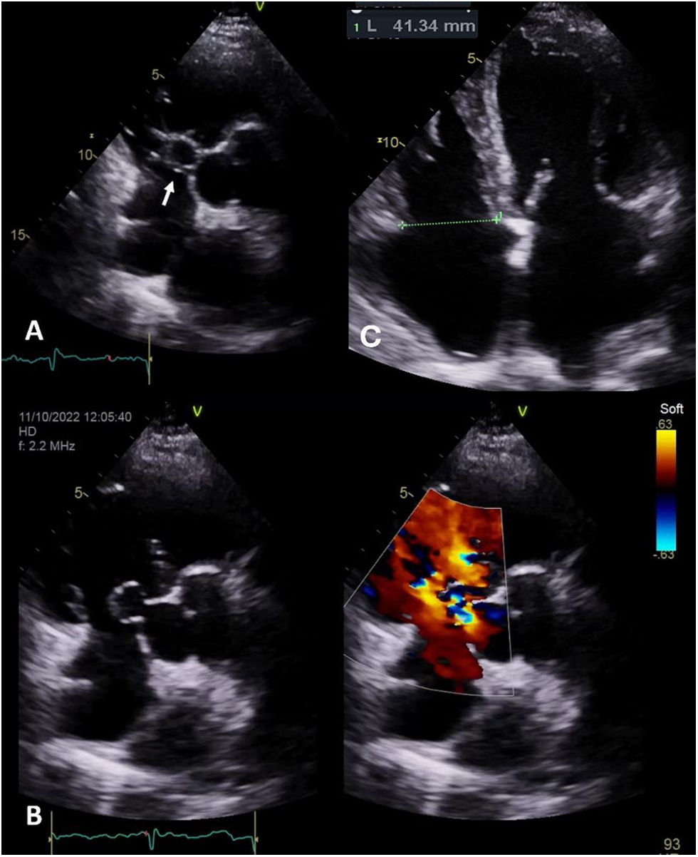
At the first evaluation, the patient was asymptomatic and maintained regular physical activity (used to run 10 km twice a week). The physical examination was unremarkable except for a slight continuous heart murmur. Transthoracic and transesophageal echocardiogram confirmed the existence of an aorto-atrial fistula with left to right cardiac shunt, caused by a rupture SOVA (Figure 1A and B; videos 1–4) with dilatation of the right heart chambers (Figure 1C).

Figure 1.

Transthoracic echocardiogram: (A) PSAX base view showing SOVA occupying the right atrium - white arrow; (B) PLAX Doppler view showing SOVA with aorta to right atrium shunt; (C) A4C view with dilatation of the right heart chambers.

(0.12MB).

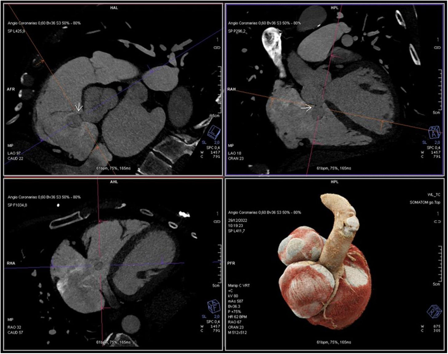
He had undergone no prior aortic surgery nor had he experienced chest trauma. Infections and connective tissue disorders capable of weakening the elastic tissue were excluded. The coronary computed tomography angiography excluded atherosclerotic coronary artery disease and showed the aneurysm again (Figure 2). Postcapillary pulmonary hypertension was confirmed by right heart catheterization and pulmonary–systemic flow ratio (Qp/Qs) was 2.1.

Figure 2.

CT angiography showing the SOVA (white arrow).

(0.18MB).

The patient underwent successful surgical repair of the fistula with direct suture.

SOVA is a rare condition among the general population (0.09%). It is a congenital or acquired discontinuity between the aortic valve annulus and the aortic media caused by weakness or degeneration of the elastic connective tissue.

SOVA may be silent and found incidentally, but the rupture of a SOVA is a devastating event that leads to symptoms of heart failure or cardiac tamponade and carries a poor prognosis if left untreated. In this case, the presence of the aorto-atrial fistula along with the evidence of right heart overload reveal signs of some chronicity, making the absence of symptoms unexpected.

Patient consent

The authors confirm that consent for submission and publication of this case has been obtained from the patient.

Funding

None declared.

Conflict of interest

The authors declare that there is no conflict of interest.

Appendix B
Supplementary data

The following are the supplementary data to this article:

(0.2MB)
(0.28MB)
(0.28MB)
(0.29MB)

Copyright © 2024. Sociedade Portuguesa de Cardiologia
Baixar PDF
Idiomas
Revista Portuguesa de Cardiologia
Opções de artigo
Ferramentas
Material Suplementar
en pt

Are you a health professional able to prescribe or dispense drugs?

Você é um profissional de saúde habilitado a prescrever ou dispensar medicamentos

Ao assinalar que é «Profissional de Saúde», declara conhecer e aceitar que a responsável pelo tratamento dos dados pessoais dos utilizadores da página de internet da Revista Portuguesa de Cardiologia (RPC), é esta entidade, com sede no Campo Grande, n.º 28, 13.º, 1700-093 Lisboa, com os telefones 217 970 685 e 217 817 630, fax 217 931 095 e com o endereço de correio eletrónico revista@spc.pt. Declaro para todos os fins, que assumo inteira responsabilidade pela veracidade e exatidão da afirmação aqui fornecida.