array:24 [
  "pii" => "S0870255114002625"
  "issn" => "08702551"
  "doi" => "10.1016/j.repc.2014.04.006"
  "estado" => "S300"
  "fechaPublicacion" => "2014-11-01"
  "aid" => "547"
  "copyright" => "Sociedade Portuguesa de Cardiologia"
  "copyrightAnyo" => "2014"
  "documento" => "article"
  "licencia" => "http://creativecommons.org/licenses/by-nc-nd/4.0/"
  "subdocumento" => "sco"
  "cita" => "Rev Port Cardiol. 2014;33:741-2"
  "abierto" => array:3 [
    "ES" => true
    "ES2" => true
    "LATM" => true
  ]
  "gratuito" => true
  "lecturas" => array:2 [
    "total" => 3508
    "formatos" => array:3 [
      "EPUB" => 202
      "HTML" => 2683
      "PDF" => 623
    ]
  ]
  "Traduccion" => array:1 [
    "en" => array:19 [
      "pii" => "S2174204914002700"
      "issn" => "21742049"
      "doi" => "10.1016/j.repce.2014.04.007"
      "estado" => "S300"
      "fechaPublicacion" => "2014-11-01"
      "aid" => "547"
      "copyright" => "Sociedade Portuguesa de Cardiologia"
      "documento" => "article"
      "licencia" => "http://creativecommons.org/licenses/by-nc-nd/4.0/"
      "subdocumento" => "sco"
      "cita" => "Rev Port Cardiol. 2014;33:741-2"
      "abierto" => array:3 [
        "ES" => true
        "ES2" => true
        "LATM" => true
      ]
      "gratuito" => true
      "lecturas" => array:2 [
        "total" => 2671
        "formatos" => array:3 [
          "EPUB" => 142
          "HTML" => 2131
          "PDF" => 398
        ]
      ]
      "en" => array:11 [
        "idiomaDefecto" => true
        "cabecera" => "<span class="elsevierStyleTextfn">Image in Cardiology</span>"
        "titulo" => "Assessment of interrupted aortic arch with magnetic resonance angiography"
        "tienePdf" => "en"
        "tieneTextoCompleto" => "en"
        "paginas" => array:1 [
          0 => array:2 [
            "paginaInicial" => "741"
            "paginaFinal" => "742"
          ]
        ]
        "titulosAlternativos" => array:1 [
          "pt" => array:1 [
            "titulo" => "A avalia&#231;&#227;o do arco a&#243;rtico interrompido por angiografia com resson&#226;ncia magn&#233;tica"
          ]
        ]
        "contieneTextoCompleto" => array:1 [
          "en" => true
        ]
        "contienePdf" => array:1 [
          "en" => true
        ]
        "resumenGrafico" => array:2 [
          "original" => 0
          "multimedia" => array:7 [
            "identificador" => "fig0005"
            "etiqueta" => "Figure 1"
            "tipo" => "MULTIMEDIAFIGURA"
            "mostrarFloat" => true
            "mostrarDisplay" => false
            "figura" => array:1 [
              0 => array:4 [
                "imagen" => "gr1.jpeg"
                "Alto" => 1806
                "Ancho" => 2167
                "Tamanyo" => 269956
              ]
            ]
            "descripcion" => array:1 [
              "en" => "<p id="spar0005" class="elsevierStyleSimplePara elsevierViewall">Three-dimensional volume rendering magnetic resonance angiography images&#58; &#40;A&#41; left posterior oblique view and &#40;B&#41; right posterior oblique view&#44; showing interruption of the descending aorta after the branching of the left subclavian artery&#46; IA&#58; interrupted aorta&#59; IMA&#58; internal mammary artery&#59; LCCA&#58; left common carotid artery&#59; LSCA&#58; left subclavian artery&#59; RCCA&#58; right common carotid artery&#59; TrBc&#58; truncus brachiocephalicus&#46;</p>"
            ]
          ]
        ]
        "autores" => array:1 [
          0 => array:2 [
            "autoresLista" => "Fahri Halit Besir, Yasemin Turker, Yasin Turker, Omer Onbas"
            "autores" => array:4 [
              0 => array:2 [
                "nombre" => "Fahri Halit"
                "apellidos" => "Besir"
              ]
              1 => array:2 [
                "nombre" => "Yasemin"
                "apellidos" => "Turker"
              ]
              2 => array:2 [
                "nombre" => "Yasin"
                "apellidos" => "Turker"
              ]
              3 => array:2 [
                "nombre" => "Omer"
                "apellidos" => "Onbas"
              ]
            ]
          ]
        ]
      ]
      "idiomaDefecto" => "en"
      "Traduccion" => array:1 [
        "en" => array:9 [
          "pii" => "S0870255114002625"
          "doi" => "10.1016/j.repc.2014.04.006"
          "estado" => "S300"
          "subdocumento" => ""
          "abierto" => array:3 [
            "ES" => true
            "ES2" => true
            "LATM" => true
          ]
          "gratuito" => true
          "lecturas" => array:1 [
            "total" => 0
          ]
          "idiomaDefecto" => "en"
          "EPUB" => "https://multimedia.elsevier.es/PublicationsMultimediaV1/item/epub/S0870255114002625?idApp=UINPBA00004E"
        ]
      ]
      "EPUB" => "https://multimedia.elsevier.es/PublicationsMultimediaV1/item/epub/S2174204914002700?idApp=UINPBA00004E"
      "url" => "/21742049/0000003300000011/v1_201412120159/S2174204914002700/v1_201412120159/en/main.assets"
    ]
  ]
  "itemSiguiente" => array:19 [
    "pii" => "S0870255114002637"
    "issn" => "08702551"
    "doi" => "10.1016/j.repc.2014.03.015"
    "estado" => "S300"
    "fechaPublicacion" => "2014-11-01"
    "aid" => "548"
    "copyright" => "Sociedade Portuguesa de Cardiologia"
    "documento" => "article"
    "licencia" => "http://creativecommons.org/licenses/by-nc-nd/4.0/"
    "subdocumento" => "sco"
    "cita" => "Rev Port Cardiol. 2014;33:743-4"
    "abierto" => array:3 [
      "ES" => true
      "ES2" => true
      "LATM" => true
    ]
    "gratuito" => true
    "lecturas" => array:2 [
      "total" => 3554
      "formatos" => array:3 [
        "EPUB" => 165
        "HTML" => 2724
        "PDF" => 665
      ]
    ]
    "en" => array:11 [
      "idiomaDefecto" => true
      "cabecera" => "<span class="elsevierStyleTextfn">Image in Cardiology</span>"
      "titulo" => "Massive calcification involving a left ventricular false tendon"
      "tienePdf" => "en"
      "tieneTextoCompleto" => "en"
      "paginas" => array:1 [
        0 => array:2 [
          "paginaInicial" => "743"
          "paginaFinal" => "744"
        ]
      ]
      "titulosAlternativos" => array:1 [
        "pt" => array:1 [
          "titulo" => "Calcifica&#231;&#227;o card&#237;aca envolvendo um falso tend&#227;o ventricular esquerdo"
        ]
      ]
      "contieneTextoCompleto" => array:1 [
        "en" => true
      ]
      "contienePdf" => array:1 [
        "en" => true
      ]
      "resumenGrafico" => array:2 [
        "original" => 0
        "multimedia" => array:7 [
          "identificador" => "fig0005"
          "etiqueta" => "Figure 1"
          "tipo" => "MULTIMEDIAFIGURA"
          "mostrarFloat" => true
          "mostrarDisplay" => false
          "figura" => array:1 [
            0 => array:4 [
              "imagen" => "gr1.jpeg"
              "Alto" => 1232
              "Ancho" => 1900
              "Tamanyo" => 225813
            ]
          ]
          "descripcion" => array:1 [
            "en" => "<p id="spar0025" class="elsevierStyleSimplePara elsevierViewall">&#40;A&#41; Two-dimensional transthoracic echocardiography&#44; apical 4-chamber view&#44; showing several nodular hyperechogenic masses with acoustic shadowing in the mid-left ventricular cavity &#40;arrows&#41;&#59; &#40;B&#41; three-dimensional transthoracic echocardiography&#44; left ventricular full-volume view depicting all of the nodular masses within the left ventricular cavity &#40;arrows&#41;&#59; &#40;C&#41; two-dimensional Doppler color flow transesophageal echocardiography&#44; mid-esophageal 3-chamber view with turbulent flow across the mid-left ventricular cavity&#59; &#40;D and E&#41; steady-state free precession cine cardiac magnetic resonance&#44; short-axis and 3-chamber long-axis views with a hypointense linear structure inside the left ventricle &#40;arrowheads&#41;&#46; LA&#58; left atrium&#59; LV&#58; left ventricle&#59; LVOT&#58; left ventricular outflow tract&#59; RV&#58; right ventricle&#46;</p>"
          ]
        ]
      ]
      "autores" => array:1 [
        0 => array:2 [
          "autoresLista" => "Rui Pl&#225;cido, Jo&#227;o Silva Marques, Maria Jos&#233; Amaro, Dulce Brito, Fausto J&#46; Pinto, Ana G&#46; Almeida"
          "autores" => array:6 [
            0 => array:2 [
              "nombre" => "Rui"
              "apellidos" => "Pl&#225;cido"
            ]
            1 => array:2 [
              "nombre" => "Jo&#227;o"
              "apellidos" => "Silva Marques"
            ]
            2 => array:2 [
              "nombre" => "Maria Jos&#233;"
              "apellidos" => "Amaro"
            ]
            3 => array:2 [
              "nombre" => "Dulce"
              "apellidos" => "Brito"
            ]
            4 => array:2 [
              "nombre" => "Fausto J&#46;"
              "apellidos" => "Pinto"
            ]
            5 => array:2 [
              "nombre" => "Ana"
              "apellidos" => "G&#46; Almeida"
            ]
          ]
        ]
      ]
    ]
    "idiomaDefecto" => "en"
    "Traduccion" => array:1 [
      "en" => array:9 [
        "pii" => "S2174204914002712"
        "doi" => "10.1016/j.repce.2014.03.015"
        "estado" => "S300"
        "subdocumento" => ""
        "abierto" => array:3 [
          "ES" => true
          "ES2" => true
          "LATM" => true
        ]
        "gratuito" => true
        "lecturas" => array:1 [
          "total" => 0
        ]
        "idiomaDefecto" => "en"
        "EPUB" => "https://multimedia.elsevier.es/PublicationsMultimediaV1/item/epub/S2174204914002712?idApp=UINPBA00004E"
      ]
    ]
    "EPUB" => "https://multimedia.elsevier.es/PublicationsMultimediaV1/item/epub/S0870255114002637?idApp=UINPBA00004E"
    "url" => "/08702551/0000003300000011/v1_201411200056/S0870255114002637/v1_201411200056/en/main.assets"
  ]
  "itemAnterior" => array:19 [
    "pii" => "S0870255114002595"
    "issn" => "08702551"
    "doi" => "10.1016/j.repc.2014.06.006"
    "estado" => "S300"
    "fechaPublicacion" => "2014-11-01"
    "aid" => "544"
    "copyright" => "Sociedade Portuguesa de Cardiologia"
    "documento" => "simple-article"
    "licencia" => "http://creativecommons.org/licenses/by-nc-nd/4.0/"
    "subdocumento" => "crp"
    "cita" => "Rev Port Cardiol. 2014;33:739&#46;e1-3"
    "abierto" => array:3 [
      "ES" => true
      "ES2" => true
      "LATM" => true
    ]
    "gratuito" => true
    "lecturas" => array:2 [
      "total" => 4175
      "formatos" => array:3 [
        "EPUB" => 192
        "HTML" => 3126
        "PDF" => 857
      ]
    ]
    "en" => array:13 [
      "idiomaDefecto" => true
      "cabecera" => "<span class="elsevierStyleTextfn">Case report</span>"
      "titulo" => "Pacemaker lead-related tricuspid stenosis successfully treated with percutaneous balloon valvuloplasty guided by 3D echocardiography"
      "tienePdf" => "en"
      "tieneTextoCompleto" => "en"
      "tieneResumen" => array:2 [
        0 => "en"
        1 => "pt"
      ]
      "paginas" => array:1 [
        0 => array:2 [
          "paginaInicial" => "739&#46;e1"
          "paginaFinal" => "739&#46;e3"
        ]
      ]
      "titulosAlternativos" => array:1 [
        "pt" => array:1 [
          "titulo" => "Estenose tric&#250;spide associada a electrocateter de <span class="elsevierStyleItalic">pacemaker</span> tratada com sucesso com valvuloplastia percut&#226;nea com bal&#227;o guiada por ecocardiografia 3D"
        ]
      ]
      "contieneResumen" => array:2 [
        "en" => true
        "pt" => true
      ]
      "contieneTextoCompleto" => array:1 [
        "en" => true
      ]
      "contienePdf" => array:1 [
        "en" => true
      ]
      "resumenGrafico" => array:2 [
        "original" => 0
        "multimedia" => array:7 [
          "identificador" => "fig0010"
          "etiqueta" => "Figure 2"
          "tipo" => "MULTIMEDIAFIGURA"
          "mostrarFloat" => true
          "mostrarDisplay" => false
          "figura" => array:1 [
            0 => array:4 [
              "imagen" => "gr2.jpeg"
              "Alto" => 525
              "Ancho" => 1599
              "Tamanyo" => 73794
            ]
          ]
          "descripcion" => array:1 [
            "en" => "<p id="spar0020" class="elsevierStyleSimplePara elsevierViewall">Angiographic images of percutaneous tricuspid valvuloplasty&#58; &#40;A&#41; pigtail catheter across the tricuspid valve&#59; &#40;B&#41; Amplatz Super Stiff guidewire across the valve&#59; &#40;C&#41; balloon inflation&#46;</p>"
          ]
        ]
      ]
      "autores" => array:1 [
        0 => array:2 [
          "autoresLista" => "Juan Carlos Rama-Merchan, Antonio Arribas-Jimenez, Javier Martin-Moreiras, Eulogio Garcia-Fernandez, Ignacio Cruz-Gonzalez"
          "autores" => array:5 [
            0 => array:2 [
              "nombre" => "Juan Carlos"
              "apellidos" => "Rama-Merchan"
            ]
            1 => array:2 [
              "nombre" => "Antonio"
              "apellidos" => "Arribas-Jimenez"
            ]
            2 => array:2 [
              "nombre" => "Javier"
              "apellidos" => "Martin-Moreiras"
            ]
            3 => array:2 [
              "nombre" => "Eulogio"
              "apellidos" => "Garcia-Fernandez"
            ]
            4 => array:2 [
              "nombre" => "Ignacio"
              "apellidos" => "Cruz-Gonzalez"
            ]
          ]
        ]
      ]
    ]
    "idiomaDefecto" => "en"
    "Traduccion" => array:1 [
      "en" => array:9 [
        "pii" => "S2174204914002694"
        "doi" => "10.1016/j.repce.2014.06.007"
        "estado" => "S300"
        "subdocumento" => ""
        "abierto" => array:3 [
          "ES" => true
          "ES2" => true
          "LATM" => true
        ]
        "gratuito" => true
        "lecturas" => array:1 [
          "total" => 0
        ]
        "idiomaDefecto" => "en"
        "EPUB" => "https://multimedia.elsevier.es/PublicationsMultimediaV1/item/epub/S2174204914002694?idApp=UINPBA00004E"
      ]
    ]
    "EPUB" => "https://multimedia.elsevier.es/PublicationsMultimediaV1/item/epub/S0870255114002595?idApp=UINPBA00004E"
    "url" => "/08702551/0000003300000011/v1_201411200056/S0870255114002595/v1_201411200056/en/main.assets"
  ]
  "en" => array:15 [
    "idiomaDefecto" => true
    "cabecera" => "<span class="elsevierStyleTextfn">Image in Cardiology</span>"
    "titulo" => "Assessment of interrupted aortic arch with magnetic resonance angiography"
    "tieneTextoCompleto" => true
    "paginas" => array:1 [
      0 => array:2 [
        "paginaInicial" => "741"
        "paginaFinal" => "742"
      ]
    ]
    "autores" => array:1 [
      0 => array:4 [
        "autoresLista" => "Fahri Halit Besir, Yasemin Turker, Yasin Turker, Omer Onbas"
        "autores" => array:4 [
          0 => array:4 [
            "nombre" => "Fahri Halit"
            "apellidos" => "Besir"
            "email" => array:1 [
              0 => "drfhbesir&#64;gmail&#46;com"
            ]
            "referencia" => array:2 [
              0 => array:2 [
                "etiqueta" => "<span class="elsevierStyleSup">a</span>"
                "identificador" => "aff0005"
              ]
              1 => array:2 [
                "etiqueta" => "<span class="elsevierStyleSup">&#42;</span>"
                "identificador" => "cor0005"
              ]
            ]
          ]
          1 => array:3 [
            "nombre" => "Yasemin"
            "apellidos" => "Turker"
            "referencia" => array:1 [
              0 => array:2 [
                "etiqueta" => "<span class="elsevierStyleSup">b</span>"
                "identificador" => "aff0010"
              ]
            ]
          ]
          2 => array:3 [
            "nombre" => "Yasin"
            "apellidos" => "Turker"
            "referencia" => array:1 [
              0 => array:2 [
                "etiqueta" => "<span class="elsevierStyleSup">c</span>"
                "identificador" => "aff0015"
              ]
            ]
          ]
          3 => array:3 [
            "nombre" => "Omer"
            "apellidos" => "Onbas"
            "referencia" => array:1 [
              0 => array:2 [
                "etiqueta" => "<span class="elsevierStyleSup">a</span>"
                "identificador" => "aff0005"
              ]
            ]
          ]
        ]
        "afiliaciones" => array:3 [
          0 => array:3 [
            "entidad" => "Duzce University School of Medicine&#44; Department of Radiology&#44; Duzce&#44; Turkey"
            "etiqueta" => "a"
            "identificador" => "aff0005"
          ]
          1 => array:3 [
            "entidad" => "Duzce Family Medicine Center&#44; Duzce&#44; Turkey"
            "etiqueta" => "b"
            "identificador" => "aff0010"
          ]
          2 => array:3 [
            "entidad" => "Duzce University&#44; Medicine School&#44; Department of Cardiology&#44; Duzce&#44; Turkey"
            "etiqueta" => "c"
            "identificador" => "aff0015"
          ]
        ]
        "correspondencia" => array:1 [
          0 => array:3 [
            "identificador" => "cor0005"
            "etiqueta" => "&#8270;"
            "correspondencia" => "Corresponding author&#46;"
          ]
        ]
      ]
    ]
    "titulosAlternativos" => array:1 [
      "pt" => array:1 [
        "titulo" => "A avalia&#231;&#227;o do arco a&#243;rtico interrompido por angiografia com resson&#226;ncia magn&#233;tica"
      ]
    ]
    "resumenGrafico" => array:2 [
      "original" => 0
      "multimedia" => array:7 [
        "identificador" => "fig0005"
        "etiqueta" => "Figure 1"
        "tipo" => "MULTIMEDIAFIGURA"
        "mostrarFloat" => true
        "mostrarDisplay" => false
        "figura" => array:1 [
          0 => array:4 [
            "imagen" => "gr1.jpeg"
            "Alto" => 1083
            "Ancho" => 1300
            "Tamanyo" => 151575
          ]
        ]
        "descripcion" => array:1 [
          "en" => "<p id="spar0005" class="elsevierStyleSimplePara elsevierViewall">Three-dimensional volume rendering magnetic resonance angiography images&#58; &#40;A&#41; left posterior oblique view and &#40;B&#41; right posterior oblique view&#44; showing interruption of the descending aorta after the branching of the left subclavian artery&#46; IA&#58; interrupted aorta&#59; IMA&#58; internal mammary artery&#59; LCCA&#58; left common carotid artery&#59; LSCA&#58; left subclavian artery&#59; RCCA&#58; right common carotid artery&#59; TrBc&#58; truncus brachiocephalicus&#46;</p>"
        ]
      ]
    ]
    "textoCompleto" => "<span class="elsevierStyleSections"><p id="par0005" class="elsevierStylePara elsevierViewall">A 22-year-old man was referred to our cardiology department for investigation of hypertension&#46; At presentation his blood pressure was 170&#47;100 mmHg in both arms&#44; and heart rate was 74 beats&#47;min&#46; The pulses were equal over both upper extremities&#44; but bilateral femoral and popliteal pulses were extremely weak and the dorsalis pedis and anterior tibial pulses were not palpable&#46; His past history was unremarkable except for hypertension&#46; There was a II&#47;VI systolic ejection murmur in the left second intercostal area and left scapular region in the back&#46; Transthoracic echocardiography showed concentric left ventricular hypertrophy with normal ventricular function&#46; Magnetic resonance &#40;MR&#41; angiography showed interruption of the descending aorta after the branching of the left subclavian artery &#40;<a class="elsevierStyleCrossRef" href="#fig0005">Figure 1</a>&#41;&#46; Dilated intercostal&#44; intramammary and thoracodorsal arteries with accompanying anastomosis between the intercostal and thoracodorsal arteries were observed &#40;<a class="elsevierStyleCrossRef" href="#fig0010">Figure 2</a>&#41;&#46;</p><elsevierMultimedia ident="fig0005"></elsevierMultimedia><elsevierMultimedia ident="fig0010"></elsevierMultimedia><p id="par0010" class="elsevierStylePara elsevierViewall">Interrupted aortic arch is defined as a complete luminal and anatomic discontinuity between the ascending and descending aorta&#46; It is a rare and severe congenital heart defect with a very poor prognosis without surgical treatment&#46; Conventional angiography is considered the gold standard and is frequently used in vascular imaging&#46; However&#44; MR imaging with excellent anatomic and functional capabilities can be performed easily&#44; and with advances in imaging technology&#44; acquiring information comparable to that of conventional angiography has become feasible&#46; MR can also be used to assess the vascular anatomy from several views with maximum intensity projection reconstructions&#46;</p><span id="sec0005" class="elsevierStyleSection elsevierViewall"><span class="elsevierStyleSectionTitle" id="sect0005">Ethical disclosures</span><span id="sec0010" class="elsevierStyleSection elsevierViewall"><span class="elsevierStyleSectionTitle" id="sect0010">Protection of human and animal subjects</span><p id="par0015" class="elsevierStylePara elsevierViewall">The authors declare that no experiments were performed on humans or animals for this study&#46;</p></span><span id="sec0015" class="elsevierStyleSection elsevierViewall"><span class="elsevierStyleSectionTitle" id="sect0015">Confidentiality of data</span><p id="par0020" class="elsevierStylePara elsevierViewall">The authors declare that no patient data appear in this article&#46;</p></span><span id="sec0020" class="elsevierStyleSection elsevierViewall"><span class="elsevierStyleSectionTitle" id="sect0020">Right to privacy and informed consent</span><p id="par0025" class="elsevierStylePara elsevierViewall">The authors declare that no patient data appear in this article&#46;</p></span></span><span id="sec0025" class="elsevierStyleSection elsevierViewall"><span class="elsevierStyleSectionTitle" id="sect0025">Conflicts of interest</span><p id="par0030" class="elsevierStylePara elsevierViewall">The authors have no conflicts of interest to declare&#46;</p></span></span>"
    "textoCompletoSecciones" => array:1 [
      "secciones" => array:2 [
        0 => array:3 [
          "identificador" => "sec0005"
          "titulo" => "Ethical disclosures"
          "secciones" => array:3 [
            0 => array:2 [
              "identificador" => "sec0010"
              "titulo" => "Protection of human and animal subjects"
            ]
            1 => array:2 [
              "identificador" => "sec0015"
              "titulo" => "Confidentiality of data"
            ]
            2 => array:2 [
              "identificador" => "sec0020"
              "titulo" => "Right to privacy and informed consent"
            ]
          ]
        ]
        1 => array:2 [
          "identificador" => "sec0025"
          "titulo" => "Conflicts of interest"
        ]
      ]
    ]
    "pdfFichero" => "main.pdf"
    "tienePdf" => true
    "fechaRecibido" => "2014-03-07"
    "fechaAceptado" => "2014-04-01"
    "multimedia" => array:2 [
      0 => array:7 [
        "identificador" => "fig0005"
        "etiqueta" => "Figure 1"
        "tipo" => "MULTIMEDIAFIGURA"
        "mostrarFloat" => true
        "mostrarDisplay" => false
        "figura" => array:1 [
          0 => array:4 [
            "imagen" => "gr1.jpeg"
            "Alto" => 1083
            "Ancho" => 1300
            "Tamanyo" => 151575
          ]
        ]
        "descripcion" => array:1 [
          "en" => "<p id="spar0005" class="elsevierStyleSimplePara elsevierViewall">Three-dimensional volume rendering magnetic resonance angiography images&#58; &#40;A&#41; left posterior oblique view and &#40;B&#41; right posterior oblique view&#44; showing interruption of the descending aorta after the branching of the left subclavian artery&#46; IA&#58; interrupted aorta&#59; IMA&#58; internal mammary artery&#59; LCCA&#58; left common carotid artery&#59; LSCA&#58; left subclavian artery&#59; RCCA&#58; right common carotid artery&#59; TrBc&#58; truncus brachiocephalicus&#46;</p>"
        ]
      ]
      1 => array:7 [
        "identificador" => "fig0010"
        "etiqueta" => "Figure 2"
        "tipo" => "MULTIMEDIAFIGURA"
        "mostrarFloat" => true
        "mostrarDisplay" => false
        "figura" => array:1 [
          0 => array:4 [
            "imagen" => "gr2.jpeg"
            "Alto" => 1007
            "Ancho" => 750
            "Tamanyo" => 105236
          ]
        ]
        "descripcion" => array:1 [
          "en" => "<p id="spar0010" class="elsevierStyleSimplePara elsevierViewall">Three-dimensional volume rendering magnetic resonance angiography images&#44; anterior coronal view&#44; showing dilated intercostal&#44; intramammary and thoracodorsal arteries with accompanying anastomosis between thoracodorsal artery branches and intercostal arteries&#46; An&#58; anastomosis&#46;</p>"
        ]
      ]
    ]
  ]
  "idiomaDefecto" => "en"
  "url" => "/08702551/0000003300000011/v1_201411200056/S0870255114002625/v1_201411200056/en/main.assets"
  "Apartado" => array:4 [
    "identificador" => "357"
    "tipo" => "SECCION"
    "pt" => array:2 [
      "titulo" => "Imagens em cardiologia"
      "idiomaDefecto" => true
    ]
    "idiomaDefecto" => "pt"
  ]
  "PDF" => "https://static.elsevier.es/multimedia/08702551/0000003300000011/v1_201411200056/S0870255114002625/v1_201411200056/en/main.pdf?idApp=UINPBA00004E&text.app=https://revportcardiol.org/"
  "EPUB" => "https://multimedia.elsevier.es/PublicationsMultimediaV1/item/epub/S0870255114002625?idApp=UINPBA00004E"
]
Partilhar
Informação da revista
Vol. 33. Núm. 11.
Páginas 741-742 (novembro 2014)
Partilhar
Partilhar
Baixar PDF
Mais opções do artigo
Vol. 33. Núm. 11.
Páginas 741-742 (novembro 2014)
Image in Cardiology
Open Access
Assessment of interrupted aortic arch with magnetic resonance angiography
A avaliação do arco aórtico interrompido por angiografia com ressonância magnética
Visitas
6244
Fahri Halit Besira,
Autor para correspondência
drfhbesir@gmail.com

Corresponding author.
, Yasemin Turkerb, Yasin Turkerc, Omer Onbasa
a Duzce University School of Medicine, Department of Radiology, Duzce, Turkey
b Duzce Family Medicine Center, Duzce, Turkey
c Duzce University, Medicine School, Department of Cardiology, Duzce, Turkey
Este item recebeu

Under a Creative Commons license
Informação do artigo
Texto Completo
Baixar PDF
Estatísticas
Figuras (2)
Texto Completo

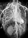
A 22-year-old man was referred to our cardiology department for investigation of hypertension. At presentation his blood pressure was 170/100 mmHg in both arms, and heart rate was 74 beats/min. The pulses were equal over both upper extremities, but bilateral femoral and popliteal pulses were extremely weak and the dorsalis pedis and anterior tibial pulses were not palpable. His past history was unremarkable except for hypertension. There was a II/VI systolic ejection murmur in the left second intercostal area and left scapular region in the back. Transthoracic echocardiography showed concentric left ventricular hypertrophy with normal ventricular function. Magnetic resonance (MR) angiography showed interruption of the descending aorta after the branching of the left subclavian artery (Figure 1). Dilated intercostal, intramammary and thoracodorsal arteries with accompanying anastomosis between the intercostal and thoracodorsal arteries were observed (Figure 2).

Figure 1.

Three-dimensional volume rendering magnetic resonance angiography images: (A) left posterior oblique view and (B) right posterior oblique view, showing interruption of the descending aorta after the branching of the left subclavian artery. IA: interrupted aorta; IMA: internal mammary artery; LCCA: left common carotid artery; LSCA: left subclavian artery; RCCA: right common carotid artery; TrBc: truncus brachiocephalicus.

(0.14MB).
Figure 2.

Three-dimensional volume rendering magnetic resonance angiography images, anterior coronal view, showing dilated intercostal, intramammary and thoracodorsal arteries with accompanying anastomosis between thoracodorsal artery branches and intercostal arteries. An: anastomosis.

(0.1MB).

Interrupted aortic arch is defined as a complete luminal and anatomic discontinuity between the ascending and descending aorta. It is a rare and severe congenital heart defect with a very poor prognosis without surgical treatment. Conventional angiography is considered the gold standard and is frequently used in vascular imaging. However, MR imaging with excellent anatomic and functional capabilities can be performed easily, and with advances in imaging technology, acquiring information comparable to that of conventional angiography has become feasible. MR can also be used to assess the vascular anatomy from several views with maximum intensity projection reconstructions.

Ethical disclosuresProtection of human and animal subjects

The authors declare that no experiments were performed on humans or animals for this study.

Confidentiality of data

The authors declare that no patient data appear in this article.

Right to privacy and informed consent

The authors declare that no patient data appear in this article.

Conflicts of interest

The authors have no conflicts of interest to declare.

Copyright © 2014. Sociedade Portuguesa de Cardiologia
Baixar PDF
Idiomas
Revista Portuguesa de Cardiologia
Opções de artigo
Ferramentas
en pt

Are you a health professional able to prescribe or dispense drugs?

Você é um profissional de saúde habilitado a prescrever ou dispensar medicamentos

Ao assinalar que é «Profissional de Saúde», declara conhecer e aceitar que a responsável pelo tratamento dos dados pessoais dos utilizadores da página de internet da Revista Portuguesa de Cardiologia (RPC), é esta entidade, com sede no Campo Grande, n.º 28, 13.º, 1700-093 Lisboa, com os telefones 217 970 685 e 217 817 630, fax 217 931 095 e com o endereço de correio eletrónico revista@spc.pt. Declaro para todos os fins, que assumo inteira responsabilidade pela veracidade e exatidão da afirmação aqui fornecida.