array:24 [
  "pii" => "S0870255113001273"
  "issn" => "08702551"
  "doi" => "10.1016/j.repc.2012.11.013"
  "estado" => "S300"
  "fechaPublicacion" => "2013-07-01"
  "aid" => "298"
  "copyright" => "Sociedade Portuguesa de Cardiologia"
  "copyrightAnyo" => "2012"
  "documento" => "article"
  "crossmark" => 0
  "licencia" => "http://creativecommons.org/licenses/by-nc-nd/4.0/"
  "subdocumento" => "sco"
  "cita" => "Rev Port Cardiol. 2013;32:619-22"
  "abierto" => array:3 [
    "ES" => true
    "ES2" => true
    "LATM" => true
  ]
  "gratuito" => true
  "lecturas" => array:2 [
    "total" => 6710
    "formatos" => array:3 [
      "EPUB" => 196
      "HTML" => 5417
      "PDF" => 1097
    ]
  ]
  "itemSiguiente" => array:19 [
    "pii" => "S0870255113001248"
    "issn" => "08702551"
    "doi" => "10.1016/j.repc.2012.11.011"
    "estado" => "S300"
    "fechaPublicacion" => "2013-07-01"
    "aid" => "295"
    "copyright" => "Sociedade Portuguesa de Cardiologia"
    "documento" => "article"
    "crossmark" => 0
    "licencia" => "http://creativecommons.org/licenses/by-nc-nd/4.0/"
    "subdocumento" => "sco"
    "cita" => "Rev Port Cardiol. 2013;32:623-7"
    "abierto" => array:3 [
      "ES" => true
      "ES2" => true
      "LATM" => true
    ]
    "gratuito" => true
    "lecturas" => array:2 [
      "total" => 10347
      "formatos" => array:3 [
        "EPUB" => 169
        "HTML" => 8963
        "PDF" => 1215
      ]
    ]
    "en" => array:13 [
      "idiomaDefecto" => true
      "cabecera" => "<span class="elsevierStyleTextfn">Case report</span>"
      "titulo" => "Retroperitoneal hematoma&#58; An unexpected complication during intervention on an occluded superficial femoral artery via a retrograde popliteal artery approach"
      "tienePdf" => "en"
      "tieneTextoCompleto" => "en"
      "tieneResumen" => array:2 [
        0 => "en"
        1 => "pt"
      ]
      "paginas" => array:1 [
        0 => array:2 [
          "paginaInicial" => "623"
          "paginaFinal" => "627"
        ]
      ]
      "titulosAlternativos" => array:1 [
        "pt" => array:1 [
          "titulo" => "Hematoma retroperitoneal&#58; uma complica&#231;&#227;o inesperada durante interven&#231;&#227;o em oclus&#227;o de art&#233;ria femoral superficial por via retr&#243;grada atrav&#233;s de abordagem popl&#237;tea"
        ]
      ]
      "contieneResumen" => array:2 [
        "en" => true
        "pt" => true
      ]
      "contieneTextoCompleto" => array:1 [
        "en" => true
      ]
      "contienePdf" => array:1 [
        "en" => true
      ]
      "resumenGrafico" => array:2 [
        "original" => 0
        "multimedia" => array:7 [
          "identificador" => "fig0025"
          "etiqueta" => "Figure 5"
          "tipo" => "MULTIMEDIAFIGURA"
          "mostrarFloat" => true
          "mostrarDisplay" => false
          "figura" => array:1 [
            0 => array:4 [
              "imagen" => "gr5.jpeg"
              "Alto" => 950
              "Ancho" => 950
              "Tamanyo" => 127239
            ]
          ]
          "descripcion" => array:1 [
            "en" => "<p id="spar0035" class="elsevierStyleSimplePara elsevierViewall">Angiogram showing retrograde filling of the proximal superficial femoral artery &#40;SFA&#41;&#44; collateral to the SFA &#40;white arrow&#41; and dissection plane in the external iliac artery &#40;short arrow&#41; with perforation into the retroperitoneal space &#40;long arrow&#41;&#46;</p>"
          ]
        ]
      ]
      "autores" => array:1 [
        0 => array:2 [
          "autoresLista" => "Nuri I&#46; Akkus, Jagan Beedupalli, Jai Varma"
          "autores" => array:3 [
            0 => array:2 [
              "nombre" => "Nuri I&#46;"
              "apellidos" => "Akkus"
            ]
            1 => array:2 [
              "nombre" => "Jagan"
              "apellidos" => "Beedupalli"
            ]
            2 => array:2 [
              "nombre" => "Jai"
              "apellidos" => "Varma"
            ]
          ]
        ]
      ]
    ]
    "idiomaDefecto" => "en"
    "EPUB" => "https://multimedia.elsevier.es/PublicationsMultimediaV1/item/epub/S0870255113001248?idApp=UINPBA00004E"
    "url" => "/08702551/0000003200000078/v1_201308220036/S0870255113001248/v1_201308220036/en/main.assets"
  ]
  "itemAnterior" => array:19 [
    "pii" => "S0870255113000413"
    "issn" => "08702551"
    "doi" => "10.1016/j.repc.2012.10.013"
    "estado" => "S300"
    "fechaPublicacion" => "2013-07-01"
    "aid" => "257"
    "copyright" => "Sociedade Portuguesa de Cardiologia"
    "documento" => "article"
    "crossmark" => 0
    "licencia" => "http://creativecommons.org/licenses/by-nc-nd/4.0/"
    "subdocumento" => "sco"
    "cita" => "Rev Port Cardiol. 2013;32:613-8"
    "abierto" => array:3 [
      "ES" => true
      "ES2" => true
      "LATM" => true
    ]
    "gratuito" => true
    "lecturas" => array:2 [
      "total" => 19764
      "formatos" => array:3 [
        "EPUB" => 228
        "HTML" => 17959
        "PDF" => 1577
      ]
    ]
    "pt" => array:13 [
      "idiomaDefecto" => true
      "cabecera" => "<span class="elsevierStyleTextfn">Caso Cl&#237;nico</span>"
      "titulo" => "Doen&#231;a coron&#225;ria n&#227;o obstrutiva documentada por tomografia computorizada card&#237;aca&#58; contraste entre a carga ateroscler&#243;tica e o risco cardiovascular"
      "tienePdf" => "pt"
      "tieneTextoCompleto" => "pt"
      "tieneResumen" => array:2 [
        0 => "pt"
        1 => "en"
      ]
      "paginas" => array:1 [
        0 => array:2 [
          "paginaInicial" => "613"
          "paginaFinal" => "618"
        ]
      ]
      "titulosAlternativos" => array:1 [
        "en" => array:1 [
          "titulo" => "Non-obstructive coronary artery disease documented by cardiac computed tomography&#58; Discrepancy between atherosclerotic burden and cardiovascular risk"
        ]
      ]
      "contieneResumen" => array:2 [
        "pt" => true
        "en" => true
      ]
      "contieneTextoCompleto" => array:1 [
        "pt" => true
      ]
      "contienePdf" => array:1 [
        "pt" => true
      ]
      "resumenGrafico" => array:2 [
        "original" => 0
        "multimedia" => array:7 [
          "identificador" => "fig0025"
          "etiqueta" => "Figura 4"
          "tipo" => "MULTIMEDIAFIGURA"
          "mostrarFloat" => true
          "mostrarDisplay" => false
          "figura" => array:1 [
            0 => array:4 [
              "imagen" => "gr4.jpeg"
              "Alto" => 1192
              "Ancho" => 1750
              "Tamanyo" => 182176
            ]
          ]
          "descripcion" => array:1 [
            "pt" => "<p id="spar0045" class="elsevierStyleSimplePara elsevierViewall">Ecocardiograma transtor&#225;cico revelando hipertrofia ventricular esquerda exc&#234;ntrica &#40;A&#41;&#44; dilata&#231;&#227;o auricular esquerda &#40;B&#41;&#44; fluxo Doppler transmitral e padr&#227;o de enchimento normais &#40;C e D&#41;&#46;</p>"
          ]
        ]
      ]
      "autores" => array:1 [
        0 => array:2 [
          "autoresLista" => "H&#233;lder Dores, Pedro de Ara&#250;jo Gon&#231;alves, Maria Salom&#233; Carvalho, Pedro Jer&#243;nimo Sousa, Hugo Marques, Nuno Cardim, Ana Aleixo, Miguel Mota Carmo, Francisco Pereira Machado, Jos&#233; Roquette"
          "autores" => array:10 [
            0 => array:2 [
              "nombre" => "H&#233;lder"
              "apellidos" => "Dores"
            ]
            1 => array:2 [
              "nombre" => "Pedro"
              "apellidos" => "de Ara&#250;jo Gon&#231;alves"
            ]
            2 => array:2 [
              "nombre" => "Maria Salom&#233;"
              "apellidos" => "Carvalho"
            ]
            3 => array:2 [
              "nombre" => "Pedro Jer&#243;nimo"
              "apellidos" => "Sousa"
            ]
            4 => array:2 [
              "nombre" => "Hugo"
              "apellidos" => "Marques"
            ]
            5 => array:2 [
              "nombre" => "Nuno"
              "apellidos" => "Cardim"
            ]
            6 => array:2 [
              "nombre" => "Ana"
              "apellidos" => "Aleixo"
            ]
            7 => array:2 [
              "nombre" => "Miguel"
              "apellidos" => "Mota Carmo"
            ]
            8 => array:2 [
              "nombre" => "Francisco"
              "apellidos" => "Pereira Machado"
            ]
            9 => array:2 [
              "nombre" => "Jos&#233;"
              "apellidos" => "Roquette"
            ]
          ]
        ]
      ]
    ]
    "idiomaDefecto" => "pt"
    "EPUB" => "https://multimedia.elsevier.es/PublicationsMultimediaV1/item/epub/S0870255113000413?idApp=UINPBA00004E"
    "url" => "/08702551/0000003200000078/v1_201308220036/S0870255113000413/v1_201308220036/pt/main.assets"
  ]
  "en" => array:20 [
    "idiomaDefecto" => true
    "cabecera" => "<span class="elsevierStyleTextfn">Case report</span>"
    "titulo" => "An unusual ST-segment elevation&#58; Apical hypertrophic cardiomyopathy shows the ace up its sleeve"
    "tieneTextoCompleto" => true
    "paginas" => array:1 [
      0 => array:2 [
        "paginaInicial" => "619"
        "paginaFinal" => "622"
      ]
    ]
    "autores" => array:1 [
      0 => array:4 [
        "autoresLista" => "Francesco de Santis, Amedeo Pergolini, Giordano Zampi, Gaetano Pero, Paolo Giuseppe Pino, Giovanni Minardi"
        "autores" => array:6 [
          0 => array:2 [
            "nombre" => "Francesco"
            "apellidos" => "de Santis"
          ]
          1 => array:2 [
            "nombre" => "Amedeo"
            "apellidos" => "Pergolini"
          ]
          2 => array:4 [
            "nombre" => "Giordano"
            "apellidos" => "Zampi"
            "email" => array:1 [
              0 => "giordano&#46;zampi&#64;alice&#46;it"
            ]
            "referencia" => array:1 [
              0 => array:2 [
                "etiqueta" => "<span class="elsevierStyleSup">¿</span>"
                "identificador" => "cor0005"
              ]
            ]
          ]
          3 => array:2 [
            "nombre" => "Gaetano"
            "apellidos" => "Pero"
          ]
          4 => array:2 [
            "nombre" => "Paolo Giuseppe"
            "apellidos" => "Pino"
          ]
          5 => array:2 [
            "nombre" => "Giovanni"
            "apellidos" => "Minardi"
          ]
        ]
        "afiliaciones" => array:1 [
          0 => array:2 [
            "entidad" => "Department of Cardiovascular Science&#44; &#8220;S&#46; Camillo-Forlanini&#8221; Hospital&#44; Rome&#44; Italy"
            "identificador" => "aff0005"
          ]
        ]
        "correspondencia" => array:1 [
          0 => array:3 [
            "identificador" => "cor0005"
            "etiqueta" => "&#8270;"
            "correspondencia" => "Corresponding author&#46;"
          ]
        ]
      ]
    ]
    "titulosAlternativos" => array:1 [
      "pt" => array:1 [
        "titulo" => "Eleva&#231;&#227;o do segment St pouco usual&#58; cardiomiopatia hipertr&#243;fica apical mostra o &#224;s na manga"
      ]
    ]
    "resumenGrafico" => array:2 [
      "original" => 0
      "multimedia" => array:7 [
        "identificador" => "fig0005"
        "etiqueta" => "Figure 1"
        "tipo" => "MULTIMEDIAFIGURA"
        "mostrarFloat" => true
        "mostrarDisplay" => false
        "figura" => array:1 [
          0 => array:4 [
            "imagen" => "gr1.jpeg"
            "Alto" => 1493
            "Ancho" => 2500
            "Tamanyo" => 433498
          ]
        ]
        "descripcion" => array:1 [
          "en" => "<p id="spar0030" class="elsevierStyleSimplePara elsevierViewall">The ECG shows an unusual 4-mm convex ST segment elevation in C4&#8211;C6 with biphasic T waves&#46;</p>"
        ]
      ]
    ]
    "textoCompleto" => "<span class="elsevierStyleSections"><span id="sec0005" class="elsevierStyleSection elsevierViewall"><span class="elsevierStyleSectionTitle" id="sect0025">Introduction</span><p id="par0005" class="elsevierStylePara elsevierViewall">Apical hypertrophic cardiomyopathy &#40;AHCM&#41; is part of the broad clinical and morphologic spectrum of hypertrophic cardiomyopathy &#40;HCM&#41;&#46;<a class="elsevierStyleCrossRef" href="#bib0005"><span class="elsevierStyleSup">1</span></a> Cases of AHCM with and without apical aneurysm mimicking myocardial infarction have occasionally been reported&#46;<a class="elsevierStyleCrossRefs" href="#bib0010"><span class="elsevierStyleSup">2&#8211;4</span></a> We report a patient with abnormalities on the electrocardiogram &#40;ECG&#41; in whom acute coronary syndrome &#40;ACS&#41; was excluded and AHCM was demonstrated by careful differential diagnosis&#46;</p></span><span id="sec0010" class="elsevierStyleSection elsevierViewall"><span class="elsevierStyleSectionTitle" id="sect0030">Case report</span><p id="par0010" class="elsevierStylePara elsevierViewall">A 77-year-old Caucasian woman was examined by a primary care physician because of dyspnea and atypical chest pain&#46; The ECG showed atrial fibrillation&#44; mean heart rate of 85 bpm&#44; right bundle branch block and 4-mm convex ST segment elevation in C4-C6 with biphasic T waves &#40;<a class="elsevierStyleCrossRef" href="#fig0005">Figure 1</a>&#41;&#46; Due to this atypical ECG&#44; similar to a previous one performed two years before&#44; rest-stress myocardial perfusion scintigraphy was prescribed&#46; This revealed a reduction of left ventricular chamber volume suggestive of HCM without signs of ischemia or previous myocardial necrosis &#40;<a class="elsevierStyleCrossRef" href="#fig0010">Figure 2</a>&#41;&#46; The patient was referred to our echo lab in order to complete the diagnostic procedure&#46; Transthoracic echocardiography &#40;TTE&#41; demonstrated AHCM&#44; showing left ventricular &#40;LV&#41; apical hypertrophy with the typical ace of spades shape &#40;<a class="elsevierStyleCrossRef" href="#fig0015">Figure 3</a>A1&#41;&#46; The ace of spades was therefore studied in depth using color Doppler &#40;<a class="elsevierStyleCrossRef" href="#fig0015">Figure 3</a>A2&#44; Video 1&#41;&#44; Sonovue contrast TTE &#40;<a class="elsevierStyleCrossRef" href="#fig0015">Figure 3</a>A3&#44; Video 2&#41; and real-time three-dimensional echocardiography &#40;<a class="elsevierStyleCrossRef" href="#fig0015">Figure 3</a>A4&#44; Video 3&#41;&#46; A small area of turbulent flow and a mid-ventricular gradient of about 63<span class="elsevierStyleHsp" style=""></span>mmHg were found &#40;<a class="elsevierStyleCrossRef" href="#fig0020">Figure 4</a>a&#41;&#46; In order to evaluate possible modification of the mid-ventricular gradient&#44; an intravenous bolus of atenolol &#40;3<span class="elsevierStyleHsp" style=""></span>mg&#41; was administered&#46; A significant decrease of about 40<span class="elsevierStyleHsp" style=""></span>mmHg was observed while the ECG was unchanged &#40;<a class="elsevierStyleCrossRef" href="#fig0020">Figure 4</a>b&#41;&#46; Beta-blocker therapy was prescribed indefinitely&#46; At six-month follow-up&#44; the patient was asymptomatic and in good clinical condition&#46;</p><elsevierMultimedia ident="fig0005"></elsevierMultimedia><elsevierMultimedia ident="fig0010"></elsevierMultimedia><elsevierMultimedia ident="fig0015"></elsevierMultimedia><elsevierMultimedia ident="fig0020"></elsevierMultimedia></span><span id="sec0015" class="elsevierStyleSection elsevierViewall"><span class="elsevierStyleSectionTitle" id="sect0035">Discussion</span><p id="par0015" class="elsevierStylePara elsevierViewall">AHCM is characterized by segmental hypertrophy confined to the distal portion of the LV chamber&#46; The prevalence of AHCM among HCM patients is 15&#37; in Japan&#44; whereas in the USA it is only 3&#37;&#46; Among AHCM patients 54&#37; are symptomatic&#44; the most common symptoms being chest pain&#44; palpitations&#44; dyspnea and syncope&#46; This condition may cause various complications including atrial fibrillation&#44; acute myocardial infarction&#44; embolic events&#44; ventricular fibrillation&#44; congestive heart failure&#44; apical aneurysm and cardiac arrest&#46; Moreover&#44; it may mimic other conditions&#44; such as cardiac tumors&#44; LV apical thrombus&#44; isolated ventricular non-compaction&#44; endomyocardial fibrosis and coronary artery disease&#46; The most common ECG findings&#44; seen in about 93&#37; of patients&#44; are negative T waves in the precordial leads &#40;&#62;10<span class="elsevierStyleHsp" style=""></span>mm deep in 47&#37;&#41;&#44; followed by signs of LV hypertrophy in 63&#37; of cases&#46; TTE usually shows LV apical hypertrophy and is thus usually the standard diagnostic tool for AHCM&#46; Symptomatic patients may be treated with verapamil&#44; beta-blockers and antiarrhythmic drugs&#44; all of which may improve the signs and symptoms of this condition&#46;</p><p id="par0020" class="elsevierStylePara elsevierViewall">In conclusion&#44; AHCM is a clinical condition that can cause various complications and simulate ACS&#46; Its diagnosis enables ACS to be excluded in patients with atypical ST-segment elevation&#46; TTE is usually sufficient to make the diagnosis&#44; enabling appropriate medical treatment&#46;</p></span><span id="sec0020" class="elsevierStyleSection elsevierViewall"><span class="elsevierStyleSectionTitle" id="sect0040">Ethical disclosures</span><span id="sec0025" class="elsevierStyleSection elsevierViewall"><span class="elsevierStyleSectionTitle" id="sect0045">Protection of human and animal subjects</span><p id="par0025" class="elsevierStylePara elsevierViewall">The authors declare that no experiments were performed on humans or animals for this study&#46;</p></span><span id="sec0030" class="elsevierStyleSection elsevierViewall"><span class="elsevierStyleSectionTitle" id="sect0050">Confidentiality of data</span><p id="par0030" class="elsevierStylePara elsevierViewall">The authors declare that no patient data appear in this article&#46;</p></span><span id="sec0035" class="elsevierStyleSection elsevierViewall"><span class="elsevierStyleSectionTitle" id="sect0055">Right to privacy and informed consent</span><p id="par0035" class="elsevierStylePara elsevierViewall">The authors declare that no patient data appear in this article&#46;</p></span></span><span id="sec0040" class="elsevierStyleSection elsevierViewall"><span class="elsevierStyleSectionTitle" id="sect0060">Conflicts of interest</span><p id="par0040" class="elsevierStylePara elsevierViewall">The authors have no conflicts of interest to declare&#46;</p></span></span>"
    "textoCompletoSecciones" => array:1 [
      "secciones" => array:10 [
        0 => array:2 [
          "identificador" => "xres257756"
          "titulo" => "Abstract"
        ]
        1 => array:2 [
          "identificador" => "xpalclavsec245338"
          "titulo" => "Keywords"
        ]
        2 => array:2 [
          "identificador" => "xres257755"
          "titulo" => "Resumo"
        ]
        3 => array:2 [
          "identificador" => "xpalclavsec245339"
          "titulo" => "Palavras-chave"
        ]
        4 => array:2 [
          "identificador" => "sec0005"
          "titulo" => "Introduction"
        ]
        5 => array:2 [
          "identificador" => "sec0010"
          "titulo" => "Case report"
        ]
        6 => array:2 [
          "identificador" => "sec0015"
          "titulo" => "Discussion"
        ]
        7 => array:3 [
          "identificador" => "sec0020"
          "titulo" => "Ethical disclosures"
          "secciones" => array:3 [
            0 => array:2 [
              "identificador" => "sec0025"
              "titulo" => "Protection of human and animal subjects"
            ]
            1 => array:2 [
              "identificador" => "sec0030"
              "titulo" => "Confidentiality of data"
            ]
            2 => array:2 [
              "identificador" => "sec0035"
              "titulo" => "Right to privacy and informed consent"
            ]
          ]
        ]
        8 => array:2 [
          "identificador" => "sec0040"
          "titulo" => "Conflicts of interest"
        ]
        9 => array:1 [
          "titulo" => "References"
        ]
      ]
    ]
    "pdfFichero" => "main.pdf"
    "tienePdf" => true
    "fechaRecibido" => "2012-09-26"
    "fechaAceptado" => "2012-11-01"
    "PalabrasClave" => array:2 [
      "en" => array:1 [
        0 => array:4 [
          "clase" => "keyword"
          "titulo" => "Keywords"
          "identificador" => "xpalclavsec245338"
          "palabras" => array:3 [
            0 => "Electrocardiogram"
            1 => "Hypertrophic cardiomyopathy"
            2 => "Congenital heart disease"
          ]
        ]
      ]
      "pt" => array:1 [
        0 => array:4 [
          "clase" => "keyword"
          "titulo" => "Palavras-chave"
          "identificador" => "xpalclavsec245339"
          "palabras" => array:3 [
            0 => "Eletrocardiograma"
            1 => "Miocardiopatia hipertr&#243;fica"
            2 => "Cardiopatias cong&#233;nitas"
          ]
        ]
      ]
    ]
    "tieneResumen" => true
    "resumen" => array:2 [
      "en" => array:2 [
        "titulo" => "Abstract"
        "resumen" => "<p id="spar0005" class="elsevierStyleSimplePara elsevierViewall">Apical hypertrophic cardiomyopathy is part of the broad clinical and morphologic spectrum of hypertrophic cardiomyopathy&#46; We report a patient with electrocardiographic abnormalities in whom acute coronary syndrome was excluded and apical hypertrophic cardiomyopathy was demonstrated by careful differential diagnosis&#46;</p>"
      ]
      "pt" => array:2 [
        "titulo" => "Resumo"
        "resumen" => "<p id="spar0010" class="elsevierStyleSimplePara elsevierViewall">A miocardiopatia hipertr&#243;fica apical faz parte do amplo espectro cl&#237;nico e morfol&#243;gico da miocardiopatia hipertr&#243;fica&#46; N&#243;s reportamos um doente com altera&#231;&#245;es electrocardiogr&#225;ficas no qual foi exclu&#237;da uma s&#237;ndrome coron&#225;ria aguda e foi demonstrada uma miocardiopatia hipertr&#243;fica apical usando um processo correto de diagn&#243;stico diferencial&#46;</p>"
      ]
    ]
    "apendice" => array:1 [
      0 => array:1 [
        "seccion" => array:1 [
          0 => array:4 [
            "apendice" => "<p id="par0050" class="elsevierStylePara elsevierViewall">The following are the supplementary data to this article&#58;<elsevierMultimedia ident="upi0005"></elsevierMultimedia><elsevierMultimedia ident="upi0010"></elsevierMultimedia><elsevierMultimedia ident="upi0015"></elsevierMultimedia></p>"
            "etiqueta" => "Appendix A"
            "titulo" => "Supplementary data"
            "identificador" => "sec0050"
          ]
        ]
      ]
    ]
    "multimedia" => array:7 [
      0 => array:7 [
        "identificador" => "fig0005"
        "etiqueta" => "Figure 1"
        "tipo" => "MULTIMEDIAFIGURA"
        "mostrarFloat" => true
        "mostrarDisplay" => false
        "figura" => array:1 [
          0 => array:4 [
            "imagen" => "gr1.jpeg"
            "Alto" => 1493
            "Ancho" => 2500
            "Tamanyo" => 433498
          ]
        ]
        "descripcion" => array:1 [
          "en" => "<p id="spar0030" class="elsevierStyleSimplePara elsevierViewall">The ECG shows an unusual 4-mm convex ST segment elevation in C4&#8211;C6 with biphasic T waves&#46;</p>"
        ]
      ]
      1 => array:7 [
        "identificador" => "fig0010"
        "etiqueta" => "Figure 2"
        "tipo" => "MULTIMEDIAFIGURA"
        "mostrarFloat" => true
        "mostrarDisplay" => false
        "figura" => array:1 [
          0 => array:4 [
            "imagen" => "gr2.jpeg"
            "Alto" => 2594
            "Ancho" => 3000
            "Tamanyo" => 841342
          ]
        ]
        "descripcion" => array:1 [
          "en" => "<p id="spar0035" class="elsevierStyleSimplePara elsevierViewall">Rest-stress myocardial perfusion scintigraphy revealing reduced left ventricular chamber volume suggestive of hypertrophic cardiomyopathy without signs of ischemia or previous myocardial necrosis&#46;</p>"
        ]
      ]
      2 => array:7 [
        "identificador" => "fig0015"
        "etiqueta" => "Figure 3"
        "tipo" => "MULTIMEDIAFIGURA"
        "mostrarFloat" => true
        "mostrarDisplay" => false
        "figura" => array:1 [
          0 => array:4 [
            "imagen" => "gr3.jpeg"
            "Alto" => 2563
            "Ancho" => 1667
            "Tamanyo" => 291285
          ]
        ]
        "descripcion" => array:1 [
          "en" => "<p id="spar0040" class="elsevierStyleSimplePara elsevierViewall">Apical hypertrophic cardiomyopathy shows the ace up its sleeve&#46; A1&#58; two-dimensional transthoracic echocardiography&#59; A2&#58; color Doppler transthoracic echocardiography&#59; A3&#58; contrast-enhanced transthoracic echocardiography&#59; A4&#58; real-time three-dimensional reconstruction&#46;</p>"
        ]
      ]
      3 => array:7 [
        "identificador" => "fig0020"
        "etiqueta" => "Figure 4"
        "tipo" => "MULTIMEDIAFIGURA"
        "mostrarFloat" => true
        "mostrarDisplay" => false
        "figura" => array:1 [
          0 => array:4 [
            "imagen" => "gr4.jpeg"
            "Alto" => 2240
            "Ancho" => 1667
            "Tamanyo" => 287952
          ]
        ]
        "descripcion" => array:1 [
          "en" => "<p id="spar0045" class="elsevierStyleSimplePara elsevierViewall">Transthoracic echocardiography&#44; apical 4-chamber view showing &#40;a&#41; a mid-ventricular gradient of about 63 mmHg and &#40;b&#41; a significant decrease in the gradient after administration of an intravenous bolus of atenolol&#46;</p>"
        ]
      ]
      4 => array:7 [
        "identificador" => "upi0005"
        "etiqueta" => "Video 1"
        "tipo" => "MULTIMEDIAECOMPONENTE"
        "mostrarFloat" => false
        "mostrarDisplay" => true
        "Ecomponente" => array:3 [
          "fichero" => "mmc1.mp4"
          "ficheroTamanyo" => 10065
          "Video" => array:2 [
            "flv" => array:5 [
              "fichero" => "mmc1.flv"
              "poster" => "mmc1.jpg"
              "tiempo" => 0
              "alto" => 0
              "ancho" => 0
            ]
            "mp4" => array:2 [
              "fichero" => "mmc1.m4v"
              "poster" => "mmc1.jpg"
            ]
          ]
        ]
        "descripcion" => array:1 [
          "en" => "<p id="spar0015" class="elsevierStyleSimplePara elsevierViewall">Transthoracic echocardiography&#44; apical four chamber view&#44; color comparison&#44; showing left ventricular apical hypertrophy with the typical spade-shaped appearance&#46;</p>"
        ]
      ]
      5 => array:7 [
        "identificador" => "upi0010"
        "etiqueta" => "Video 2"
        "tipo" => "MULTIMEDIAECOMPONENTE"
        "mostrarFloat" => false
        "mostrarDisplay" => true
        "Ecomponente" => array:3 [
          "fichero" => "mmc2.mp4"
          "ficheroTamanyo" => 9108
          "Video" => array:2 [
            "flv" => array:5 [
              "fichero" => "mmc2.flv"
              "poster" => "mmc2.jpg"
              "tiempo" => 0
              "alto" => 0
              "ancho" => 0
            ]
            "mp4" => array:2 [
              "fichero" => "mmc2.m4v"
              "poster" => "mmc2.jpg"
            ]
          ]
        ]
        "descripcion" => array:1 [
          "en" => "<p id="spar0020" class="elsevierStyleSimplePara elsevierViewall">Transthoracic echocardiography&#44; apical four chamber view&#44; echocontrastography with Sonovue improving the definition of the endorcardial border&#46;</p>"
        ]
      ]
      6 => array:7 [
        "identificador" => "upi0015"
        "etiqueta" => "Video 3"
        "tipo" => "MULTIMEDIAECOMPONENTE"
        "mostrarFloat" => false
        "mostrarDisplay" => true
        "Ecomponente" => array:3 [
          "fichero" => "mmc3.mp4"
          "ficheroTamanyo" => 6354
          "Video" => array:2 [
            "flv" => array:5 [
              "fichero" => "mmc3.flv"
              "poster" => "mmc3.jpg"
              "tiempo" => 0
              "alto" => 0
              "ancho" => 0
            ]
            "mp4" => array:2 [
              "fichero" => "mmc3.m4v"
              "poster" => "mmc3.jpg"
            ]
          ]
        ]
        "descripcion" => array:1 [
          "en" => "<p id="spar0025" class="elsevierStyleSimplePara elsevierViewall">Off-line three dimensional reconstruction&#46;</p>"
        ]
      ]
    ]
    "bibliografia" => array:2 [
      "titulo" => "References"
      "seccion" => array:1 [
        0 => array:2 [
          "identificador" => "bibs0005"
          "bibliografiaReferencia" => array:4 [
            0 => array:3 [
              "identificador" => "bib0005"
              "etiqueta" => "1"
              "referencia" => array:1 [
                0 => array:2 [
                  "contribucion" => array:1 [
                    0 => array:2 [
                      "titulo" => "Hypertrophic cardiomyopathy with ventricular septal hypertrophy localized to the apical region of the left ventricle &#40;apical hypertrophic cardiomyopathy&#41;"
                      "autores" => array:1 [
                        0 => array:2 [
                          "etal" => true
                          "autores" => array:3 [
                            0 => "B&#46;J&#46; Maron"
                            1 => "R&#46;O&#46; Bonow"
                            2 => "T&#46;N&#46; Seshagiri"
                          ]
                        ]
                      ]
                    ]
                  ]
                  "host" => array:1 [
                    0 => array:1 [
                      "Revista" => array:6 [
                        "tituloSerie" => "Am J Cardiol"
                        "fecha" => "1982"
                        "volumen" => "49"
                        "paginaInicial" => "1838"
                        "paginaFinal" => "1848"
                        "link" => array:1 [
                          0 => array:2 [
                            "url" => "https://www.ncbi.nlm.nih.gov/pubmed/6211078"
                            "web" => "Medline"
                          ]
                        ]
                      ]
                    ]
                  ]
                ]
              ]
            ]
            1 => array:3 [
              "identificador" => "bib0010"
              "etiqueta" => "2"
              "referencia" => array:1 [
                0 => array:2 [
                  "contribucion" => array:1 [
                    0 => array:2 [
                      "titulo" => "Apical hypertrophic cardiomyopathy mimicking acute myocardial infarction"
                      "autores" => array:1 [
                        0 => array:2 [
                          "etal" => false
                          "autores" => array:3 [
                            0 => "C&#46;S&#46; Lin"
                            1 => "C&#46;H&#46; Chen"
                            2 => "P&#46;Y&#46; Ding"
                          ]
                        ]
                      ]
                    ]
                  ]
                  "host" => array:1 [
                    0 => array:1 [
                      "Revista" => array:6 [
                        "tituloSerie" => "Int J Cardiol"
                        "fecha" => "1998"
                        "volumen" => "64"
                        "paginaInicial" => "305"
                        "paginaFinal" => "307"
                        "link" => array:1 [
                          0 => array:2 [
                            "url" => "https://www.ncbi.nlm.nih.gov/pubmed/9672414"
                            "web" => "Medline"
                          ]
                        ]
                      ]
                    ]
                  ]
                ]
              ]
            ]
            2 => array:3 [
              "identificador" => "bib0015"
              "etiqueta" => "3"
              "referencia" => array:1 [
                0 => array:2 [
                  "contribucion" => array:1 [
                    0 => array:2 [
                      "titulo" => "The electrocardiogram in apical hypertrophic myocardiopathy&#46; A case report with unique manifestations"
                      "autores" => array:1 [
                        0 => array:2 [
                          "etal" => true
                          "autores" => array:3 [
                            0 => "L&#46;M&#46; Penas"
                            1 => "P&#46;I&#46; Mosquera"
                            2 => "Z&#46;B&#46; Bouzas"
                          ]
                        ]
                      ]
                    ]
                  ]
                  "host" => array:1 [
                    0 => array:1 [
                      "Revista" => array:6 [
                        "tituloSerie" => "Rev Esp Cardiol"
                        "fecha" => "1999"
                        "volumen" => "52"
                        "paginaInicial" => "1148"
                        "paginaFinal" => "1150"
                        "link" => array:1 [
                          0 => array:2 [
                            "url" => "https://www.ncbi.nlm.nih.gov/pubmed/10659661"
                            "web" => "Medline"
                          ]
                        ]
                      ]
                    ]
                  ]
                ]
              ]
            ]
            3 => array:3 [
              "identificador" => "bib0020"
              "etiqueta" => "4"
              "referencia" => array:1 [
                0 => array:2 [
                  "contribucion" => array:1 [
                    0 => array:2 [
                      "titulo" => "Apical hypertrophic cardiomyopathy mimics acute coronary syndrome"
                      "autores" => array:1 [
                        0 => array:2 [
                          "etal" => false
                          "autores" => array:3 [
                            0 => "T&#46; Say&#305;n"
                            1 => "T&#46; Kocum"
                            2 => "C&#46; Kervanc&#305;oglu"
                          ]
                        ]
                      ]
                    ]
                  ]
                  "host" => array:1 [
                    0 => array:1 [
                      "Revista" => array:6 [
                        "tituloSerie" => "Int J Cardiol"
                        "fecha" => "2001"
                        "volumen" => "80"
                        "paginaInicial" => "77"
                        "paginaFinal" => "79"
                        "link" => array:1 [
                          0 => array:2 [
                            "url" => "https://www.ncbi.nlm.nih.gov/pubmed/11575264"
                            "web" => "Medline"
                          ]
                        ]
                      ]
                    ]
                  ]
                ]
              ]
            ]
          ]
        ]
      ]
    ]
  ]
  "idiomaDefecto" => "en"
  "url" => "/08702551/0000003200000078/v1_201308220036/S0870255113001273/v1_201308220036/en/main.assets"
  "Apartado" => array:4 [
    "identificador" => "362"
    "tipo" => "SECCION"
    "pt" => array:2 [
      "titulo" => "Casos cl&#237;nicos"
      "idiomaDefecto" => true
    ]
    "idiomaDefecto" => "pt"
  ]
  "PDF" => "https://static.elsevier.es/multimedia/08702551/0000003200000078/v1_201308220036/S0870255113001273/v1_201308220036/en/main.pdf?idApp=UINPBA00004E&text.app=https://revportcardiol.org/"
  "EPUB" => "https://multimedia.elsevier.es/PublicationsMultimediaV1/item/epub/S0870255113001273?idApp=UINPBA00004E"
]
Partilhar
Informação da revista
Vol. 32. Núm. 7 - 8.
Páginas 619-622 (julho - agosto 2013)
Partilhar
Partilhar
Baixar PDF
Mais opções do artigo
Visitas
10539
Vol. 32. Núm. 7 - 8.
Páginas 619-622 (julho - agosto 2013)
Case report
Open Access
An unusual ST-segment elevation: Apical hypertrophic cardiomyopathy shows the ace up its sleeve
Elevação do segment St pouco usual: cardiomiopatia hipertrófica apical mostra o às na manga
Visitas
10539
Francesco de Santis, Amedeo Pergolini, Giordano Zampi
Autor para correspondência
giordano.zampi@alice.it

Corresponding author.
, Gaetano Pero, Paolo Giuseppe Pino, Giovanni Minardi
Department of Cardiovascular Science, “S. Camillo-Forlanini” Hospital, Rome, Italy
Este item recebeu

Under a Creative Commons license
Informação do artigo
Resume
Texto Completo
Bibliografia
Baixar PDF
Estatísticas
Figuras (4)
Mostrar maisMostrar menos
Material adicional (3)
Abstract

Apical hypertrophic cardiomyopathy is part of the broad clinical and morphologic spectrum of hypertrophic cardiomyopathy. We report a patient with electrocardiographic abnormalities in whom acute coronary syndrome was excluded and apical hypertrophic cardiomyopathy was demonstrated by careful differential diagnosis.

Keywords:
Electrocardiogram
Hypertrophic cardiomyopathy
Congenital heart disease
Resumo

A miocardiopatia hipertrófica apical faz parte do amplo espectro clínico e morfológico da miocardiopatia hipertrófica. Nós reportamos um doente com alterações electrocardiográficas no qual foi excluída uma síndrome coronária aguda e foi demonstrada uma miocardiopatia hipertrófica apical usando um processo correto de diagnóstico diferencial.

Palavras-chave:
Eletrocardiograma
Miocardiopatia hipertrófica
Cardiopatias congénitas
Texto Completo
Introduction

Apical hypertrophic cardiomyopathy (AHCM) is part of the broad clinical and morphologic spectrum of hypertrophic cardiomyopathy (HCM).1 Cases of AHCM with and without apical aneurysm mimicking myocardial infarction have occasionally been reported.2–4 We report a patient with abnormalities on the electrocardiogram (ECG) in whom acute coronary syndrome (ACS) was excluded and AHCM was demonstrated by careful differential diagnosis.

Case report

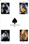
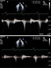
A 77-year-old Caucasian woman was examined by a primary care physician because of dyspnea and atypical chest pain. The ECG showed atrial fibrillation, mean heart rate of 85 bpm, right bundle branch block and 4-mm convex ST segment elevation in C4-C6 with biphasic T waves (Figure 1). Due to this atypical ECG, similar to a previous one performed two years before, rest-stress myocardial perfusion scintigraphy was prescribed. This revealed a reduction of left ventricular chamber volume suggestive of HCM without signs of ischemia or previous myocardial necrosis (Figure 2). The patient was referred to our echo lab in order to complete the diagnostic procedure. Transthoracic echocardiography (TTE) demonstrated AHCM, showing left ventricular (LV) apical hypertrophy with the typical ace of spades shape (Figure 3A1). The ace of spades was therefore studied in depth using color Doppler (Figure 3A2, Video 1), Sonovue contrast TTE (Figure 3A3, Video 2) and real-time three-dimensional echocardiography (Figure 3A4, Video 3). A small area of turbulent flow and a mid-ventricular gradient of about 63mmHg were found (Figure 4a). In order to evaluate possible modification of the mid-ventricular gradient, an intravenous bolus of atenolol (3mg) was administered. A significant decrease of about 40mmHg was observed while the ECG was unchanged (Figure 4b). Beta-blocker therapy was prescribed indefinitely. At six-month follow-up, the patient was asymptomatic and in good clinical condition.

Figure 1.

The ECG shows an unusual 4-mm convex ST segment elevation in C4–C6 with biphasic T waves.

(0.41MB).
Figure 2.

Rest-stress myocardial perfusion scintigraphy revealing reduced left ventricular chamber volume suggestive of hypertrophic cardiomyopathy without signs of ischemia or previous myocardial necrosis.

(0.8MB).
Figure 3.

Apical hypertrophic cardiomyopathy shows the ace up its sleeve. A1: two-dimensional transthoracic echocardiography; A2: color Doppler transthoracic echocardiography; A3: contrast-enhanced transthoracic echocardiography; A4: real-time three-dimensional reconstruction.

(0.28MB).
Figure 4.

Transthoracic echocardiography, apical 4-chamber view showing (a) a mid-ventricular gradient of about 63 mmHg and (b) a significant decrease in the gradient after administration of an intravenous bolus of atenolol.

(0.27MB).
Discussion

AHCM is characterized by segmental hypertrophy confined to the distal portion of the LV chamber. The prevalence of AHCM among HCM patients is 15% in Japan, whereas in the USA it is only 3%. Among AHCM patients 54% are symptomatic, the most common symptoms being chest pain, palpitations, dyspnea and syncope. This condition may cause various complications including atrial fibrillation, acute myocardial infarction, embolic events, ventricular fibrillation, congestive heart failure, apical aneurysm and cardiac arrest. Moreover, it may mimic other conditions, such as cardiac tumors, LV apical thrombus, isolated ventricular non-compaction, endomyocardial fibrosis and coronary artery disease. The most common ECG findings, seen in about 93% of patients, are negative T waves in the precordial leads (>10mm deep in 47%), followed by signs of LV hypertrophy in 63% of cases. TTE usually shows LV apical hypertrophy and is thus usually the standard diagnostic tool for AHCM. Symptomatic patients may be treated with verapamil, beta-blockers and antiarrhythmic drugs, all of which may improve the signs and symptoms of this condition.

In conclusion, AHCM is a clinical condition that can cause various complications and simulate ACS. Its diagnosis enables ACS to be excluded in patients with atypical ST-segment elevation. TTE is usually sufficient to make the diagnosis, enabling appropriate medical treatment.

Ethical disclosuresProtection of human and animal subjects

The authors declare that no experiments were performed on humans or animals for this study.

Confidentiality of data

The authors declare that no patient data appear in this article.

Right to privacy and informed consent

The authors declare that no patient data appear in this article.

Conflicts of interest

The authors have no conflicts of interest to declare.

Appendix A
Supplementary data

The following are the supplementary data to this article:

(0.01MB)

Transthoracic echocardiography, apical four chamber view, color comparison, showing left ventricular apical hypertrophy with the typical spade-shaped appearance.

(0.01MB)

Transthoracic echocardiography, apical four chamber view, echocontrastography with Sonovue improving the definition of the endorcardial border.

(0.01MB)

Off-line three dimensional reconstruction.

References
[1]
B.J. Maron, R.O. Bonow, T.N. Seshagiri, et al.
Hypertrophic cardiomyopathy with ventricular septal hypertrophy localized to the apical region of the left ventricle (apical hypertrophic cardiomyopathy).
Am J Cardiol, 49 (1982), pp. 1838-1848
[2]
C.S. Lin, C.H. Chen, P.Y. Ding.
Apical hypertrophic cardiomyopathy mimicking acute myocardial infarction.
Int J Cardiol, 64 (1998), pp. 305-307
[3]
L.M. Penas, P.I. Mosquera, Z.B. Bouzas, et al.
The electrocardiogram in apical hypertrophic myocardiopathy. A case report with unique manifestations.
Rev Esp Cardiol, 52 (1999), pp. 1148-1150
[4]
T. Sayın, T. Kocum, C. Kervancıoglu.
Apical hypertrophic cardiomyopathy mimics acute coronary syndrome.
Int J Cardiol, 80 (2001), pp. 77-79
Copyright © 2012. Sociedade Portuguesa de Cardiologia
Baixar PDF
Idiomas
Revista Portuguesa de Cardiologia
Opções de artigo
Ferramentas
Material Suplementar
en pt

Are you a health professional able to prescribe or dispense drugs?

Você é um profissional de saúde habilitado a prescrever ou dispensar medicamentos

Ao assinalar que é «Profissional de Saúde», declara conhecer e aceitar que a responsável pelo tratamento dos dados pessoais dos utilizadores da página de internet da Revista Portuguesa de Cardiologia (RPC), é esta entidade, com sede no Campo Grande, n.º 28, 13.º, 1700-093 Lisboa, com os telefones 217 970 685 e 217 817 630, fax 217 931 095 e com o endereço de correio eletrónico revista@spc.pt. Declaro para todos os fins, que assumo inteira responsabilidade pela veracidade e exatidão da afirmação aqui fornecida.