array:24 [
  "pii" => "S0870255114002881"
  "issn" => "08702551"
  "doi" => "10.1016/j.repc.2014.07.005"
  "estado" => "S300"
  "fechaPublicacion" => "2014-12-01"
  "aid" => "558"
  "copyright" => "Sociedade Portuguesa de Cardiologia"
  "copyrightAnyo" => "2014"
  "documento" => "article"
  "licencia" => "http://creativecommons.org/licenses/by-nc-nd/4.0/"
  "subdocumento" => "sco"
  "cita" => "Rev Port Cardiol. 2014;33:813-5"
  "abierto" => array:3 [
    "ES" => true
    "ES2" => true
    "LATM" => true
  ]
  "gratuito" => true
  "lecturas" => array:2 [
    "total" => 2975
    "formatos" => array:3 [
      "EPUB" => 190
      "HTML" => 2198
      "PDF" => 587
    ]
  ]
  "Traduccion" => array:1 [
    "en" => array:19 [
      "pii" => "S217420491400292X"
      "issn" => "21742049"
      "doi" => "10.1016/j.repce.2014.07.006"
      "estado" => "S300"
      "fechaPublicacion" => "2014-12-01"
      "aid" => "558"
      "copyright" => "Sociedade Portuguesa de Cardiologia"
      "documento" => "article"
      "licencia" => "http://creativecommons.org/licenses/by-nc-nd/4.0/"
      "subdocumento" => "sco"
      "cita" => "Rev Port Cardiol. 2014;33:813-5"
      "abierto" => array:3 [
        "ES" => true
        "ES2" => true
        "LATM" => true
      ]
      "gratuito" => true
      "lecturas" => array:2 [
        "total" => 2451
        "formatos" => array:3 [
          "EPUB" => 155
          "HTML" => 1798
          "PDF" => 498
        ]
      ]
      "en" => array:11 [
        "idiomaDefecto" => true
        "cabecera" => "<span class="elsevierStyleTextfn">Image in Cardiology</span>"
        "titulo" => "Acute aortic dissection complicated by aorto-right ventricular fistula"
        "tienePdf" => "en"
        "tieneTextoCompleto" => "en"
        "paginas" => array:1 [
          0 => array:2 [
            "paginaInicial" => "813"
            "paginaFinal" => "815"
          ]
        ]
        "titulosAlternativos" => array:1 [
          "pt" => array:1 [
            "titulo" => "Disse&#231;&#227;o aguda da aorta complicada por f&#237;stula aorta-ventr&#237;culo direita"
          ]
        ]
        "contieneTextoCompleto" => array:1 [
          "en" => true
        ]
        "contienePdf" => array:1 [
          "en" => true
        ]
        "resumenGrafico" => array:2 [
          "original" => 0
          "multimedia" => array:7 [
            "identificador" => "fig0015"
            "etiqueta" => "Figure 3"
            "tipo" => "MULTIMEDIAFIGURA"
            "mostrarFloat" => true
            "mostrarDisplay" => false
            "figura" => array:1 [
              0 => array:4 [
                "imagen" => "gr3.jpeg"
                "Alto" => 953
                "Ancho" => 953
                "Tamanyo" => 157921
              ]
            ]
            "descripcion" => array:1 [
              "en" => "<p id="spar0015" class="elsevierStyleSimplePara elsevierViewall">Computed tomography angiography demonstrating aortic root dissection and a fistula arising from the false lumen&#46;</p>"
            ]
          ]
        ]
        "autores" => array:1 [
          0 => array:2 [
            "autoresLista" => "Paulo Fonseca, Adelaide Dias, Nuno Bettencourt, Nuno Ferreira, Concei&#231;&#227;o Fonseca, Vasco Gama"
            "autores" => array:6 [
              0 => array:2 [
                "nombre" => "Paulo"
                "apellidos" => "Fonseca"
              ]
              1 => array:2 [
                "nombre" => "Adelaide"
                "apellidos" => "Dias"
              ]
              2 => array:2 [
                "nombre" => "Nuno"
                "apellidos" => "Bettencourt"
              ]
              3 => array:2 [
                "nombre" => "Nuno"
                "apellidos" => "Ferreira"
              ]
              4 => array:2 [
                "nombre" => "Concei&#231;&#227;o"
                "apellidos" => "Fonseca"
              ]
              5 => array:2 [
                "nombre" => "Vasco"
                "apellidos" => "Gama"
              ]
            ]
          ]
        ]
      ]
      "idiomaDefecto" => "en"
      "Traduccion" => array:1 [
        "en" => array:9 [
          "pii" => "S0870255114002881"
          "doi" => "10.1016/j.repc.2014.07.005"
          "estado" => "S300"
          "subdocumento" => ""
          "abierto" => array:3 [
            "ES" => true
            "ES2" => true
            "LATM" => true
          ]
          "gratuito" => true
          "lecturas" => array:1 [
            "total" => 0
          ]
          "idiomaDefecto" => "en"
          "EPUB" => "https://multimedia.elsevier.es/PublicationsMultimediaV1/item/epub/S0870255114002881?idApp=UINPBA00004E"
        ]
      ]
      "EPUB" => "https://multimedia.elsevier.es/PublicationsMultimediaV1/item/epub/S217420491400292X?idApp=UINPBA00004E"
      "url" => "/21742049/0000003300000012/v1_201501081043/S217420491400292X/v1_201501081043/en/main.assets"
    ]
  ]
  "itemSiguiente" => array:19 [
    "pii" => "S0870255114002613"
    "issn" => "08702551"
    "doi" => "10.1016/j.repc.2014.08.003"
    "estado" => "S300"
    "fechaPublicacion" => "2014-12-01"
    "aid" => "546"
    "copyright" => "Sociedade Portuguesa de Cardiologia"
    "documento" => "simple-article"
    "licencia" => "http://creativecommons.org/licenses/by-nc-nd/4.0/"
    "subdocumento" => "cor"
    "cita" => "Rev Port Cardiol. 2014;33:817-8"
    "abierto" => array:3 [
      "ES" => true
      "ES2" => true
      "LATM" => true
    ]
    "gratuito" => true
    "lecturas" => array:2 [
      "total" => 2559
      "formatos" => array:3 [
        "EPUB" => 157
        "HTML" => 1803
        "PDF" => 599
      ]
    ]
    "pt" => array:11 [
      "idiomaDefecto" => true
      "cabecera" => "<span class="elsevierStyleTextfn">Carta ao Editor</span>"
      "titulo" => "Lutar contra a mortalidade por doen&#231;as cardiovasculares&#58; um desafio para a sociedade &#8211; apenas depois dos profissionais de sa&#250;de definirem o seu rumo"
      "tienePdf" => "pt"
      "tieneTextoCompleto" => "pt"
      "paginas" => array:1 [
        0 => array:2 [
          "paginaInicial" => "817"
          "paginaFinal" => "818"
        ]
      ]
      "titulosAlternativos" => array:1 [
        "en" => array:1 [
          "titulo" => "Fighting mortality from cardiovascular disease&#58; A challenge to society &#8211; but only after health professionals have shown the way"
        ]
      ]
      "contieneTextoCompleto" => array:1 [
        "pt" => true
      ]
      "contienePdf" => array:1 [
        "pt" => true
      ]
      "resumenGrafico" => array:2 [
        "original" => 0
        "multimedia" => array:7 [
          "identificador" => "fig0005"
          "etiqueta" => "Figura 1"
          "tipo" => "MULTIMEDIAFIGURA"
          "mostrarFloat" => true
          "mostrarDisplay" => false
          "figura" => array:1 [
            0 => array:4 [
              "imagen" => "gr1.jpeg"
              "Alto" => 1024
              "Ancho" => 1653
              "Tamanyo" => 78182
            ]
          ]
          "descripcion" => array:1 [
            "pt" => "<p id="spar0005" class="elsevierStyleSimplePara elsevierViewall">Evolu&#231;&#227;o do n&#250;mero de tromb&#243;lises endovenosas realizadas no per&#237;odo&#46;</p>"
          ]
        ]
      ]
      "autores" => array:1 [
        0 => array:2 [
          "autoresLista" => "Miguel Soares&#8208;Oliveira"
          "autores" => array:1 [
            0 => array:2 [
              "nombre" => "Miguel"
              "apellidos" => "Soares&#8208;Oliveira"
            ]
          ]
        ]
      ]
    ]
    "idiomaDefecto" => "pt"
    "Traduccion" => array:1 [
      "en" => array:9 [
        "pii" => "S2174204914002621"
        "doi" => "10.1016/j.repce.2014.08.004"
        "estado" => "S300"
        "subdocumento" => ""
        "abierto" => array:3 [
          "ES" => true
          "ES2" => true
          "LATM" => true
        ]
        "gratuito" => true
        "lecturas" => array:1 [
          "total" => 0
        ]
        "idiomaDefecto" => "en"
        "EPUB" => "https://multimedia.elsevier.es/PublicationsMultimediaV1/item/epub/S2174204914002621?idApp=UINPBA00004E"
      ]
    ]
    "EPUB" => "https://multimedia.elsevier.es/PublicationsMultimediaV1/item/epub/S0870255114002613?idApp=UINPBA00004E"
    "url" => "/08702551/0000003300000012/v1_201412140112/S0870255114002613/v1_201412140112/pt/main.assets"
  ]
  "itemAnterior" => array:19 [
    "pii" => "S0870255114002868"
    "issn" => "08702551"
    "doi" => "10.1016/j.repc.2014.06.008"
    "estado" => "S300"
    "fechaPublicacion" => "2014-12-01"
    "aid" => "556"
    "copyright" => "Sociedade Portuguesa de Cardiologia"
    "documento" => "article"
    "licencia" => "http://creativecommons.org/licenses/by-nc-nd/4.0/"
    "subdocumento" => "sco"
    "cita" => "Rev Port Cardiol. 2014;33:811-2"
    "abierto" => array:3 [
      "ES" => true
      "ES2" => true
      "LATM" => true
    ]
    "gratuito" => true
    "lecturas" => array:2 [
      "total" => 3328
      "formatos" => array:3 [
        "EPUB" => 150
        "HTML" => 2556
        "PDF" => 622
      ]
    ]
    "en" => array:11 [
      "idiomaDefecto" => true
      "cabecera" => "<span class="elsevierStyleTextfn">Image in Cardiology</span>"
      "titulo" => "Abdominal aortic aneurysm &#8211; Characterization by three-dimensional computed tomography angiography"
      "tienePdf" => "en"
      "tieneTextoCompleto" => "en"
      "paginas" => array:1 [
        0 => array:2 [
          "paginaInicial" => "811"
          "paginaFinal" => "812"
        ]
      ]
      "titulosAlternativos" => array:1 [
        "pt" => array:1 [
          "titulo" => "Aneurisma da aorta abdominal &#8211; Caracteriza&#231;&#227;o por angiotomografia computadorizada tridimensional"
        ]
      ]
      "contieneTextoCompleto" => array:1 [
        "en" => true
      ]
      "contienePdf" => array:1 [
        "en" => true
      ]
      "resumenGrafico" => array:2 [
        "original" => 0
        "multimedia" => array:7 [
          "identificador" => "fig0005"
          "etiqueta" => "Figure 1"
          "tipo" => "MULTIMEDIAFIGURA"
          "mostrarFloat" => true
          "mostrarDisplay" => false
          "figura" => array:1 [
            0 => array:4 [
              "imagen" => "gr1.jpeg"
              "Alto" => 995
              "Ancho" => 1500
              "Tamanyo" => 179218
            ]
          ]
          "descripcion" => array:1 [
            "en" => "<p id="spar0005" class="elsevierStyleSimplePara elsevierViewall">Contrast-enhanced computed tomography image of the abdominal aortic aneurysm in coronal view &#40;A&#41; and sagittal view &#40;B&#41;&#46;</p>"
          ]
        ]
      ]
      "autores" => array:1 [
        0 => array:2 [
          "autoresLista" => "Jo&#227;o Caiano Gil, Jorge Martins, Ant&#243;nio Furtado"
          "autores" => array:3 [
            0 => array:2 [
              "nombre" => "Jo&#227;o"
              "apellidos" => "Caiano Gil"
            ]
            1 => array:2 [
              "nombre" => "Jorge"
              "apellidos" => "Martins"
            ]
            2 => array:2 [
              "nombre" => "Ant&#243;nio"
              "apellidos" => "Furtado"
            ]
          ]
        ]
      ]
    ]
    "idiomaDefecto" => "en"
    "Traduccion" => array:1 [
      "en" => array:9 [
        "pii" => "S2174204914002918"
        "doi" => "10.1016/j.repce.2014.06.009"
        "estado" => "S300"
        "subdocumento" => ""
        "abierto" => array:3 [
          "ES" => true
          "ES2" => true
          "LATM" => true
        ]
        "gratuito" => true
        "lecturas" => array:1 [
          "total" => 0
        ]
        "idiomaDefecto" => "en"
        "EPUB" => "https://multimedia.elsevier.es/PublicationsMultimediaV1/item/epub/S2174204914002918?idApp=UINPBA00004E"
      ]
    ]
    "EPUB" => "https://multimedia.elsevier.es/PublicationsMultimediaV1/item/epub/S0870255114002868?idApp=UINPBA00004E"
    "url" => "/08702551/0000003300000012/v1_201412140112/S0870255114002868/v1_201412140112/en/main.assets"
  ]
  "en" => array:15 [
    "idiomaDefecto" => true
    "cabecera" => "<span class="elsevierStyleTextfn">Image in Cardiology</span>"
    "titulo" => "Acute aortic dissection complicated by aorto-right ventricular fistula"
    "tieneTextoCompleto" => true
    "paginas" => array:1 [
      0 => array:2 [
        "paginaInicial" => "813"
        "paginaFinal" => "815"
      ]
    ]
    "autores" => array:1 [
      0 => array:4 [
        "autoresLista" => "Paulo Fonseca, Adelaide Dias, Nuno Bettencourt, Nuno Ferreira, Concei&#231;&#227;o Fonseca, Vasco Gama"
        "autores" => array:6 [
          0 => array:4 [
            "nombre" => "Paulo"
            "apellidos" => "Fonseca"
            "email" => array:1 [
              0 => "paulobarbosafonseca&#64;hotmail&#46;com"
            ]
            "referencia" => array:1 [
              0 => array:2 [
                "etiqueta" => "<span class="elsevierStyleSup">&#42;</span>"
                "identificador" => "cor0005"
              ]
            ]
          ]
          1 => array:2 [
            "nombre" => "Adelaide"
            "apellidos" => "Dias"
          ]
          2 => array:2 [
            "nombre" => "Nuno"
            "apellidos" => "Bettencourt"
          ]
          3 => array:2 [
            "nombre" => "Nuno"
            "apellidos" => "Ferreira"
          ]
          4 => array:2 [
            "nombre" => "Concei&#231;&#227;o"
            "apellidos" => "Fonseca"
          ]
          5 => array:2 [
            "nombre" => "Vasco"
            "apellidos" => "Gama"
          ]
        ]
        "afiliaciones" => array:1 [
          0 => array:2 [
            "entidad" => "Servi&#231;o de Cardiologia&#44; Centro Hospitalar de Vila Nova de Gaia&#47;Espinho&#44; Vila Nova de Gaia&#44; Portugal"
            "identificador" => "aff0005"
          ]
        ]
        "correspondencia" => array:1 [
          0 => array:3 [
            "identificador" => "cor0005"
            "etiqueta" => "&#8270;"
            "correspondencia" => "Corresponding author&#46;"
          ]
        ]
      ]
    ]
    "titulosAlternativos" => array:1 [
      "pt" => array:1 [
        "titulo" => "Disse&#231;&#227;o aguda da aorta complicada por f&#237;stula aorta-ventr&#237;culo direita"
      ]
    ]
    "resumenGrafico" => array:2 [
      "original" => 0
      "multimedia" => array:7 [
        "identificador" => "fig0020"
        "etiqueta" => "Figure 4"
        "tipo" => "MULTIMEDIAFIGURA"
        "mostrarFloat" => true
        "mostrarDisplay" => false
        "figura" => array:1 [
          0 => array:4 [
            "imagen" => "gr4.jpeg"
            "Alto" => 1585
            "Ancho" => 1583
            "Tamanyo" => 356494
          ]
        ]
        "descripcion" => array:1 [
          "en" => "<p id="spar0020" class="elsevierStyleSimplePara elsevierViewall">Three-dimensional computed tomography angiography showing a fistula between the aortic root and right ventricle&#46;</p>"
        ]
      ]
    ]
    "textoCompleto" => "<span class="elsevierStyleSections"><p id="par0005" class="elsevierStylePara elsevierViewall">A 68-year-old man with a history of systemic hypertension and chronic renal failure was admitted to our emergency department with chest pain and dyspnea&#46; He had undergone tube graft replacement of the ascending aorta due to an acute type A aortic dissection 10 years previously&#46; On admission he was hypertensive and tachycardic with a continuous murmur at the left upper sternal border&#46; The electrocardiogram revealed nonspecific repolarization abnormalities and the transthoracic echocardiogram showed a severely dilated aortic root with an intimal flap and continuous turbulent flow from the aortic root toward the right ventricle &#40;<a class="elsevierStyleCrossRefs" href="#fig0005">Figures 1 and 2</a>&#41;&#46; There was moderate aortic regurgitation caused by poor leaflet coaptation&#46; The right chambers were slightly dilated and both ventricles had normal systolic function&#46; Chest computed tomography confirmed aortic root dissection with a fistula between the aortic false lumen and right ventricle &#40;<a class="elsevierStyleCrossRefs" href="#fig0015">Figures 3 and 4</a>&#41;&#46; The coronary arteries were not involved&#46; The patient was rejected for surgery due to very high surgical risk and died a few days later after sudden hypotension&#46;</p><elsevierMultimedia ident="fig0005"></elsevierMultimedia><elsevierMultimedia ident="fig0010"></elsevierMultimedia><elsevierMultimedia ident="fig0015"></elsevierMultimedia><elsevierMultimedia ident="fig0020"></elsevierMultimedia><p id="par0010" class="elsevierStylePara elsevierViewall">Aorto-right ventricular fistula is a very rare complication of aortic dissection&#46; A significant proportion of patients with aortic dissection complicated by a fistula to the cardiac chambers have a history of prior cardiac surgery&#44; predominantly aortic valve surgery&#46; Postoperative pericardial adhesions may prevent free aortic rupture into the pericardial space&#44; increasing the predisposition to penetrate into a neighboring cardiac chamber&#46;</p><span id="sec0005" class="elsevierStyleSection elsevierViewall"><span class="elsevierStyleSectionTitle" id="sect0005">Ethical disclosures</span><span id="sec0010" class="elsevierStyleSection elsevierViewall"><span class="elsevierStyleSectionTitle" id="sect0010">Protection of human and animal subjects</span><p id="par0015" class="elsevierStylePara elsevierViewall">The authors declare that no experiments were performed on humans or animals for this study&#46;</p></span><span id="sec0015" class="elsevierStyleSection elsevierViewall"><span class="elsevierStyleSectionTitle" id="sect0015">Confidentiality of data</span><p id="par0020" class="elsevierStylePara elsevierViewall">The authors declare that they have followed the protocols of their work center on the publication of patient data&#46;</p></span><span id="sec0020" class="elsevierStyleSection elsevierViewall"><span class="elsevierStyleSectionTitle" id="sect0020">Right to privacy and informed consent</span><p id="par0025" class="elsevierStylePara elsevierViewall">The authors have obtained the written informed consent of the patients or subjects mentioned in the article&#46; The corresponding author is in possession of this document&#46;</p></span></span><span id="sec0025" class="elsevierStyleSection elsevierViewall"><span class="elsevierStyleSectionTitle" id="sect0025">Conflicts of interest</span><p id="par0030" class="elsevierStylePara elsevierViewall">The authors have no conflicts of interest to declare&#46;</p></span></span>"
    "textoCompletoSecciones" => array:1 [
      "secciones" => array:2 [
        0 => array:3 [
          "identificador" => "sec0005"
          "titulo" => "Ethical disclosures"
          "secciones" => array:3 [
            0 => array:2 [
              "identificador" => "sec0010"
              "titulo" => "Protection of human and animal subjects"
            ]
            1 => array:2 [
              "identificador" => "sec0015"
              "titulo" => "Confidentiality of data"
            ]
            2 => array:2 [
              "identificador" => "sec0020"
              "titulo" => "Right to privacy and informed consent"
            ]
          ]
        ]
        1 => array:2 [
          "identificador" => "sec0025"
          "titulo" => "Conflicts of interest"
        ]
      ]
    ]
    "pdfFichero" => "main.pdf"
    "tienePdf" => true
    "fechaRecibido" => "2014-06-02"
    "fechaAceptado" => "2014-07-31"
    "multimedia" => array:4 [
      0 => array:7 [
        "identificador" => "fig0005"
        "etiqueta" => "Figure 1"
        "tipo" => "MULTIMEDIAFIGURA"
        "mostrarFloat" => true
        "mostrarDisplay" => false
        "figura" => array:1 [
          0 => array:4 [
            "imagen" => "gr1.jpeg"
            "Alto" => 1560
            "Ancho" => 1650
            "Tamanyo" => 211393
          ]
        ]
        "descripcion" => array:1 [
          "en" => "<p id="spar0005" class="elsevierStyleSimplePara elsevierViewall">Transthoracic echocardiography&#44; parasternal short-axis view with color Doppler&#44; demonstrating an aortic intimal flap and turbulent flow from the aortic root toward the right ventricle&#46;</p>"
        ]
      ]
      1 => array:7 [
        "identificador" => "fig0010"
        "etiqueta" => "Figure 2"
        "tipo" => "MULTIMEDIAFIGURA"
        "mostrarFloat" => true
        "mostrarDisplay" => false
        "figura" => array:1 [
          0 => array:4 [
            "imagen" => "gr2.jpeg"
            "Alto" => 1755
            "Ancho" => 2500
            "Tamanyo" => 508166
          ]
        ]
        "descripcion" => array:1 [
          "en" => "<p id="spar0010" class="elsevierStyleSimplePara elsevierViewall">Continuous Doppler echocardiography showing continuous flow between the aortic root and right ventricle&#46;</p>"
        ]
      ]
      2 => array:7 [
        "identificador" => "fig0015"
        "etiqueta" => "Figure 3"
        "tipo" => "MULTIMEDIAFIGURA"
        "mostrarFloat" => true
        "mostrarDisplay" => false
        "figura" => array:1 [
          0 => array:4 [
            "imagen" => "gr3.jpeg"
            "Alto" => 1583
            "Ancho" => 1583
            "Tamanyo" => 253361
          ]
        ]
        "descripcion" => array:1 [
          "en" => "<p id="spar0015" class="elsevierStyleSimplePara elsevierViewall">Computed tomography angiography demonstrating aortic root dissection and a fistula arising from the false lumen&#46;</p>"
        ]
      ]
      3 => array:7 [
        "identificador" => "fig0020"
        "etiqueta" => "Figure 4"
        "tipo" => "MULTIMEDIAFIGURA"
        "mostrarFloat" => true
        "mostrarDisplay" => false
        "figura" => array:1 [
          0 => array:4 [
            "imagen" => "gr4.jpeg"
            "Alto" => 1585
            "Ancho" => 1583
            "Tamanyo" => 356494
          ]
        ]
        "descripcion" => array:1 [
          "en" => "<p id="spar0020" class="elsevierStyleSimplePara elsevierViewall">Three-dimensional computed tomography angiography showing a fistula between the aortic root and right ventricle&#46;</p>"
        ]
      ]
    ]
  ]
  "idiomaDefecto" => "en"
  "url" => "/08702551/0000003300000012/v1_201412140112/S0870255114002881/v1_201412140112/en/main.assets"
  "Apartado" => array:4 [
    "identificador" => "357"
    "tipo" => "SECCION"
    "pt" => array:2 [
      "titulo" => "Imagens em cardiologia"
      "idiomaDefecto" => true
    ]
    "idiomaDefecto" => "pt"
  ]
  "PDF" => "https://static.elsevier.es/multimedia/08702551/0000003300000012/v1_201412140112/S0870255114002881/v1_201412140112/en/main.pdf?idApp=UINPBA00004E&text.app=https://revportcardiol.org/"
  "EPUB" => "https://multimedia.elsevier.es/PublicationsMultimediaV1/item/epub/S0870255114002881?idApp=UINPBA00004E"
]
Partilhar
Informação da revista
Vol. 33. Núm. 12.
Páginas 813-815 (dezembro 2014)
Partilhar
Partilhar
Baixar PDF
Mais opções do artigo
Vol. 33. Núm. 12.
Páginas 813-815 (dezembro 2014)
Image in Cardiology
Open Access
Acute aortic dissection complicated by aorto-right ventricular fistula
Disseção aguda da aorta complicada por fístula aorta-ventrículo direita
Visitas
6086
Paulo Fonseca
Autor para correspondência
paulobarbosafonseca@hotmail.com

Corresponding author.
, Adelaide Dias, Nuno Bettencourt, Nuno Ferreira, Conceição Fonseca, Vasco Gama
Serviço de Cardiologia, Centro Hospitalar de Vila Nova de Gaia/Espinho, Vila Nova de Gaia, Portugal
Este item recebeu

Under a Creative Commons license
Informação do artigo
Texto Completo
Baixar PDF
Estatísticas
Figuras (4)
Mostrar maisMostrar menos
Texto Completo

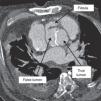
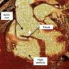
A 68-year-old man with a history of systemic hypertension and chronic renal failure was admitted to our emergency department with chest pain and dyspnea. He had undergone tube graft replacement of the ascending aorta due to an acute type A aortic dissection 10 years previously. On admission he was hypertensive and tachycardic with a continuous murmur at the left upper sternal border. The electrocardiogram revealed nonspecific repolarization abnormalities and the transthoracic echocardiogram showed a severely dilated aortic root with an intimal flap and continuous turbulent flow from the aortic root toward the right ventricle (Figures 1 and 2). There was moderate aortic regurgitation caused by poor leaflet coaptation. The right chambers were slightly dilated and both ventricles had normal systolic function. Chest computed tomography confirmed aortic root dissection with a fistula between the aortic false lumen and right ventricle (Figures 3 and 4). The coronary arteries were not involved. The patient was rejected for surgery due to very high surgical risk and died a few days later after sudden hypotension.

Figure 1.

Transthoracic echocardiography, parasternal short-axis view with color Doppler, demonstrating an aortic intimal flap and turbulent flow from the aortic root toward the right ventricle.

(0.2MB).
Figure 2.

Continuous Doppler echocardiography showing continuous flow between the aortic root and right ventricle.

(0.48MB).
Figure 3.

Computed tomography angiography demonstrating aortic root dissection and a fistula arising from the false lumen.

(0.24MB).
Figure 4.

Three-dimensional computed tomography angiography showing a fistula between the aortic root and right ventricle.

(0.34MB).

Aorto-right ventricular fistula is a very rare complication of aortic dissection. A significant proportion of patients with aortic dissection complicated by a fistula to the cardiac chambers have a history of prior cardiac surgery, predominantly aortic valve surgery. Postoperative pericardial adhesions may prevent free aortic rupture into the pericardial space, increasing the predisposition to penetrate into a neighboring cardiac chamber.

Ethical disclosuresProtection of human and animal subjects

The authors declare that no experiments were performed on humans or animals for this study.

Confidentiality of data

The authors declare that they have followed the protocols of their work center on the publication of patient data.

Right to privacy and informed consent

The authors have obtained the written informed consent of the patients or subjects mentioned in the article. The corresponding author is in possession of this document.

Conflicts of interest

The authors have no conflicts of interest to declare.

Copyright © 2014. Sociedade Portuguesa de Cardiologia
Baixar PDF
Idiomas
Revista Portuguesa de Cardiologia
Opções de artigo
Ferramentas
en pt

Are you a health professional able to prescribe or dispense drugs?

Você é um profissional de saúde habilitado a prescrever ou dispensar medicamentos

Ao assinalar que é «Profissional de Saúde», declara conhecer e aceitar que a responsável pelo tratamento dos dados pessoais dos utilizadores da página de internet da Revista Portuguesa de Cardiologia (RPC), é esta entidade, com sede no Campo Grande, n.º 28, 13.º, 1700-093 Lisboa, com os telefones 217 970 685 e 217 817 630, fax 217 931 095 e com o endereço de correio eletrónico revista@spc.pt. Declaro para todos os fins, que assumo inteira responsabilidade pela veracidade e exatidão da afirmação aqui fornecida.