


Epicardial fat most commonly produces a hypoechogenic image on the transthoracic echocardiogram (TTE), nevertheless it can be hyperechogenic if very thick. Epicardial lipomatosis is a condition in which this fat layer is highly enhanced due to adipocyte hyperplasia. Its appearance on TTE can be misleading, as it can mimic other conditions, but cardiovascular magnetic resonance (CMR) allows clear identification of this entity. We present a case that demonstrates the important role CMR plays in establishing a diagnosis of epicardial lipomatosis.
A 79-year-old woman was referred for emergency cardiac surgery due to myocardial infarction of undetermined age, with echocardiographic suspicion of left ventricular (LV) free wall rupture.
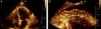
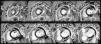
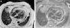
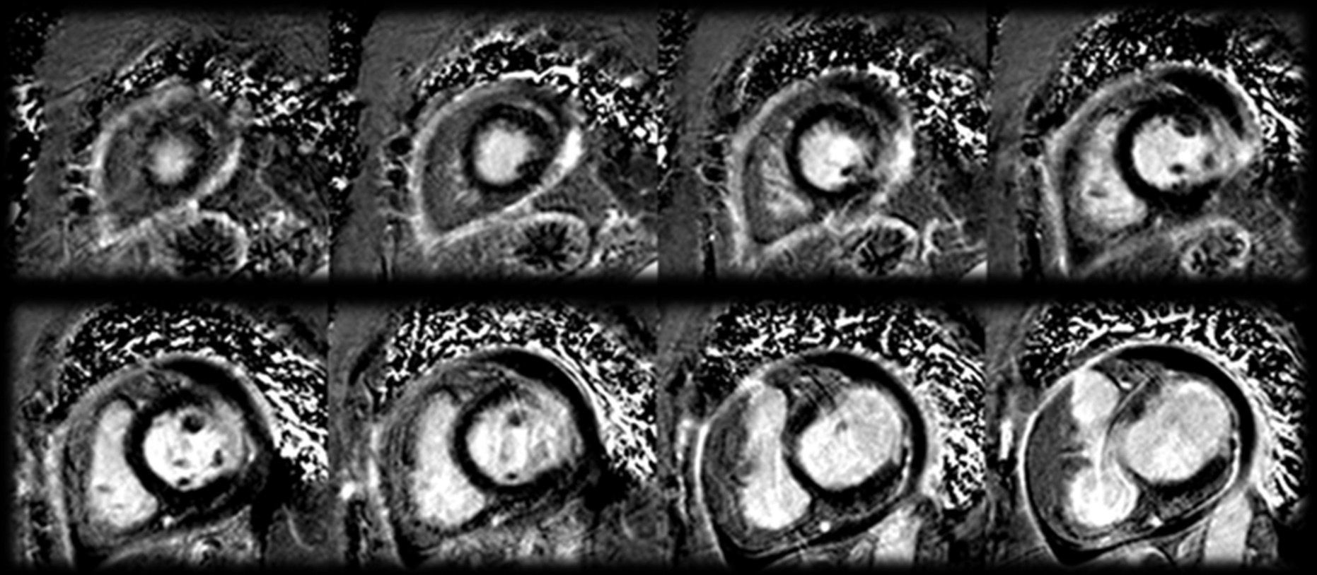
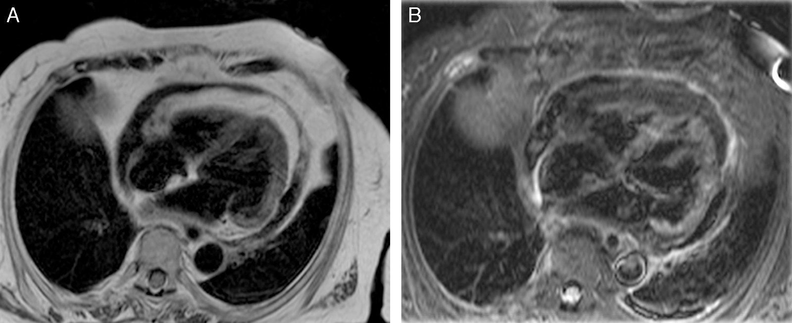
On admission, TTE was repeated, showing a moderate circumferential pericardial effusion, with a hyperechogenic image suggestive of thrombus or fibrin, extending from the LV apex to the posterior and lateral walls, without contrast leakage to the pericardium (Figure 1). Cardiac catheterization revealed angiographically normal coronary arteries. CMR was performed that confirmed the moderate pericardial effusion and showed mild to moderate depression of LV systolic function with akinesia and transmural late gadolinium enhancement in the basal and mid lateral wall segments with prominent epicardial lipomatosis, corresponding to the thrombus/fibrin image on TTE (Figures 2 and 3). This information ruled out the initial suspicion and the patient was treated conservatively. Hospital stay was uneventful and she was discharged 15 days later with a diagnosis of lateral wall myocardial infarction with angiographically normal coronary arteries.

The authors declare that no experiments were performed on humans or animals for this investigation.
Confidentiality of dataThe authors declare that no patient data appears in this article.
Right to privacy and informed consent.The authors declare that no patient data appears in this article.
Conflicts of interestThe authors have no conflicts of interest to declare.
Please cite this article as: Magalhães P, Bettencourt N, Sampaio F, et al. O que não mata engorda!. Rev Port Cardiol. 2015;34:295–296.
