


A 61-year-old patient underwent transthoracic echocardiography which showed left ventricular (LV) hypertrophy and a structure with similar density to the myocardium, located at the level of the LV mid segments (Figure 1A and B, red arrows), dividing the ventricle into two parts and communicating via a small flow (Figure 1C, orange arrow). The basal half of the LV was hypertrophied and trabeculated and presented preserved myocardial contraction, while the apical half was aneurysmatic. A thrombus was detected on follow-up echocardiography (Figure 1D).
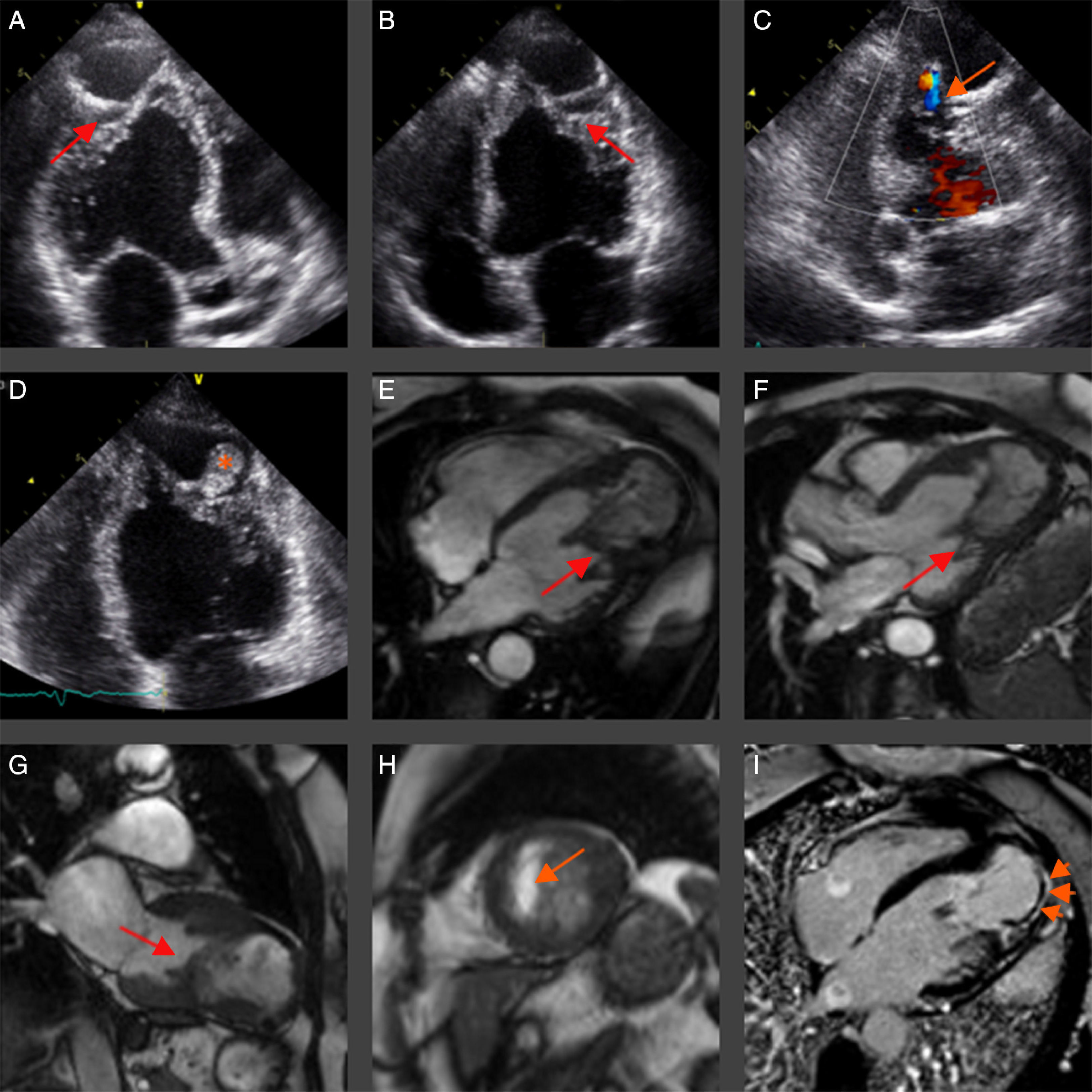
Cardiac magnetic resonance showed LV hypertrophy with maximum wall thickness of 17 mm at the inferoseptal wall mid segment. The posteromedial papillary muscle presented abnormal insertion at the inferolateral and anteroseptal walls and fusion with the anterolateral papillary muscle, forming a muscular band which divided the LV cavity (Figures 1E–H). The apical segments presented heterogeneous intramural late gadolinium enhancement (Figure 1I, arrowheads).
Hypertrophic cardiomyopathy has an estimated prevalence of 1 in 500 individuals. However, the presence of a ventricular aneurysm is quite rare, described in only 2% of these patients, and carries a poor prognosis. Overt heart failure, embolic events and ventricular dysrhythmia are possible complications that often cause morbidity and mortality.
This clinical case stands out from previous cases described in the literature, because the apical aneurysm is very large and the displacement of papillary muscles is very pronounced, giving the appearance of separating the ventricle into two chambers.
Ethical disclosuresProtection of human and animal subjectsThe authors declare that no experiments were performed on humans or animals for this study.
Confidentiality of dataThe authors declare that no patient data appear in this article.
Right to privacy and informed consentThe authors declare that no patient data appear in this article.
Conflicts of interestThe authors have no conflicts of interest to declare.
