array:23 [
  "pii" => "S2174204915000288"
  "issn" => "21742049"
  "doi" => "10.1016/j.repce.2015.02.002"
  "estado" => "S300"
  "fechaPublicacion" => "2015-03-01"
  "aid" => "594"
  "copyright" => "Sociedade Portuguesa de Cardiologia"
  "copyrightAnyo" => "2014"
  "documento" => "article"
  "licencia" => "http://creativecommons.org/licenses/by-nc-nd/4.0/"
  "subdocumento" => "sco"
  "cita" => "Rev Port Cardiol. 2015;34:221-2"
  "abierto" => array:3 [
    "ES" => true
    "ES2" => true
    "LATM" => true
  ]
  "gratuito" => true
  "lecturas" => array:2 [
    "total" => 3028
    "formatos" => array:3 [
      "EPUB" => 146
      "HTML" => 2278
      "PDF" => 604
    ]
  ]
  "Traduccion" => array:1 [
    "pt" => array:19 [
      "pii" => "S0870255115000141"
      "issn" => "08702551"
      "doi" => "10.1016/j.repc.2014.11.004"
      "estado" => "S300"
      "fechaPublicacion" => "2015-03-01"
      "aid" => "594"
      "copyright" => "Sociedade Portuguesa de Cardiologia"
      "documento" => "article"
      "licencia" => "http://creativecommons.org/licenses/by-nc-nd/4.0/"
      "subdocumento" => "sco"
      "cita" => "Rev Port Cardiol. 2015;34:221-2"
      "abierto" => array:3 [
        "ES" => true
        "ES2" => true
        "LATM" => true
      ]
      "gratuito" => true
      "lecturas" => array:2 [
        "total" => 3312
        "formatos" => array:3 [
          "EPUB" => 185
          "HTML" => 2392
          "PDF" => 735
        ]
      ]
      "pt" => array:11 [
        "idiomaDefecto" => true
        "cabecera" => "<span class="elsevierStyleTextfn">Imagem em Cardiologia</span>"
        "titulo" => "Utiliza&#231;&#227;o de sistema de extra&#231;&#227;o <span class="elsevierStyleItalic">Snare</span> para implanta&#231;&#227;o de eletrocateter ventricular esquerdo na ressincroniza&#231;&#227;o card&#237;aca"
        "tienePdf" => "pt"
        "tieneTextoCompleto" => "pt"
        "paginas" => array:1 [
          0 => array:2 [
            "paginaInicial" => "221"
            "paginaFinal" => "222"
          ]
        ]
        "titulosAlternativos" => array:1 [
          "en" => array:1 [
            "titulo" => "Snare system for left ventricular lead placement in cardiac resynchronization therapy"
          ]
        ]
        "contieneTextoCompleto" => array:1 [
          "pt" => true
        ]
        "contienePdf" => array:1 [
          "pt" => true
        ]
        "resumenGrafico" => array:2 [
          "original" => 0
          "multimedia" => array:7 [
            "identificador" => "fig0020"
            "etiqueta" => "Figura 4"
            "tipo" => "MULTIMEDIAFIGURA"
            "mostrarFloat" => true
            "mostrarDisplay" => false
            "figura" => array:1 [
              0 => array:4 [
                "imagen" => "gr4.jpeg"
                "Alto" => 851
                "Ancho" => 900
                "Tamanyo" => 54935
              ]
            ]
            "descripcion" => array:1 [
              "pt" => "<p id="spar0020" class="elsevierStyleSimplePara elsevierViewall">Posicionamento final dos eletrocateteres em sistema de ressincroniza&#231;&#227;o card&#237;aca com fun&#231;&#227;o de desfibrilha&#231;&#227;o&#46;</p>"
            ]
          ]
        ]
        "autores" => array:1 [
          0 => array:2 [
            "autoresLista" => "Andreia Magalh&#227;es, Miguel Menezes, Nuno Cortez&#8208;Dias, Jo&#227;o de Sousa, Pedro Marques"
            "autores" => array:5 [
              0 => array:2 [
                "nombre" => "Andreia"
                "apellidos" => "Magalh&#227;es"
              ]
              1 => array:2 [
                "nombre" => "Miguel"
                "apellidos" => "Menezes"
              ]
              2 => array:2 [
                "nombre" => "Nuno"
                "apellidos" => "Cortez&#8208;Dias"
              ]
              3 => array:2 [
                "nombre" => "Jo&#227;o"
                "apellidos" => "de Sousa"
              ]
              4 => array:2 [
                "nombre" => "Pedro"
                "apellidos" => "Marques"
              ]
            ]
          ]
        ]
      ]
      "idiomaDefecto" => "pt"
      "Traduccion" => array:1 [
        "en" => array:9 [
          "pii" => "S2174204915000288"
          "doi" => "10.1016/j.repce.2015.02.002"
          "estado" => "S300"
          "subdocumento" => ""
          "abierto" => array:3 [
            "ES" => true
            "ES2" => true
            "LATM" => true
          ]
          "gratuito" => true
          "lecturas" => array:1 [
            "total" => 0
          ]
          "idiomaDefecto" => "en"
          "EPUB" => "https://multimedia.elsevier.es/PublicationsMultimediaV1/item/epub/S2174204915000288?idApp=UINPBA00004E"
        ]
      ]
      "EPUB" => "https://multimedia.elsevier.es/PublicationsMultimediaV1/item/epub/S0870255115000141?idApp=UINPBA00004E"
      "url" => "/08702551/0000003400000003/v4_201503200652/S0870255115000141/v4_201503200652/pt/main.assets"
    ]
  ]
  "itemAnterior" => array:19 [
    "pii" => "S2174204915000276"
    "issn" => "21742049"
    "doi" => "10.1016/j.repce.2015.02.001"
    "estado" => "S300"
    "fechaPublicacion" => "2015-03-01"
    "aid" => "597"
    "copyright" => "Sociedade Portuguesa de Cardiologia"
    "documento" => "article"
    "licencia" => "http://creativecommons.org/licenses/by-nc-nd/4.0/"
    "subdocumento" => "sco"
    "cita" => "Rev Port Cardiol. 2015;34:219-20"
    "abierto" => array:3 [
      "ES" => true
      "ES2" => true
      "LATM" => true
    ]
    "gratuito" => true
    "lecturas" => array:2 [
      "total" => 3364
      "formatos" => array:3 [
        "EPUB" => 158
        "HTML" => 2546
        "PDF" => 660
      ]
    ]
    "en" => array:11 [
      "idiomaDefecto" => true
      "cabecera" => "<span class="elsevierStyleTextfn">Image in Cardiology</span>"
      "titulo" => "Cardiac calcification&#58; An incidental finding"
      "tienePdf" => "en"
      "tieneTextoCompleto" => "en"
      "paginas" => array:1 [
        0 => array:2 [
          "paginaInicial" => "219"
          "paginaFinal" => "220"
        ]
      ]
      "titulosAlternativos" => array:1 [
        "pt" => array:1 [
          "titulo" => "Calcifica&#231;&#227;o card&#237;aca&#58; um achado acidental"
        ]
      ]
      "contieneTextoCompleto" => array:1 [
        "en" => true
      ]
      "contienePdf" => array:1 [
        "en" => true
      ]
      "resumenGrafico" => array:2 [
        "original" => 0
        "multimedia" => array:7 [
          "identificador" => "fig0010"
          "etiqueta" => "Figure 2"
          "tipo" => "MULTIMEDIAFIGURA"
          "mostrarFloat" => true
          "mostrarDisplay" => false
          "figura" => array:1 [
            0 => array:4 [
              "imagen" => "gr2.jpeg"
              "Alto" => 1435
              "Ancho" => 1900
              "Tamanyo" => 289476
            ]
          ]
          "descripcion" => array:1 [
            "en" => "<p id="spar0010" class="elsevierStyleSimplePara elsevierViewall">Echocardiogram showing the lesion&#46; AE&#58; left atrium&#59; VE&#58; left ventricle&#46;</p>"
          ]
        ]
      ]
      "autores" => array:1 [
        0 => array:2 [
          "autoresLista" => "Ana Jorge Lopes, Lu&#237;s Ferreira Santos, Pedro Gama"
          "autores" => array:3 [
            0 => array:2 [
              "nombre" => "Ana Jorge"
              "apellidos" => "Lopes"
            ]
            1 => array:2 [
              "nombre" => "Lu&#237;s"
              "apellidos" => "Ferreira Santos"
            ]
            2 => array:2 [
              "nombre" => "Pedro"
              "apellidos" => "Gama"
            ]
          ]
        ]
      ]
    ]
    "idiomaDefecto" => "en"
    "Traduccion" => array:1 [
      "pt" => array:9 [
        "pii" => "S0870255115000177"
        "doi" => "10.1016/j.repc.2014.10.003"
        "estado" => "S300"
        "subdocumento" => ""
        "abierto" => array:3 [
          "ES" => true
          "ES2" => true
          "LATM" => true
        ]
        "gratuito" => true
        "lecturas" => array:1 [
          "total" => 0
        ]
        "idiomaDefecto" => "pt"
        "EPUB" => "https://multimedia.elsevier.es/PublicationsMultimediaV1/item/epub/S0870255115000177?idApp=UINPBA00004E"
      ]
    ]
    "EPUB" => "https://multimedia.elsevier.es/PublicationsMultimediaV1/item/epub/S2174204915000276?idApp=UINPBA00004E"
    "url" => "/21742049/0000003400000003/v2_201504010220/S2174204915000276/v2_201504010220/en/main.assets"
  ]
  "en" => array:16 [
    "idiomaDefecto" => true
    "cabecera" => "<span class="elsevierStyleTextfn">Image in Cardiology</span>"
    "titulo" => "Snare system for left ventricular lead placement in cardiac resynchronization therapy"
    "tieneTextoCompleto" => true
    "paginas" => array:1 [
      0 => array:2 [
        "paginaInicial" => "221"
        "paginaFinal" => "222"
      ]
    ]
    "autores" => array:1 [
      0 => array:4 [
        "autoresLista" => "Andreia Magalh&#227;es, Miguel Menezes, Nuno Cortez-Dias, Jo&#227;o de Sousa, Pedro Marques"
        "autores" => array:5 [
          0 => array:4 [
            "nombre" => "Andreia"
            "apellidos" => "Magalh&#227;es"
            "email" => array:1 [
              0 => "andreia&#95;sm&#64;msn&#46;com"
            ]
            "referencia" => array:1 [
              0 => array:2 [
                "etiqueta" => "<span class="elsevierStyleSup">&#42;</span>"
                "identificador" => "cor0005"
              ]
            ]
          ]
          1 => array:2 [
            "nombre" => "Miguel"
            "apellidos" => "Menezes"
          ]
          2 => array:2 [
            "nombre" => "Nuno"
            "apellidos" => "Cortez-Dias"
          ]
          3 => array:2 [
            "nombre" => "Jo&#227;o"
            "apellidos" => "de Sousa"
          ]
          4 => array:2 [
            "nombre" => "Pedro"
            "apellidos" => "Marques"
          ]
        ]
        "afiliaciones" => array:1 [
          0 => array:2 [
            "entidad" => "Servi&#231;o de Cardiologia&#44; Centro Hospitalar Lisboa Norte&#44; Hospital de Santa Maria&#44; Lisboa&#44; Portugal"
            "identificador" => "aff0005"
          ]
        ]
        "correspondencia" => array:1 [
          0 => array:3 [
            "identificador" => "cor0005"
            "etiqueta" => "&#8270;"
            "correspondencia" => "Corresponding author&#46;"
          ]
        ]
      ]
    ]
    "titulosAlternativos" => array:1 [
      "pt" => array:1 [
        "titulo" => "Utiliza&#231;&#227;o de sistema de extra&#231;&#227;o <span class="elsevierStyleItalic">Snare</span> para implanta&#231;&#227;o de eletrocateter ventricular esquerdo na ressincroniza&#231;&#227;o card&#237;aca"
      ]
    ]
    "resumenGrafico" => array:2 [
      "original" => 0
      "multimedia" => array:7 [
        "identificador" => "fig0010"
        "etiqueta" => "Figure 2"
        "tipo" => "MULTIMEDIAFIGURA"
        "mostrarFloat" => true
        "mostrarDisplay" => false
        "figura" => array:1 [
          0 => array:4 [
            "imagen" => "gr2.jpeg"
            "Alto" => 1638
            "Ancho" => 1505
            "Tamanyo" => 121078
          ]
        ]
        "descripcion" => array:1 [
          "en" => "<p id="spar0010" class="elsevierStyleSimplePara elsevierViewall">Advancing the guide wire from the lateral vein via a posterior collateral vein up to the coronary sinus ostium &#40;arrows&#41;&#46;</p>"
        ]
      ]
    ]
    "textoCompleto" => "<span class="elsevierStyleSections"><p id="par0005" class="elsevierStylePara elsevierViewall">A 79-year-old man&#44; previously diagnosed with ischemic heart disease and valve disease&#44; who had undergone coronary artery bypass grafting and aortic valve replacement&#44; was referred for implantation of a cardiac resynchronization system with defibrillator &#40;CRT-D&#41;&#46; He was in NYHA functional class III under optimized medical therapy&#44; with left ventricular ejection fraction of 25&#37;&#44; mean QRS of 160 ms&#44; and left bundle branch block&#46;</p><p id="par0010" class="elsevierStylePara elsevierViewall">After the coronary sinus was accessed&#44; venography showed the target lateral vein &#40;<a class="elsevierStyleCrossRef" href="#fig0005">Figure 1</a>&#41;&#46; The guide wire was introduced up to the distal portion of the vein and the left ventricular lead was advanced&#44; but the proximal region of the vessel could not be crossed&#46; It was decided to advance the guide wire from the lateral vein via a posterior collateral vessel up to the coronary sinus ostium &#40;<a class="elsevierStyleCrossRef" href="#fig0010">Figure 2</a>&#41;&#46; A snare system was introduced through a sheath via the left subclavian vein up to the atrium &#40;<a class="elsevierStyleCrossRef" href="#fig0015">Figure 3</a>A&#41; and the end of the guide wire was gradually pulled towards the skin surface&#46; Thus&#44; both ends of the guide wire were under the operator&#39;s control &#40;<a class="elsevierStyleCrossRef" href="#fig0015">Figure 3</a>B&#41; and the lead could be advanced to the desired position by pulling the guide wire &#40;<a class="elsevierStyleCrossRef" href="#fig0020">Figure 4</a>&#41;&#46; CRT parameters were satisfactory without diaphragmatic stimulation&#46; There were no complications&#46;</p><elsevierMultimedia ident="fig0005"></elsevierMultimedia><elsevierMultimedia ident="fig0010"></elsevierMultimedia><elsevierMultimedia ident="fig0015"></elsevierMultimedia><elsevierMultimedia ident="fig0020"></elsevierMultimedia><p id="par0015" class="elsevierStylePara elsevierViewall">Correct positioning of the left ventricular lead is crucial to a favorable response to CRT&#44; but venous anatomy often hinders this goal&#44; and alternative and sometimes technically challenging approaches must be adopted to obtain the greatest clinical benefit&#44; as described here&#46;</p><span id="sec0025" class="elsevierStyleSection elsevierViewall"><span class="elsevierStyleSectionTitle" id="sect0005">Conflicts of interest</span><p id="par0035" class="elsevierStylePara elsevierViewall">The authors have no conflicts of interest to declare&#46;</p></span><span id="sec0005" class="elsevierStyleSection elsevierViewall"><span class="elsevierStyleSectionTitle" id="sect0010">Ethical disclosures</span><span id="sec0010" class="elsevierStyleSection elsevierViewall"><span class="elsevierStyleSectionTitle" id="sect0015">Protection of human and animal subjects</span><p id="par0020" class="elsevierStylePara elsevierViewall">The authors declare that no experiments were performed on humans or animals for this study&#46;</p></span><span id="sec0015" class="elsevierStyleSection elsevierViewall"><span class="elsevierStyleSectionTitle" id="sect0020">Confidentiality of data</span><p id="par0025" class="elsevierStylePara elsevierViewall">The authors declare that no patient data appear in this article&#46;</p></span><span id="sec0020" class="elsevierStyleSection elsevierViewall"><span class="elsevierStyleSectionTitle" id="sect0025">Right to privacy and informed consent</span><p id="par0030" class="elsevierStylePara elsevierViewall">The authors declare that no patient data appear in this article&#46;</p></span></span></span>"
    "textoCompletoSecciones" => array:1 [
      "secciones" => array:2 [
        0 => array:2 [
          "identificador" => "sec0025"
          "titulo" => "Conflicts of interest"
        ]
        1 => array:3 [
          "identificador" => "sec0005"
          "titulo" => "Ethical disclosures"
          "secciones" => array:3 [
            0 => array:2 [
              "identificador" => "sec0010"
              "titulo" => "Protection of human and animal subjects"
            ]
            1 => array:2 [
              "identificador" => "sec0015"
              "titulo" => "Confidentiality of data"
            ]
            2 => array:2 [
              "identificador" => "sec0020"
              "titulo" => "Right to privacy and informed consent"
            ]
          ]
        ]
      ]
    ]
    "pdfFichero" => "main.pdf"
    "tienePdf" => true
    "fechaRecibido" => "2014-08-30"
    "fechaAceptado" => "2014-11-15"
    "NotaPie" => array:1 [
      0 => array:2 [
        "etiqueta" => "&#9734;"
        "nota" => "<p class="elsevierStyleNotepara" id="npar0005">Please cite this article as&#58; Magalh&#227;es A&#44; Menezes M&#44; Cortez-Dias N&#44; de Sousa J&#44; Marques P&#46; Utiliza&#231;&#227;o de sistema de extra&#231;&#227;o <span class="elsevierStyleItalic">Snare</span> para implanta&#231;&#227;o de eletrocateter ventricular esquerdo na ressincroniza&#231;&#227;o card&#237;aca&#46; Rev Port Cardiol&#46; 2015&#59;34&#58;221&#8211;222&#46;</p>"
      ]
    ]
    "multimedia" => array:4 [
      0 => array:7 [
        "identificador" => "fig0005"
        "etiqueta" => "Figure 1"
        "tipo" => "MULTIMEDIAFIGURA"
        "mostrarFloat" => true
        "mostrarDisplay" => false
        "figura" => array:1 [
          0 => array:4 [
            "imagen" => "gr1.jpeg"
            "Alto" => 1445
            "Ancho" => 1503
            "Tamanyo" => 132388
          ]
        ]
        "descripcion" => array:1 [
          "en" => "<p id="spar0005" class="elsevierStyleSimplePara elsevierViewall">Venography showing the target lateral vein&#46; SC&#58; coronary sinus&#59; Veia alvo&#58; target vein&#46;</p>"
        ]
      ]
      1 => array:7 [
        "identificador" => "fig0010"
        "etiqueta" => "Figure 2"
        "tipo" => "MULTIMEDIAFIGURA"
        "mostrarFloat" => true
        "mostrarDisplay" => false
        "figura" => array:1 [
          0 => array:4 [
            "imagen" => "gr2.jpeg"
            "Alto" => 1638
            "Ancho" => 1505
            "Tamanyo" => 121078
          ]
        ]
        "descripcion" => array:1 [
          "en" => "<p id="spar0010" class="elsevierStyleSimplePara elsevierViewall">Advancing the guide wire from the lateral vein via a posterior collateral vein up to the coronary sinus ostium &#40;arrows&#41;&#46;</p>"
        ]
      ]
      2 => array:7 [
        "identificador" => "fig0015"
        "etiqueta" => "Figure 3"
        "tipo" => "MULTIMEDIAFIGURA"
        "mostrarFloat" => true
        "mostrarDisplay" => false
        "figura" => array:1 [
          0 => array:4 [
            "imagen" => "gr3.jpeg"
            "Alto" => 959
            "Ancho" => 2508
            "Tamanyo" => 207071
          ]
        ]
        "descripcion" => array:1 [
          "en" => "<p id="spar0015" class="elsevierStyleSimplePara elsevierViewall">&#40;A&#41; The snare system inside the atrium capturing the end of the guide wire&#59; &#40;B&#41; both ends of the guide wire on the skin surface in two sheaths&#46; Bainha no SC&#58; sheath in the coronary sinus&#59; Fio guia&#58; guide wire&#46;</p>"
        ]
      ]
      3 => array:7 [
        "identificador" => "fig0020"
        "etiqueta" => "Figure 4"
        "tipo" => "MULTIMEDIAFIGURA"
        "mostrarFloat" => true
        "mostrarDisplay" => false
        "figura" => array:1 [
          0 => array:4 [
            "imagen" => "gr4.jpeg"
            "Alto" => 1421
            "Ancho" => 1504
            "Tamanyo" => 110491
          ]
        ]
        "descripcion" => array:1 [
          "en" => "<p id="spar0020" class="elsevierStyleSimplePara elsevierViewall">Final position of the leads of the cardiac resynchronization system with defibrillator&#46;</p>"
        ]
      ]
    ]
  ]
  "idiomaDefecto" => "en"
  "url" => "/21742049/0000003400000003/v2_201504010220/S2174204915000288/v2_201504010220/en/main.assets"
  "Apartado" => array:4 [
    "identificador" => "9915"
    "tipo" => "SECCION"
    "en" => array:2 [
      "titulo" => "Images in cardiology"
      "idiomaDefecto" => true
    ]
    "idiomaDefecto" => "en"
  ]
  "PDF" => "https://static.elsevier.es/multimedia/21742049/0000003400000003/v2_201504010220/S2174204915000288/v2_201504010220/en/main.pdf?idApp=UINPBA00004E&text.app=https://revportcardiol.org/"
  "EPUB" => "https://multimedia.elsevier.es/PublicationsMultimediaV1/item/epub/S2174204915000288?idApp=UINPBA00004E"
]
Share
Journal Information
Vol. 34. Issue 3.
Pages 221-222 (March 2015)
Vol. 34. Issue 3.
Pages 221-222 (March 2015)
Image in Cardiology
Open Access
Snare system for left ventricular lead placement in cardiac resynchronization therapy
Utilização de sistema de extração Snare para implantação de eletrocateter ventricular esquerdo na ressincronização cardíaca
Visits
5419
Andreia Magalhães
Corresponding author
andreia_sm@msn.com

Corresponding author.
, Miguel Menezes, Nuno Cortez-Dias, João de Sousa, Pedro Marques
Serviço de Cardiologia, Centro Hospitalar Lisboa Norte, Hospital de Santa Maria, Lisboa, Portugal
This item has received

Under a Creative Commons license
Article information
Full Text
Download PDF
Statistics
Figures (4)
Show moreShow less
Full Text

A 79-year-old man, previously diagnosed with ischemic heart disease and valve disease, who had undergone coronary artery bypass grafting and aortic valve replacement, was referred for implantation of a cardiac resynchronization system with defibrillator (CRT-D). He was in NYHA functional class III under optimized medical therapy, with left ventricular ejection fraction of 25%, mean QRS of 160 ms, and left bundle branch block.

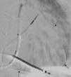
After the coronary sinus was accessed, venography showed the target lateral vein (Figure 1). The guide wire was introduced up to the distal portion of the vein and the left ventricular lead was advanced, but the proximal region of the vessel could not be crossed. It was decided to advance the guide wire from the lateral vein via a posterior collateral vessel up to the coronary sinus ostium (Figure 2). A snare system was introduced through a sheath via the left subclavian vein up to the atrium (Figure 3A) and the end of the guide wire was gradually pulled towards the skin surface. Thus, both ends of the guide wire were under the operator's control (Figure 3B) and the lead could be advanced to the desired position by pulling the guide wire (Figure 4). CRT parameters were satisfactory without diaphragmatic stimulation. There were no complications.

Figure 1.

Venography showing the target lateral vein. SC: coronary sinus; Veia alvo: target vein.

(0.13MB).
Figure 2.

Advancing the guide wire from the lateral vein via a posterior collateral vein up to the coronary sinus ostium (arrows).

(0.12MB).
Figure 3.

(A) The snare system inside the atrium capturing the end of the guide wire; (B) both ends of the guide wire on the skin surface in two sheaths. Bainha no SC: sheath in the coronary sinus; Fio guia: guide wire.

(0.2MB).
Figure 4.

Final position of the leads of the cardiac resynchronization system with defibrillator.

(0.11MB).

Correct positioning of the left ventricular lead is crucial to a favorable response to CRT, but venous anatomy often hinders this goal, and alternative and sometimes technically challenging approaches must be adopted to obtain the greatest clinical benefit, as described here.

Conflicts of interest

The authors have no conflicts of interest to declare.

Ethical disclosuresProtection of human and animal subjects

The authors declare that no experiments were performed on humans or animals for this study.

Confidentiality of data

The authors declare that no patient data appear in this article.

Right to privacy and informed consent

The authors declare that no patient data appear in this article.

Please cite this article as: Magalhães A, Menezes M, Cortez-Dias N, de Sousa J, Marques P. Utilização de sistema de extração Snare para implantação de eletrocateter ventricular esquerdo na ressincronização cardíaca. Rev Port Cardiol. 2015;34:221–222.

Copyright © 2014. Sociedade Portuguesa de Cardiologia
Download PDF
Idiomas
Revista Portuguesa de Cardiologia (English edition)
Article options
Tools
en pt

Are you a health professional able to prescribe or dispense drugs?

Você é um profissional de saúde habilitado a prescrever ou dispensar medicamentos

By checking that you are a health professional, you are stating that you are aware and accept that the Portuguese Journal of Cardiology (RPC) is the Data Controller that processes the personal information of users of its website, with its registered office at Campo Grande, n.º 28, 13.º, 1700-093 Lisbon, telephone 217 970 685 and 217 817 630, fax 217 931 095, and email revista@spc.pt. I declare for all purposes that the information provided herein is accurate and correct.