array:25 [
  "pii" => "S2174204916300423"
  "issn" => "21742049"
  "doi" => "10.1016/j.repce.2015.11.027"
  "estado" => "S300"
  "fechaPublicacion" => "2016-05-01"
  "aid" => "811"
  "copyright" => "Sociedade Portuguesa de Cardiologia"
  "copyrightAnyo" => "2016"
  "documento" => "article"
  "crossmark" => 1
  "licencia" => "http://creativecommons.org/licenses/by-nc-nd/4.0/"
  "subdocumento" => "sco"
  "cita" => "Rev Port Cardiol. 2016;35:315-6"
  "abierto" => array:3 [
    "ES" => true
    "ES2" => true
    "LATM" => true
  ]
  "gratuito" => true
  "lecturas" => array:2 [
    "total" => 1981
    "formatos" => array:3 [
      "EPUB" => 158
      "HTML" => 1479
      "PDF" => 344
    ]
  ]
  "Traduccion" => array:1 [
    "en" => array:20 [
      "pii" => "S0870255116300099"
      "issn" => "08702551"
      "doi" => "10.1016/j.repc.2015.11.019"
      "estado" => "S300"
      "fechaPublicacion" => "2016-05-01"
      "aid" => "811"
      "copyright" => "Sociedade Portuguesa de Cardiologia"
      "documento" => "article"
      "crossmark" => 1
      "licencia" => "http://creativecommons.org/licenses/by-nc-nd/4.0/"
      "subdocumento" => "sco"
      "cita" => "Rev Port Cardiol. 2016;35:315-6"
      "abierto" => array:3 [
        "ES" => true
        "ES2" => true
        "LATM" => true
      ]
      "gratuito" => true
      "lecturas" => array:2 [
        "total" => 2096
        "formatos" => array:3 [
          "EPUB" => 192
          "HTML" => 1276
          "PDF" => 628
        ]
      ]
      "en" => array:11 [
        "idiomaDefecto" => true
        "cabecera" => "<span class="elsevierStyleTextfn">Image in Cardiology</span>"
        "titulo" => "Hoarseness"
        "tienePdf" => "en"
        "tieneTextoCompleto" => "en"
        "paginas" => array:1 [
          0 => array:2 [
            "paginaInicial" => "315"
            "paginaFinal" => "316"
          ]
        ]
        "titulosAlternativos" => array:1 [
          "pt" => array:1 [
            "titulo" => "Disfonia"
          ]
        ]
        "contieneTextoCompleto" => array:1 [
          "en" => true
        ]
        "contienePdf" => array:1 [
          "en" => true
        ]
        "resumenGrafico" => array:2 [
          "original" => 0
          "multimedia" => array:7 [
            "identificador" => "fig0005"
            "etiqueta" => "Figure 1"
            "tipo" => "MULTIMEDIAFIGURA"
            "mostrarFloat" => true
            "mostrarDisplay" => false
            "figura" => array:1 [
              0 => array:4 [
                "imagen" => "gr1.jpeg"
                "Alto" => 672
                "Ancho" => 750
                "Tamanyo" => 43904
              ]
            ]
            "descripcion" => array:1 [
              "en" => "<p id="spar0005" class="elsevierStyleSimplePara elsevierViewall">Chest X-ray&#44; showing a slight mediastinal bulge adjacent to the aortic knuckle&#46;</p>"
            ]
          ]
        ]
        "autores" => array:1 [
          0 => array:2 [
            "autoresLista" => "Sofia L&#225;zaro Mendes, Domingos Ramos, Mariano P&#234;go"
            "autores" => array:3 [
              0 => array:2 [
                "nombre" => "Sofia L&#225;zaro"
                "apellidos" => "Mendes"
              ]
              1 => array:2 [
                "nombre" => "Domingos"
                "apellidos" => "Ramos"
              ]
              2 => array:2 [
                "nombre" => "Mariano"
                "apellidos" => "P&#234;go"
              ]
            ]
          ]
        ]
      ]
      "idiomaDefecto" => "en"
      "Traduccion" => array:1 [
        "en" => array:9 [
          "pii" => "S2174204916300423"
          "doi" => "10.1016/j.repce.2015.11.027"
          "estado" => "S300"
          "subdocumento" => ""
          "abierto" => array:3 [
            "ES" => true
            "ES2" => true
            "LATM" => true
          ]
          "gratuito" => true
          "lecturas" => array:1 [
            "total" => 0
          ]
          "idiomaDefecto" => "en"
          "EPUB" => "https://multimedia.elsevier.es/PublicationsMultimediaV1/item/epub/S2174204916300423?idApp=UINPBA00004E"
        ]
      ]
      "EPUB" => "https://multimedia.elsevier.es/PublicationsMultimediaV1/item/epub/S0870255116300099?idApp=UINPBA00004E"
      "url" => "/08702551/0000003500000005/v3_201702080059/S0870255116300099/v3_201702080059/en/main.assets"
    ]
  ]
  "itemSiguiente" => array:19 [
    "pii" => "S2174204916300447"
    "issn" => "21742049"
    "doi" => "10.1016/j.repce.2016.05.004"
    "estado" => "S300"
    "fechaPublicacion" => "2016-05-01"
    "aid" => "800"
    "documento" => "simple-article"
    "crossmark" => 0
    "licencia" => "http://creativecommons.org/licenses/by-nc-nd/4.0/"
    "subdocumento" => "prv"
    "cita" => "Rev Port Cardiol. 2016;35:317-8"
    "abierto" => array:3 [
      "ES" => true
      "ES2" => true
      "LATM" => true
    ]
    "gratuito" => true
    "lecturas" => array:2 [
      "total" => 1976
      "formatos" => array:3 [
        "EPUB" => 145
        "HTML" => 1429
        "PDF" => 402
      ]
    ]
    "en" => array:10 [
      "idiomaDefecto" => true
      "cabecera" => "<span class="elsevierStyleTextfn">Recommended article of the Month</span>"
      "titulo" => "Comment on &#8220;Variables measured during cardiopulmonary exercise testing as predictors of mortality in chronic systolic heart failure&#8221;"
      "tienePdf" => "en"
      "tieneTextoCompleto" => "en"
      "paginas" => array:1 [
        0 => array:2 [
          "paginaInicial" => "317"
          "paginaFinal" => "318"
        ]
      ]
      "titulosAlternativos" => array:1 [
        "pt" => array:1 [
          "titulo" => "Coment&#225;rio a &#171;Vari&#225;veis medidas durante prova de esfor&#231;o cardiorrespirat&#243;ria como preditoras de mortalidade na insufici&#234;ncia card&#237;aca cr&#243;nica com disfun&#231;&#227;o sist&#243;lica&#187;"
        ]
      ]
      "contieneTextoCompleto" => array:1 [
        "en" => true
      ]
      "contienePdf" => array:1 [
        "en" => true
      ]
      "autores" => array:1 [
        0 => array:2 [
          "autoresLista" => "Ana Abreu"
          "autores" => array:1 [
            0 => array:2 [
              "nombre" => "Ana"
              "apellidos" => "Abreu"
            ]
          ]
        ]
      ]
    ]
    "idiomaDefecto" => "en"
    "Traduccion" => array:1 [
      "pt" => array:9 [
        "pii" => "S0870255116000925"
        "doi" => "10.1016/j.repc.2016.03.001"
        "estado" => "S300"
        "subdocumento" => ""
        "abierto" => array:3 [
          "ES" => true
          "ES2" => true
          "LATM" => true
        ]
        "gratuito" => true
        "lecturas" => array:1 [
          "total" => 0
        ]
        "idiomaDefecto" => "pt"
        "EPUB" => "https://multimedia.elsevier.es/PublicationsMultimediaV1/item/epub/S0870255116000925?idApp=UINPBA00004E"
      ]
    ]
    "EPUB" => "https://multimedia.elsevier.es/PublicationsMultimediaV1/item/epub/S2174204916300447?idApp=UINPBA00004E"
    "url" => "/21742049/0000003500000005/v3_201704120129/S2174204916300447/v3_201704120129/en/main.assets"
  ]
  "itemAnterior" => array:20 [
    "pii" => "S2174204916300411"
    "issn" => "21742049"
    "doi" => "10.1016/j.repce.2015.11.026"
    "estado" => "S300"
    "fechaPublicacion" => "2016-05-01"
    "aid" => "810"
    "copyright" => "Sociedade Portuguesa de Cardiologia"
    "documento" => "article"
    "crossmark" => 1
    "licencia" => "http://creativecommons.org/licenses/by-nc-nd/4.0/"
    "subdocumento" => "sco"
    "cita" => "Rev Port Cardiol. 2016;35:313-4"
    "abierto" => array:3 [
      "ES" => true
      "ES2" => true
      "LATM" => true
    ]
    "gratuito" => true
    "lecturas" => array:2 [
      "total" => 2078
      "formatos" => array:3 [
        "EPUB" => 152
        "HTML" => 1460
        "PDF" => 466
      ]
    ]
    "en" => array:11 [
      "idiomaDefecto" => true
      "cabecera" => "<span class="elsevierStyleTextfn">Image in Cardiology</span>"
      "titulo" => "Left heart failure&#44; a challenging diagnosis in cardiology"
      "tienePdf" => "en"
      "tieneTextoCompleto" => "en"
      "paginas" => array:1 [
        0 => array:2 [
          "paginaInicial" => "313"
          "paginaFinal" => "314"
        ]
      ]
      "titulosAlternativos" => array:1 [
        "pt" => array:1 [
          "titulo" => "Insufici&#234;ncia card&#237;aca esquerda&#44; um desafio diagn&#243;stico em cardiologia"
        ]
      ]
      "contieneTextoCompleto" => array:1 [
        "en" => true
      ]
      "contienePdf" => array:1 [
        "en" => true
      ]
      "resumenGrafico" => array:2 [
        "original" => 0
        "multimedia" => array:7 [
          "identificador" => "fig0010"
          "etiqueta" => "Figure 2"
          "tipo" => "MULTIMEDIAFIGURA"
          "mostrarFloat" => true
          "mostrarDisplay" => false
          "figura" => array:1 [
            0 => array:4 [
              "imagen" => "gr2.jpeg"
              "Alto" => 465
              "Ancho" => 1517
              "Tamanyo" => 68614
            ]
          ]
          "descripcion" => array:1 [
            "en" => "<p id="spar0010" class="elsevierStyleSimplePara elsevierViewall">Fluoroscopy during percutaneous patent ductus arteriosus closure&#58; left&#44; a continuous flow can be seen from the aorta to the pulmonary artery &#40;yellow arrow&#41;&#59; center&#44; during device placement &#40;yellow arrow&#41;&#59; right&#44; after the procedure the device is properly positioned with no residual shunt &#40;yellow arrow&#41;&#46;</p>"
          ]
        ]
      ]
      "autores" => array:1 [
        0 => array:2 [
          "autoresLista" => "Miryan Cassandra, In&#234;s Almeida, Rog&#233;rio Teixeira, Joana Trigo, Marco Costa, Lino Gon&#231;alves"
          "autores" => array:6 [
            0 => array:2 [
              "nombre" => "Miryan"
              "apellidos" => "Cassandra"
            ]
            1 => array:2 [
              "nombre" => "In&#234;s"
              "apellidos" => "Almeida"
            ]
            2 => array:2 [
              "nombre" => "Rog&#233;rio"
              "apellidos" => "Teixeira"
            ]
            3 => array:2 [
              "nombre" => "Joana"
              "apellidos" => "Trigo"
            ]
            4 => array:2 [
              "nombre" => "Marco"
              "apellidos" => "Costa"
            ]
            5 => array:2 [
              "nombre" => "Lino"
              "apellidos" => "Gon&#231;alves"
            ]
          ]
        ]
      ]
    ]
    "idiomaDefecto" => "en"
    "Traduccion" => array:1 [
      "en" => array:9 [
        "pii" => "S0870255116300087"
        "doi" => "10.1016/j.repc.2015.11.018"
        "estado" => "S300"
        "subdocumento" => ""
        "abierto" => array:3 [
          "ES" => true
          "ES2" => true
          "LATM" => true
        ]
        "gratuito" => true
        "lecturas" => array:1 [
          "total" => 0
        ]
        "idiomaDefecto" => "en"
        "EPUB" => "https://multimedia.elsevier.es/PublicationsMultimediaV1/item/epub/S0870255116300087?idApp=UINPBA00004E"
      ]
    ]
    "EPUB" => "https://multimedia.elsevier.es/PublicationsMultimediaV1/item/epub/S2174204916300411?idApp=UINPBA00004E"
    "url" => "/21742049/0000003500000005/v3_201704120129/S2174204916300411/v3_201704120129/en/main.assets"
  ]
  "en" => array:15 [
    "idiomaDefecto" => true
    "cabecera" => "<span class="elsevierStyleTextfn">Image in Cardiology</span>"
    "titulo" => "Hoarseness"
    "tieneTextoCompleto" => true
    "paginas" => array:1 [
      0 => array:2 [
        "paginaInicial" => "315"
        "paginaFinal" => "316"
      ]
    ]
    "autores" => array:1 [
      0 => array:4 [
        "autoresLista" => "Sofia L&#225;zaro Mendes, Domingos Ramos, Mariano P&#234;go"
        "autores" => array:3 [
          0 => array:4 [
            "nombre" => "Sofia L&#225;zaro"
            "apellidos" => "Mendes"
            "email" => array:1 [
              0 => "sofialazaromendes&#64;gmail&#46;com"
            ]
            "referencia" => array:1 [
              0 => array:2 [
                "etiqueta" => "<span class="elsevierStyleSup">&#42;</span>"
                "identificador" => "cor0005"
              ]
            ]
          ]
          1 => array:2 [
            "nombre" => "Domingos"
            "apellidos" => "Ramos"
          ]
          2 => array:2 [
            "nombre" => "Mariano"
            "apellidos" => "P&#234;go"
          ]
        ]
        "afiliaciones" => array:1 [
          0 => array:2 [
            "entidad" => "Cardiology Department&#44; Coimbra University Hospital and Medical School&#44; Coimbra&#44; Portugal"
            "identificador" => "aff0005"
          ]
        ]
        "correspondencia" => array:1 [
          0 => array:3 [
            "identificador" => "cor0005"
            "etiqueta" => "&#8270;"
            "correspondencia" => "Corresponding author&#46;"
          ]
        ]
      ]
    ]
    "titulosAlternativos" => array:1 [
      "pt" => array:1 [
        "titulo" => "Disfonia"
      ]
    ]
    "resumenGrafico" => array:2 [
      "original" => 0
      "multimedia" => array:7 [
        "identificador" => "fig0010"
        "etiqueta" => "Figure 2"
        "tipo" => "MULTIMEDIAFIGURA"
        "mostrarFloat" => true
        "mostrarDisplay" => false
        "figura" => array:1 [
          0 => array:4 [
            "imagen" => "gr2.jpeg"
            "Alto" => 601
            "Ancho" => 697
            "Tamanyo" => 65546
          ]
        ]
        "descripcion" => array:1 [
          "en" => "<p id="spar0010" class="elsevierStyleSimplePara elsevierViewall">Contrast-enhanced thoracic computed tomography with frontal reconstruction&#44; showing a saccular aneurysm of the aortic arch&#46;</p>"
        ]
      ]
    ]
    "textoCompleto" => "<span class="elsevierStyleSections"><p id="par0005" class="elsevierStylePara elsevierViewall">A 63-year-old man presented to the hospital with gradual-onset hoarseness of three months&#8217; duration&#46; He had a history of smoking assessed at 58 pack-years&#44; and no other symptoms such as cough&#44; dyspnea or weight loss&#46; His heart rate was 60 beats&#47;min and blood pressure was 159&#47;67 mmHg&#59; the cardiac exam revealed a diastolic murmur in the aortic area&#46; Indirect laryngoscopy revealed a paralyzed left vocal cord in paramedian position&#46; The chest X-ray &#40;<a class="elsevierStyleCrossRef" href="#fig0005">Figure 1</a>&#41; showed a slight mediastinal bulge adjacent to the aortic knuckle but no pulmonary mass&#46;</p><elsevierMultimedia ident="fig0005"></elsevierMultimedia><p id="par0010" class="elsevierStylePara elsevierViewall">Contrast-enhanced thoracic computed tomography with frontal and sagittal reconstruction &#40;<a class="elsevierStyleCrossRefs" href="#fig0010">Figures 2 and 3</a>&#41; was performed and revealed a saccular aneurysm of the aortic arch with dilatation up to 6&#46;9 cm and partial thrombosis of the wall&#46;</p><elsevierMultimedia ident="fig0010"></elsevierMultimedia><elsevierMultimedia ident="fig0015"></elsevierMultimedia><p id="par0015" class="elsevierStylePara elsevierViewall">There was no tumor along the intrathoracic route of the left recurrent laryngeal nerve &#40;RLN&#41;&#46; The transthoracic echocardiography was normal&#46;</p><p id="par0020" class="elsevierStylePara elsevierViewall">The patient underwent aneurysmectomy and his postoperative course was excellent and uncomplicated&#44; but 12 months after surgery he has not yet recovered from RLN palsy&#46;</p><p id="par0025" class="elsevierStylePara elsevierViewall">Thoracic saccular aortic aneurysm is an important consideration in the differential diagnosis of RLN palsy&#46;</p><span id="sec0005" class="elsevierStyleSection elsevierViewall"><span class="elsevierStyleSectionTitle" id="sect0005">Ethical disclosures</span><span id="sec0010" class="elsevierStyleSection elsevierViewall"><span class="elsevierStyleSectionTitle" id="sect0010">Protection of human and animal subjects</span><p id="par0030" class="elsevierStylePara elsevierViewall">The authors declare that no experiments were performed on humans or animals for this study&#46;</p></span><span id="sec0015" class="elsevierStyleSection elsevierViewall"><span class="elsevierStyleSectionTitle" id="sect0015">Confidentiality of data</span><p id="par0035" class="elsevierStylePara elsevierViewall">The authors declare that they have followed the protocols of their work center on the publication of patient data&#46;</p></span><span id="sec0020" class="elsevierStyleSection elsevierViewall"><span class="elsevierStyleSectionTitle" id="sect0020">Right to privacy and informed consent</span><p id="par0040" class="elsevierStylePara elsevierViewall">The authors declare that no patient data appear in this article&#46;</p></span></span><span id="sec0025" class="elsevierStyleSection elsevierViewall"><span class="elsevierStyleSectionTitle" id="sect0025">Conflicts of interest</span><p id="par0045" class="elsevierStylePara elsevierViewall">The authors have no conflicts of interest to declare&#46;</p></span></span>"
    "textoCompletoSecciones" => array:1 [
      "secciones" => array:2 [
        0 => array:3 [
          "identificador" => "sec0005"
          "titulo" => "Ethical disclosures"
          "secciones" => array:3 [
            0 => array:2 [
              "identificador" => "sec0010"
              "titulo" => "Protection of human and animal subjects"
            ]
            1 => array:2 [
              "identificador" => "sec0015"
              "titulo" => "Confidentiality of data"
            ]
            2 => array:2 [
              "identificador" => "sec0020"
              "titulo" => "Right to privacy and informed consent"
            ]
          ]
        ]
        1 => array:2 [
          "identificador" => "sec0025"
          "titulo" => "Conflicts of interest"
        ]
      ]
    ]
    "pdfFichero" => "main.pdf"
    "tienePdf" => true
    "fechaRecibido" => "2015-07-29"
    "fechaAceptado" => "2015-11-21"
    "multimedia" => array:3 [
      0 => array:7 [
        "identificador" => "fig0005"
        "etiqueta" => "Figure 1"
        "tipo" => "MULTIMEDIAFIGURA"
        "mostrarFloat" => true
        "mostrarDisplay" => false
        "figura" => array:1 [
          0 => array:4 [
            "imagen" => "gr1.jpeg"
            "Alto" => 600
            "Ancho" => 670
            "Tamanyo" => 45100
          ]
        ]
        "descripcion" => array:1 [
          "en" => "<p id="spar0005" class="elsevierStyleSimplePara elsevierViewall">Chest X-ray&#44; showing a slight mediastinal bulge adjacent to the aortic knuckle&#46;</p>"
        ]
      ]
      1 => array:7 [
        "identificador" => "fig0010"
        "etiqueta" => "Figure 2"
        "tipo" => "MULTIMEDIAFIGURA"
        "mostrarFloat" => true
        "mostrarDisplay" => false
        "figura" => array:1 [
          0 => array:4 [
            "imagen" => "gr2.jpeg"
            "Alto" => 601
            "Ancho" => 697
            "Tamanyo" => 65546
          ]
        ]
        "descripcion" => array:1 [
          "en" => "<p id="spar0010" class="elsevierStyleSimplePara elsevierViewall">Contrast-enhanced thoracic computed tomography with frontal reconstruction&#44; showing a saccular aneurysm of the aortic arch&#46;</p>"
        ]
      ]
      2 => array:7 [
        "identificador" => "fig0015"
        "etiqueta" => "Figure 3"
        "tipo" => "MULTIMEDIAFIGURA"
        "mostrarFloat" => true
        "mostrarDisplay" => false
        "figura" => array:1 [
          0 => array:4 [
            "imagen" => "gr3.jpeg"
            "Alto" => 608
            "Ancho" => 602
            "Tamanyo" => 50643
          ]
        ]
        "descripcion" => array:1 [
          "en" => "<p id="spar0015" class="elsevierStyleSimplePara elsevierViewall">Contrast-enhanced thoracic computed tomography with sagittal reconstruction&#44; showing a saccular aneurysm of the aortic arch&#46;</p>"
        ]
      ]
    ]
  ]
  "idiomaDefecto" => "en"
  "url" => "/21742049/0000003500000005/v3_201704120129/S2174204916300423/v3_201704120129/en/main.assets"
  "Apartado" => array:4 [
    "identificador" => "9915"
    "tipo" => "SECCION"
    "en" => array:2 [
      "titulo" => "Images in cardiology"
      "idiomaDefecto" => true
    ]
    "idiomaDefecto" => "en"
  ]
  "PDF" => "https://static.elsevier.es/multimedia/21742049/0000003500000005/v3_201704120129/S2174204916300423/v3_201704120129/en/main.pdf?idApp=UINPBA00004E&text.app=https://revportcardiol.org/"
  "EPUB" => "https://multimedia.elsevier.es/PublicationsMultimediaV1/item/epub/S2174204916300423?idApp=UINPBA00004E"
]
Share
Journal Information
Vol. 35. Issue 5.
Pages 315-316 (May 2016)
Share
Share
Download PDF
More article options
Vol. 35. Issue 5.
Pages 315-316 (May 2016)
Image in Cardiology
Open Access
Hoarseness
Disfonia
Visits
4478
Sofia Lázaro Mendes
Corresponding author
sofialazaromendes@gmail.com

Corresponding author.
, Domingos Ramos, Mariano Pêgo
Cardiology Department, Coimbra University Hospital and Medical School, Coimbra, Portugal
This item has received

Under a Creative Commons license
Article information
Full Text
Download PDF
Statistics
Figures (3)
Show moreShow less
Full Text

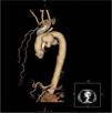
A 63-year-old man presented to the hospital with gradual-onset hoarseness of three months’ duration. He had a history of smoking assessed at 58 pack-years, and no other symptoms such as cough, dyspnea or weight loss. His heart rate was 60 beats/min and blood pressure was 159/67 mmHg; the cardiac exam revealed a diastolic murmur in the aortic area. Indirect laryngoscopy revealed a paralyzed left vocal cord in paramedian position. The chest X-ray (Figure 1) showed a slight mediastinal bulge adjacent to the aortic knuckle but no pulmonary mass.

Figure 1.

Chest X-ray, showing a slight mediastinal bulge adjacent to the aortic knuckle.

(0.04MB).

Contrast-enhanced thoracic computed tomography with frontal and sagittal reconstruction (Figures 2 and 3) was performed and revealed a saccular aneurysm of the aortic arch with dilatation up to 6.9 cm and partial thrombosis of the wall.

Figure 2.

Contrast-enhanced thoracic computed tomography with frontal reconstruction, showing a saccular aneurysm of the aortic arch.

(0.06MB).
Figure 3.

Contrast-enhanced thoracic computed tomography with sagittal reconstruction, showing a saccular aneurysm of the aortic arch.

(0.05MB).

There was no tumor along the intrathoracic route of the left recurrent laryngeal nerve (RLN). The transthoracic echocardiography was normal.

The patient underwent aneurysmectomy and his postoperative course was excellent and uncomplicated, but 12 months after surgery he has not yet recovered from RLN palsy.

Thoracic saccular aortic aneurysm is an important consideration in the differential diagnosis of RLN palsy.

Ethical disclosuresProtection of human and animal subjects

The authors declare that no experiments were performed on humans or animals for this study.

Confidentiality of data

The authors declare that they have followed the protocols of their work center on the publication of patient data.

Right to privacy and informed consent

The authors declare that no patient data appear in this article.

Conflicts of interest

The authors have no conflicts of interest to declare.

Copyright © 2016. Sociedade Portuguesa de Cardiologia
Download PDF
Idiomas
Revista Portuguesa de Cardiologia (English edition)
Article options
Tools
en pt

Are you a health professional able to prescribe or dispense drugs?

Você é um profissional de saúde habilitado a prescrever ou dispensar medicamentos

By checking that you are a health professional, you are stating that you are aware and accept that the Portuguese Journal of Cardiology (RPC) is the Data Controller that processes the personal information of users of its website, with its registered office at Campo Grande, n.º 28, 13.º, 1700-093 Lisbon, telephone 217 970 685 and 217 817 630, fax 217 931 095, and email revista@spc.pt. I declare for all purposes that the information provided herein is accurate and correct.