


An 82-year-old woman with a history of mild hypertension reported fatigue on strenuous exertion since adolescence that was not investigated. The fatigue had worsened progressively until it was triggered by minimal exertion over the previous year.
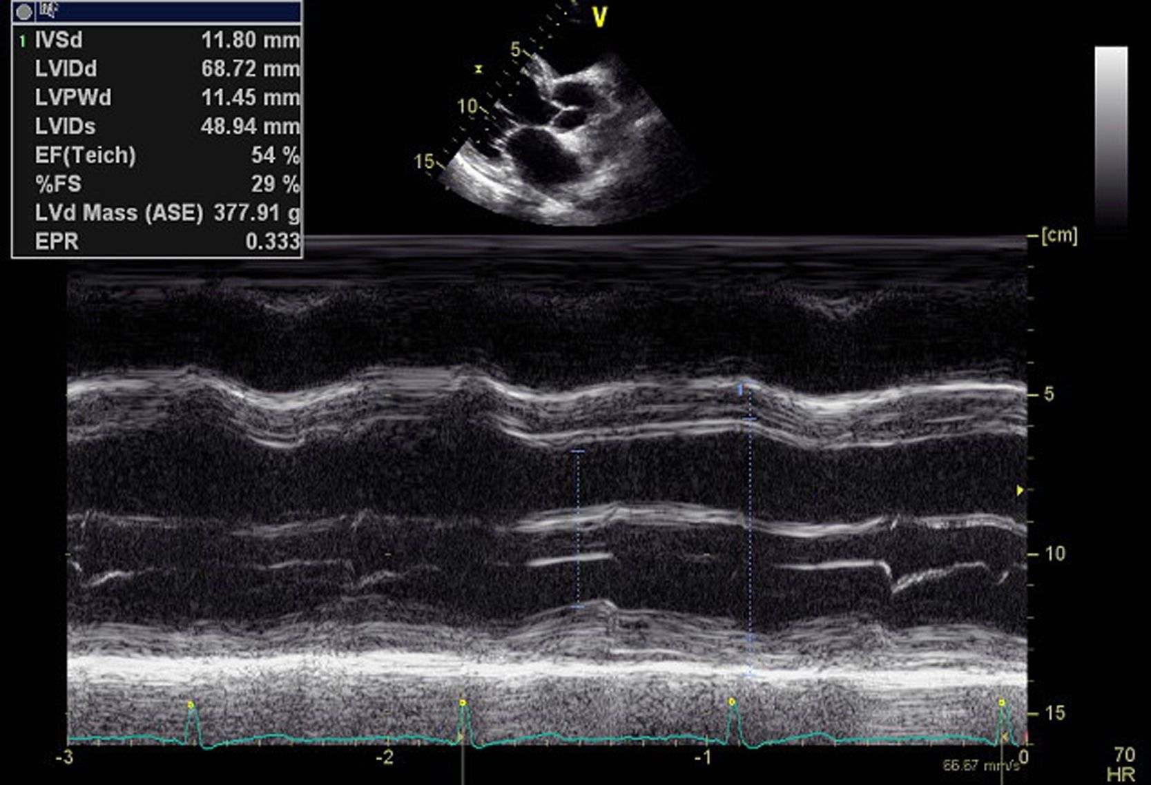
Echocardiography performed at another center revealed a dilated left ventricle with non-hypertrophied walls, global systolic dysfunction (ejection fraction 37%) but no wall motion abnormalities, mild mitral regurgitation and left atrial dilatation. She also presented moderate tricuspid regurgitation, moderate pulmonary hypertension (estimated at 49 mmHg) and marked dilatation of the pulmonary artery (47 mm).
The patient was referred to our department to exclude ischemic etiology; cardiac catheterization showed no apparent coronary disease.
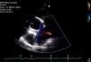
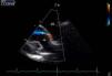
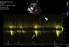
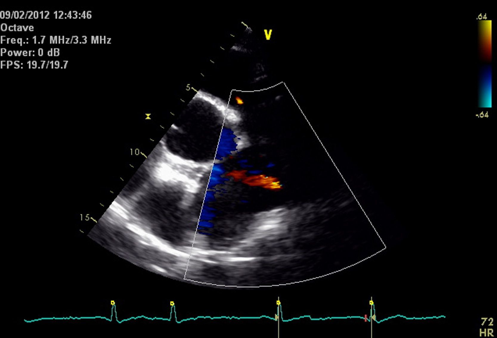
A grade II/VI systolic–diastolic murmur was audible at the upper left sternal border that was not explained by previous studies. Transthoracic echocardiography was repeated (Figure 1), which confirmed the previous findings, but also showed continuous turbulent flow at the level of the pulmonary artery bifurcation, from the aorta towards the trunk and right branch of the pulmonary artery; it was also visible in suprasternal view (Figures 2–4). A diagnosis was made of patent ductus arteriosus (PDA).
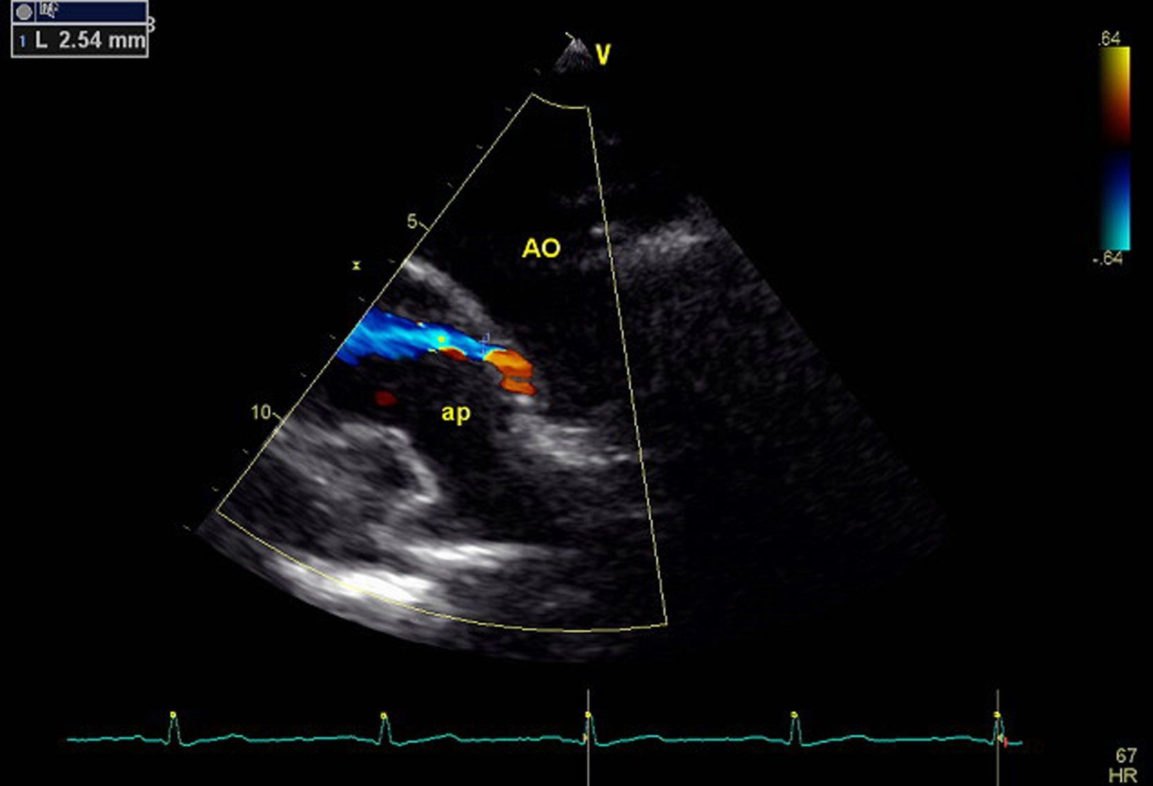
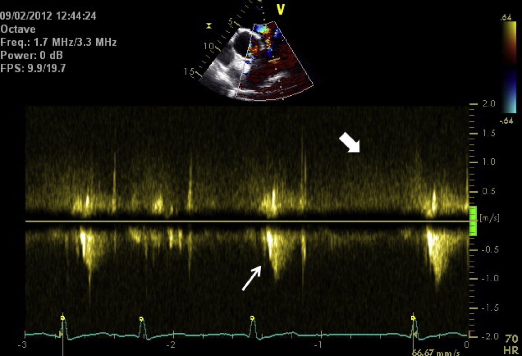
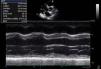
Echocardiogram, left parasternal short-axis view at the pulmonary valve level. Pulsed Doppler study immediately distal to the pulmonary valve guided by color Doppler (above), showing systolic flow through the pulmonary valve (narrow arrow) against a background of systolic–diastolic flow from aorta to pulmonary artery (wide arrow).
In view of the patient's age, it was decided to optimize medical therapy. Her condition improved and she now has fatigue on moderate exertion.
This case of PDA is unusual for being diagnosed so late in life. The patient's left ventricular dysfunction was probably due to increased flow caused by the PDA, which was the only pathological condition identified apart from mild hypertension. The elevated pulmonary artery pressure resulting from the PDA probably reduced the shunt, which is now weak and turbulent.
Ethical responsibilitiesProtection of human and animal subjectsThe authors declare that the procedures followed were in accordance with the regulations of the relevant clinical research ethics committee and with those of the Code of Ethics of the World Medical Association (Declaration of Helsinki).
Confidentiality of dataThe authors declare that they have followed the protocols of their work center on the publication of patient data and that all the patients included in the study received sufficient information and gave their written informed consent to participate in the study.
Right to privacy and informed consentThe authors have obtained the written informed consent of the patients or subjects mentioned in the article. The corresponding author is in possession of this document.
Conflicts of interestThe authors have no conflicts of interest to declare.
Please cite this article as: Pereira-da-Silva T, et al. Causa inesperada de disfunção sistólica em octogenária. Rev Port Cardiol. 2013. http://dx.doi.org/10.1016/j.repc.2012.10.007.
