


Persistent left superior vena cava (PLSVC) is the most common thoracic congenital malformation and its diagnosis usually occurs by chance. Demonstrative clinical cases of dual-chamber (DDD) pacemaker implantation, implantable cardioverter defibrillators and cardiac resynchronization systems through PLSVC access have already been described in the literature. However, in most of the cases presented, the positioning of the lead in the right ventricle (RV) was restricted to the RV apex. To date, the literature reports only four cases of RV lead positioning in the right ventricular outflow tract (RVOT) via the PLSVC. The authors present the case of a 79-year-old female patient, diagnosed with syncope and left branch bundle block, in whom implantation of a DDD pacemaker was proposed. During the procedure, venous access was obtained via the left cephalic vein, and the lead was found to progress to the left of the spinal column, suggesting the presence of PLSVC. Through this access, it was possible to sequentially insert the RV lead in the RVOT and the atrial lead in the right atrial appendage, both with an active fixation system (Figure 1).
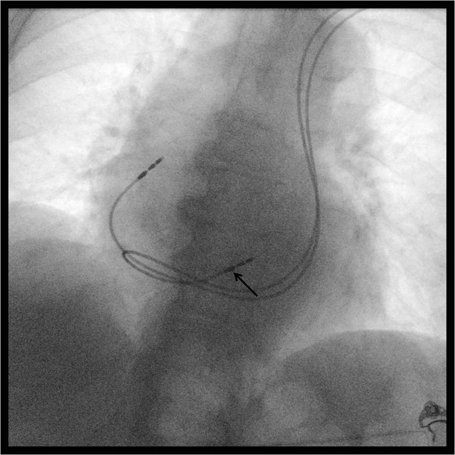
In conclusion, although PLSVC makes implantation of these types of cardiac devices difficult, it is not a limiting factor for the optimal positioning of the RV lead in the RVOT. Studies have shown that this placement is beneficial and is associated with less ventricular dyssynchrony, narrower QRS and higher cardiac output than apical right ventricular pacing.
Conflicts of interestThe authors have no conflicts of interest to declare.
Please cite this article as: Guimarães T, Bernardes A, de Sousa J, Marques P. Veia cava superior esquerda persistente – Um acesso vascular sem limitações. Rev Port Cardiol. 2018;37:627–628.
