


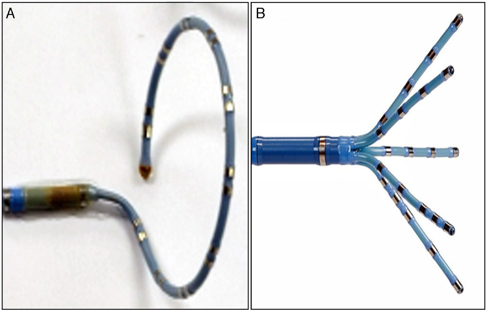
Ablation of atrial fibrillation (AF) through electrical isolation of the pulmonary veins (PVs) is usually performed using circular multipolar catheters. We describe our initial experience with the PentaRay® catheter (Biosense Webster, Inc.), which has five highly flexible branches and a total of 20 poles (Figure 1). This was used for ablation of persistent AF in a 36-year-old female patient, in conjunction with the CARTO® navigation system (Biosense Webster, Inc.), to create a three-dimensional reconstruction of the left atrium (volume 142 ml) and the PVs, which was then integrated with images obtained by computed tomography angiography (Figure 2). The PentaRay® catheter enabled rapid acquisition of a large number of points to obtain a voltage map with high signal resolution. The five-branched design, with highly flexible arms, fits easily inside the PV ostia (Figure 3). The PentaRay® is also much less likely to become wedged in the mitral valve apparatus.

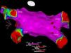
Voltage map in sinus rhythm, visualized from the left atrial posterior wall using the CARTO® system. VPID: right inferior pulmonary vein; VPIE: left inferior pulmonary vein; VPSD: right superior pulmonary vein; VPSE: left superior pulmonary vein; purple area: normal voltage; red area: low voltage.

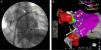
Fluoroscopy with PentaRay® catheter positioned in the left superior pulmonary vein (A); image from the CARTO® system showing the PentaRay® catheter in the left inferior pulmonary vein (B). VPIE: left inferior pulmonary vein; VPSE: left superior pulmonary vein; yellow arrow: PentaRay® catheter; white arrow: ablation catheter; black arrow: decapolar catheter in the coronary sinus.
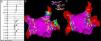
In the case presented, no low-voltage areas were identified in the atrium during pre-ablation mapping and potentials were seen in all PVs (Figure 4). Antral ablation lines were created in all four PVs. At the end of the procedure, remapping with the PentaRay® catheter showed low voltage (<0.15 mV) in the PVs with the antral ablation lines (Figure 4), which was confirmed by pacing maneuvers inside and outside the PVs.

Pre-ablation electrogram (A) showing potentials in the right superior pulmonary vein (arrows); voltage maps before (B) and after (C) ablation visualized from the left atrial roof, showing the sites of radiofrequency application (red and white circles). VPID: right inferior pulmonary vein; VPIE: left inferior pulmonary vein; VPSD: right superior pulmonary vein; VPSE: left superior pulmonary vein.
The authors declare that no experiments were performed on humans or animals for this study.
Confidentiality of dataThe authors declare that no patient data appear in this article.
Right to privacy and informed consentThe authors declare that no patient data appear in this article.
Conflicts of interestThe authors have no conflicts of interest to declare.
Please cite this article as: Teixeira PP, Cunha PS, Delgado AS, Pimenta R, Oliveira MM, Ferreira RC. Cateter PentaRay na ablação de fibrilhação auricular persistente. Rev Port Cardiol. 2016;35:121–123.
