


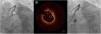
A 75-year-old male underwent primary angioplasty of a subocclusive proximal to medial left anterior descending artery (LAD) stenosis, involving the origin of the first diagonal (D1). A provisional stenting technique was used, with two guidewires in the distal LAD and D1 and implantation of 3 mm×30 mm and 2.75 mm×28 mm drug-eluting stents (DES) in the LAD. Withdrawal of the jailed D1 guidewire caused a deep intubation of the guide catheter into the LAD. The subsequent angiogram was notable for a suspicious haziness proximal to the stents (Figure 1A, arrow). We decided to perform optical coherence tomography (OCT) of the LAD. However, the catheter did not pass further than the proximal segment, showing a conspicuous circular image (Figure 1B). Meanwhile, the intravascular ultrasound catheter was able to progress and did not show the image, only malapposed struts. Angioplasty proximal to the previous stents was performed with implantation of a 3.5 mm×18 mm DES, with a good final angiographic result (Figure 1C). In subsequent OCT images the circular image had disappeared.
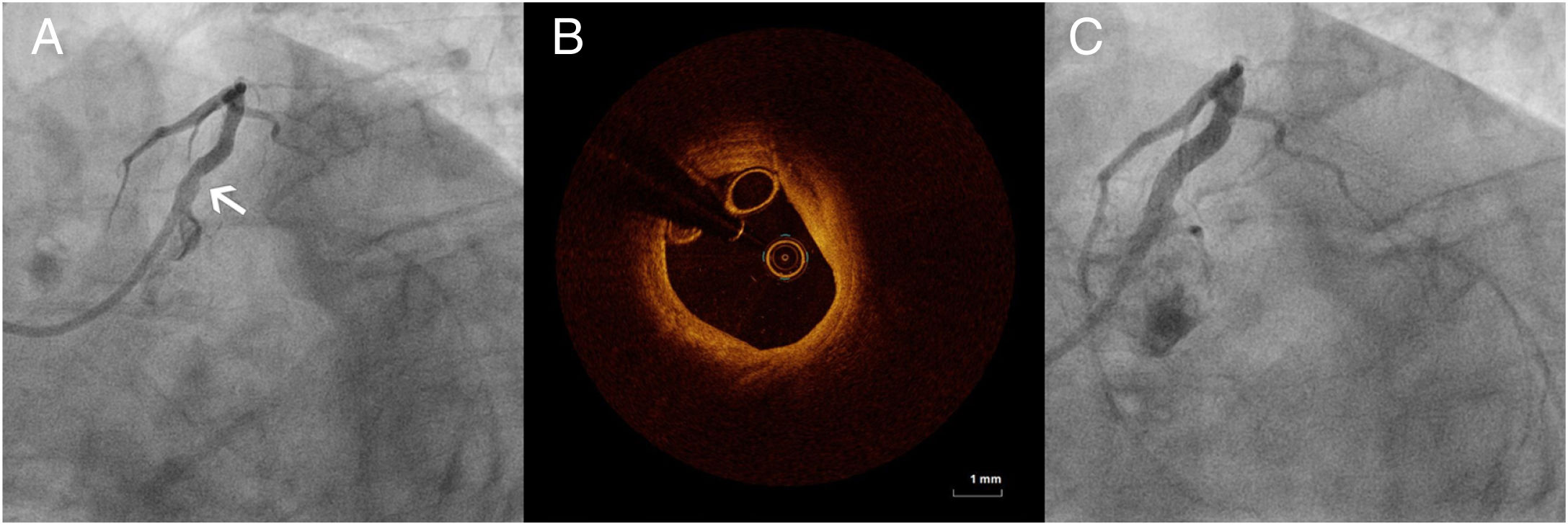
(A) Angiography showing a suspicious haziness proximal to the stents (arrow); (B) optical coherence tomography image of the proximal left anterior descending artery (proximal to the stent), with a suspicious circular image; (C) final angiographic result, after angioplasty with a 3.5 mm×18 mm drug-eluting stent proximal to the previous stents.
We postulate that the intubation of the guide catheter during retrieval of the jailed guidewire damaged the proximal stent struts. Thus, the circular OCT image was of the OCT catheter itself, which was folded by attempts to pass the catheter through the damaged stent. Our case illustrates an unusual image that can lead to uncertainties during OCT-guided angioplasty. The report of this peculiarity could help the interpretation of doubtful OCT images obtained in daily practice in the cath lab.
Conflicts of interestThe authors have no conflicts of interest to declare.
