Informação da revista
Vol. 32. Núm. 3.
Páginas 265-267 (março 2013)
Partilhar
Partilhar
Baixar PDF
Mais opções do artigo
Vol. 32. Núm. 3.
Páginas 265-267 (março 2013)
Images in cardiology
Open Access
Left ventricular apical hypoplasia: An unusual diagnosis
Hipoplasia apical do ventrículo esquerdo: um diagnóstico raro
Visitas
8261
Alexandra Sousaa,
Autor para correspondência
xanasousa81@gmail.com

Corresponding author.
, Teresa Pinhoa, Pedro Almeidaa, António Madureirab, Filipe Macedoa, Maria Júlia Maciela
a Cardiology Department, Centro Hospitalar de São João, Porto, Portugal
b Radiology Department, Centro Hospitalar de São João, Porto, Portugal
Este item recebeu

Under a Creative Commons license
Informação do artigo
Texto Completo
Baixar PDF
Estatísticas
Figuras (4)
Mostrar maisMostrar menos
Material adicional (8)
Texto Completo

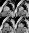
A 36-year-old woman was referred for medical evaluation prior to non-cardiac surgery. She was healthy and only complained of sporadic palpitations. The 12-lead electrocardiogram showed sinus rhythm and left bundle branch block. Transthoracic echocardiography revealed a normally functioning, slightly dilated left ventricle (LV), with the interventricular septum bulging toward the right ventricle (RV), which was elongated and wrapped around the LV (Figure 1; Supplementary material Videos S1 and S2). The papillary muscles were structurally abnormal and presented an apical origin, findings better visualized in contrast images (Figure 2; Videos S3–S5). Cardiac magnetic resonance confirmed the presence of a truncated LV, a complex network of papillary muscles of apical origin, and a banana-shaped, normally functioning RV (Figures 3 and 4; Videos S6–S8). There was no evidence of fatty infiltration or late enhancement with gadolinium.

Figure 1.

Transthoracic echocardiogram. (A) Apical 4-chamber view; (B) apical 2-chamber view. Solid arrow: right ventricle. Dashed arrow: network of papillary muscles.

(0.14MB).
Figure 2.

(A) Transthoracic echocardiogram, parasternal short-axis view. Contrast transthoracic echocardiogram: (B) parasternal short-axis view; (C) apical 4-chamber view; (D) apical 3-chamber view.

(0.13MB).
Figure 3.

Cardiac magnetic resonance. Steady-state free precession images in horizontal long-axis view (A) and vertical long-axis view (B). Solid arrow: right ventricle. Dashed arrow: network of papillary muscles.

(0.17MB).
Figure 4.

Cardiac magnetic resonance. Sequence of steady-state free precession images, in short-axis view, from base to mid-ventricular level (A–D), showing the complex network of papillary muscles.

(0.16MB).

During a three-year follow-up, the patient remained asymptomatic with no evidence of arrhythmias in serial Holter monitoring and with no signs of heart failure.

These findings are consistent with LV apical hypoplasia, a recently recognized cardiomyopathy, with only 15 cases reported since its first description in 2004. The age of diagnosis (3 months to 63 years) and clinical presentation vary widely, with most individuals remaining asymptomatic or mildly symptomatic for variable periods of time. However, a case of a 19-year-old male with a rapidly fatal presentation has been described. The precise pathophysiological basis and natural history of this entity remain unknown.

To our knowledge, this is the first case described in the Portuguese population.

Ethical disclosuresProtection of human and animal subjects

The authors declare that no experiments were performed on humans or animals for this study.

Confidentiality of data

The authors declare that they have followed the protocols of their work center on the publication of patient data and that all the patients included in the study received sufficient information and gave their written informed consent to participate in the study.

Right to privacy and informed consent

The authors declare that no patient data appear in this article.

Conflicts of interest

The authors have no conflicts of interest to declare.

Appendix A
Supplementary data

The following are the supplementary data to this article:

(0.86MB)
(0.85MB)
(0.76MB)
(0.41MB)
(0.43MB)
(0.04MB)
(0.1MB)
(1.15MB)

Copyright © 2012. Sociedade Portuguesa de Cardiologia
Baixar PDF
Idiomas
Revista Portuguesa de Cardiologia
Opções de artigo
Ferramentas
Material Suplementar
en pt

Are you a health professional able to prescribe or dispense drugs?

Você é um profissional de saúde habilitado a prescrever ou dispensar medicamentos

Ao assinalar que é «Profissional de Saúde», declara conhecer e aceitar que a responsável pelo tratamento dos dados pessoais dos utilizadores da página de internet da Revista Portuguesa de Cardiologia (RPC), é esta entidade, com sede no Campo Grande, n.º 28, 13.º, 1700-093 Lisboa, com os telefones 217 970 685 e 217 817 630, fax 217 931 095 e com o endereço de correio eletrónico revista@spc.pt. Declaro para todos os fins, que assumo inteira responsabilidade pela veracidade e exatidão da afirmação aqui fornecida.