Informação da revista
Vol. 41. Núm. 3.
Páginas 263-264 (Março 2022)
Partilhar
Partilhar
Baixar PDF
Mais opções do artigo
Vol. 41. Núm. 3.
Páginas 263-264 (Março 2022)
Image in Cardiology
Open Access
Left atrial appendage thrombosis discovered by transthoracic echocardiography
Trombose de apêndice auricular esquerdo detetada por ecocardiografia transtorácica
Visitas
1907
Amedeo Pergolinia, Giordano Zampib,
Autor para correspondência
giordano.zampi@alice.it

Corresponding author.
, Daniele Pontilloc
a Department of Cardiovascular Science “S. Camillo-Forlanini” Hospital, Rome, Italy
b Department of Cardiology, Belcolle Hospital, Viterbo, Italy
c Cardiac Intensive Care Unit, Belcolle Hospital, Viterbo, Italy
Este item recebeu

Under a Creative Commons license
Informação do artigo
Texto Completo
Baixar PDF
Estatísticas
Figuras (3)
Mostrar maisMostrar menos
Material adicional (4)
Texto Completo

A 65-year-old Caucasian female with a prior history of mitral valve replacement with a mechanical prosthesis was admitted to the emergency room of our hospital complaining of exertional dyspnea and left eye amaurosis fugax. A laboratory panel showed her international normalized ratio to be suboptimal (1.8).

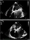
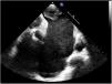
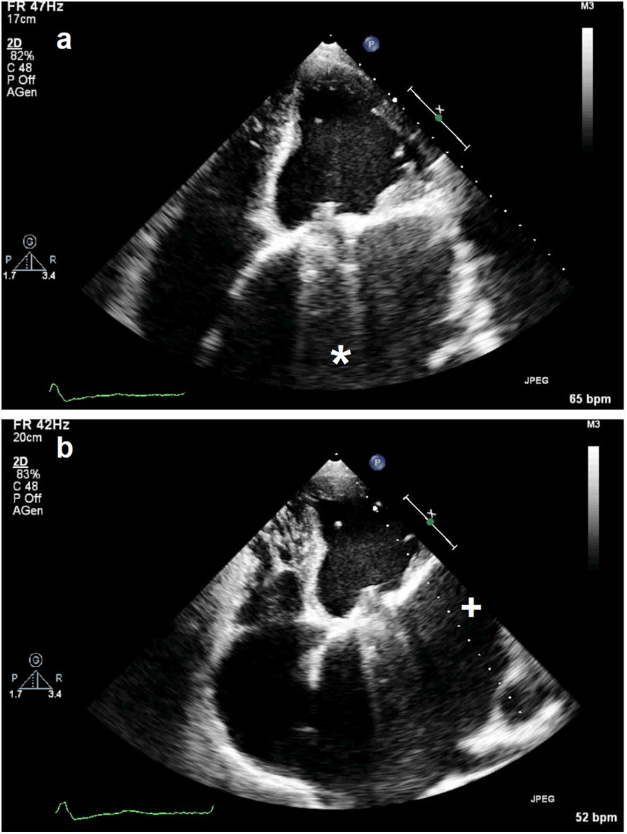
A transthoracic echocardiogram (TTE) showed a normally functioning prosthetic mitral valve and a huge left atrium (volume approximately 500 ml) (Figure 1a, *); moving from the apex to the midaxillary line from an off-axis view (Figure 1b, +), a giant left atrial appendage (LAA) appeared presenting severe enlargement with smoke and a massive thrombosis of the fundus (Figure 2).

Figure 1.

Transthoracic echocardiogram: (a) 4-chamber view showing a huge left atrium (*); (b) giant left atrial appendage (+).

(0,11MB).
Figure 2.

Transthoracic echocardiogram: 4-chamber off-axis view, showing severe left atrial appendage enlargement with smoke and a massive thrombosis of the fundus.

(0,06MB).

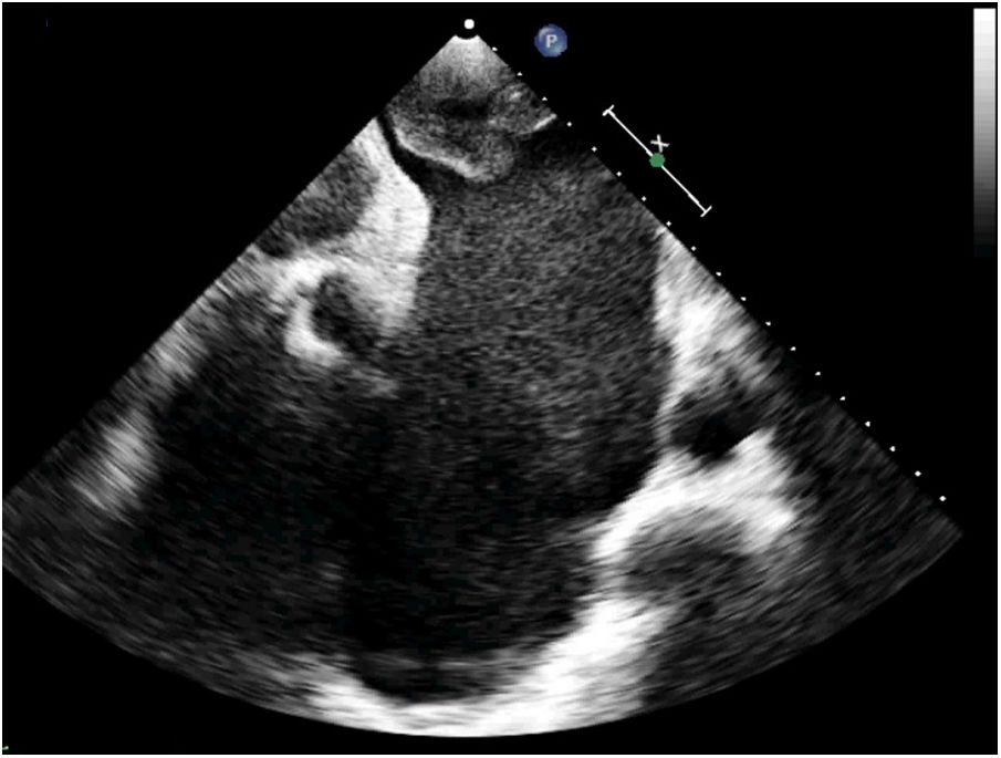
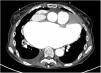
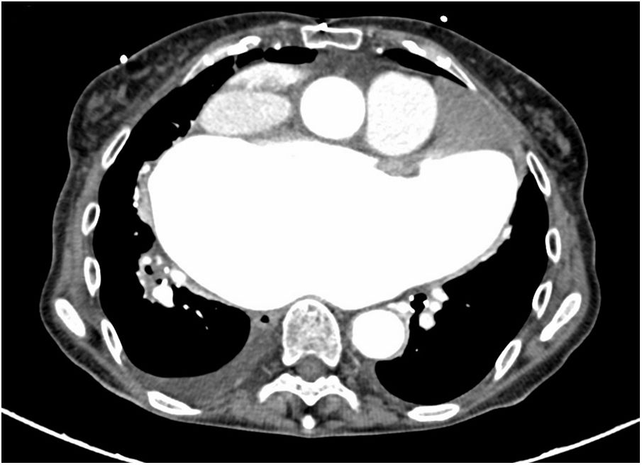
Computed tomography of the brain and of the chest was performed soon after to exclude ischemic stroke or pulmonary embolism, with a negative result and confirming the TTE findings (Figure 3).

Figure 3.

Computed tomography of the chest, axial view, confirming the transthoracic echocardiographic findings.

(0,06MB).

The patient was diagnosed with a transient ischemic attack and LAA thrombosis and admitted to our intensive care unit to be started on heparin infusion.

Conflicts of interest

The authors have no conflicts of interest to declare.

Appendix A
Supplementary material

The following are the supplementary material to this article:

(1,11MB)
(1,15MB)
(1,04MB)
(2,61MB)

Copyright © 2021. Sociedade Portuguesa de Cardiologia
Idiomas
Revista Portuguesa de Cardiologia
Opções de artigo
Ferramentas
Material Suplementar
en pt

Are you a health professional able to prescribe or dispense drugs?

Você é um profissional de saúde habilitado a prescrever ou dispensar medicamentos

Ao assinalar que é «Profissional de Saúde», declara conhecer e aceitar que a responsável pelo tratamento dos dados pessoais dos utilizadores da página de internet da Revista Portuguesa de Cardiologia (RPC), é esta entidade, com sede no Campo Grande, n.º 28, 13.º, 1700-093 Lisboa, com os telefones 217 970 685 e 217 817 630, fax 217 931 095 e com o endereço de correio eletrónico revista@spc.pt. Declaro para todos os fins, que assumo inteira responsabilidade pela veracidade e exatidão da afirmação aqui fornecida.