


Inferior vena cava (IVC) syndrome is a condition due to thrombus, extension of a tumor, extrinsic compression or intrinsic caval disease. Tumor invasion of the IVC includes renal carcinoma, adrenal carcinoma, pheochromocytoma and retroperitoneal metastatic disease from ovarian, cervical or prostate carcinoma. Computed tomography, magnetic resonance and cardiac ultrasound are often necessary to make a diagnosis.
A 45-year-old female with no past medical history presented to the hospital with six months of dry cough, associated with diffuse abdominal pain and lower extremity edema. On physical examination, grade two lower extremity edema and ascites were found. The results of the laboratory tests demonstrated elevated Ca-125. Abdominal tomography was performed showing left complex adnexal mass and lobulated hepatic mass with compression and displacement of inferior vena cava and right atrial extension (Figure 1A and 1B). Transthoracic echocardiography documented echo-dense mass of 32 mm×31 mm, area 9.8 cm2, suggestive of intracavitary right atrial thrombus (Figure 2A, 2B and 2C, Video 1A-1C in supplemental material), left ventricular ejection fraction and diastolic function were normal and the valves had no pathologic changes. Cardiac magnetic resonance was not performed. Additionally, Holter electrocardiogram recorded atrial tachycardia. Anticoagulation was initiated. Left oophorectomy was performed based on results of diagnosis of serous ovarian carcinoma. Liver, neck and bone metastatic disease was observed. Chemotherapy with carboplatin and paclitaxel was started. Currently, patient is receiving the second cycle of chemotherapy, symptoms have been relieved without cardiovascular events. Control images will be performed after chemotherapy has been completed to assess therapy response.
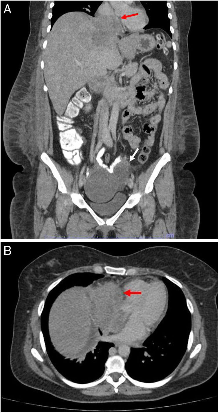
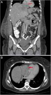
A: Coronal thoracic computed tomography showing left complex adnexal mass (white arrow) and lobulated hepatic mass with compression and displacement of inferior vena cava and right atrial extension (red arrow). B: Axial thoracic computed tomography showing mass with right atrial extension (red arrow).
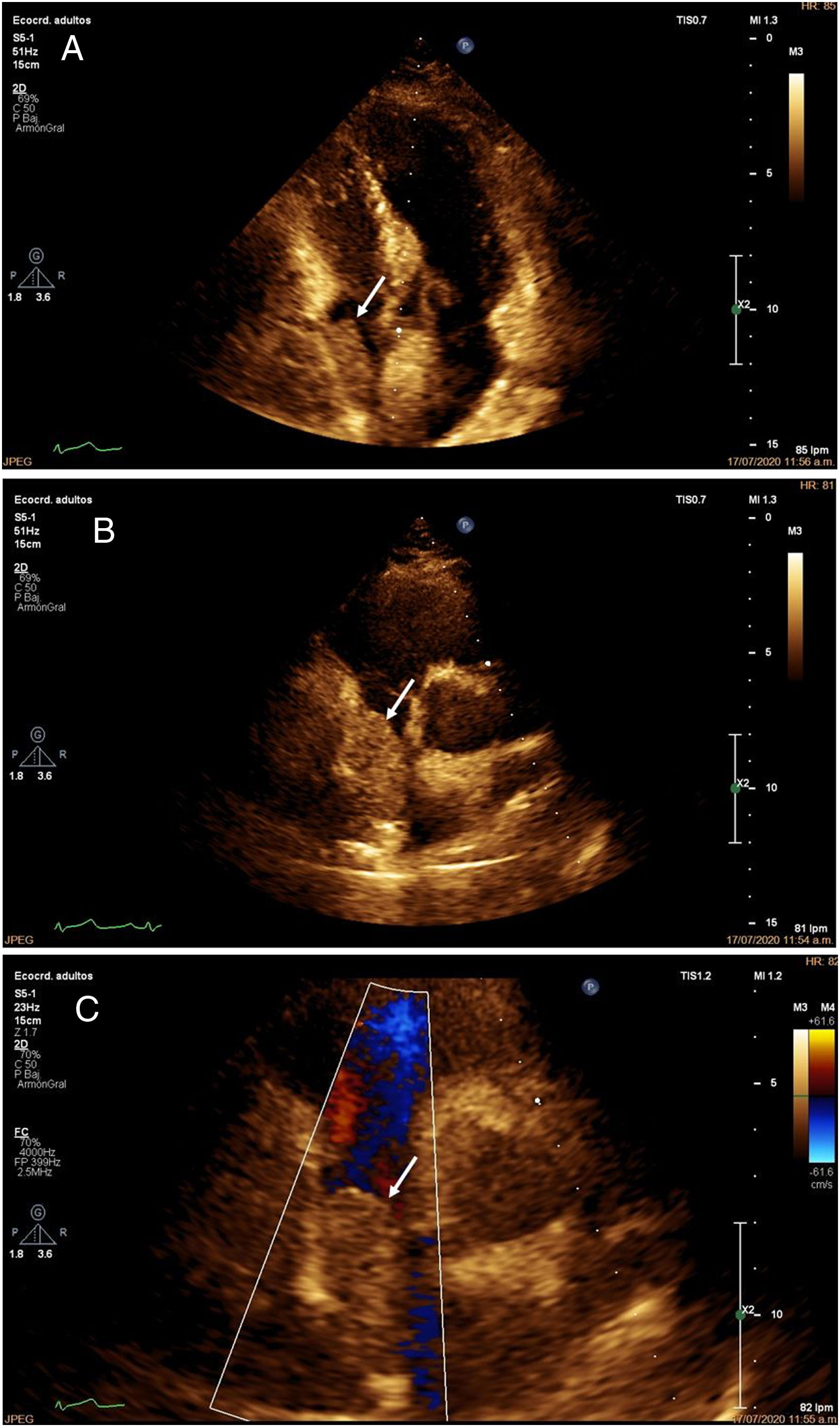
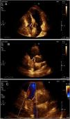
A: Transthoracic echocardiography showing echo-dense mass of 32 mm×31 mm, area 9.8 cm2 within right atrium suggestive of mass or intracavitary thrombus (white arrow). C: Transthoracic echocardiography showing mass within right atrium (white arrow). B: Transthoracic echocardiography showing restriction to color flow Doppler (white arrow).
Treatment of inferior vena cava syndrome has not been clearly described, but presumably it involves treating the underlying cause. Anticoagulation with dalteparin continues. This case is important because imaging and clinical symptoms can lead to detection of malignancy.
FundingNo funding was used in support of this manuscript.
Authors’ contributionsAll authors contributed significantly to the manuscript. All authors provided critique and feedback on the manuscript. All authors read and approved the final version of the manuscript.
Conflicts of interestThe authors have no conflicts of interest to declare.
