array:24 [
  "pii" => "S0870255122004802"
  "issn" => "08702551"
  "doi" => "10.1016/j.repc.2022.09.002"
  "estado" => "S300"
  "fechaPublicacion" => "2023-01-01"
  "aid" => "2063"
  "copyright" => "Sociedade Portuguesa de Cardiologia"
  "copyrightAnyo" => "2022"
  "documento" => "article"
  "crossmark" => 1
  "licencia" => "http://creativecommons.org/licenses/by-nc-nd/4.0/"
  "subdocumento" => "sco"
  "cita" => "Rev Port Cardiol. 2023;42:75-6"
  "abierto" => array:3 [
    "ES" => true
    "ES2" => true
    "LATM" => true
  ]
  "gratuito" => true
  "lecturas" => array:1 [
    "total" => 0
  ]
  "itemSiguiente" => array:19 [
    "pii" => "S0870255122004814"
    "issn" => "08702551"
    "doi" => "10.1016/j.repc.2022.01.010"
    "estado" => "S300"
    "fechaPublicacion" => "2023-01-01"
    "aid" => "2062"
    "copyright" => "Sociedade Portuguesa de Cardiologia"
    "documento" => "article"
    "crossmark" => 1
    "licencia" => "http://creativecommons.org/licenses/by-nc-nd/4.0/"
    "subdocumento" => "sco"
    "cita" => "Rev Port Cardiol. 2023;42:77-8"
    "abierto" => array:3 [
      "ES" => true
      "ES2" => true
      "LATM" => true
    ]
    "gratuito" => true
    "lecturas" => array:1 [
      "total" => 0
    ]
    "en" => array:11 [
      "idiomaDefecto" => true
      "cabecera" => "<span class="elsevierStyleTextfn">Image in Cardiology</span>"
      "titulo" => "An unthinkable cause of syncope"
      "tienePdf" => "en"
      "tieneTextoCompleto" => "en"
      "paginas" => array:1 [
        0 => array:2 [
          "paginaInicial" => "77"
          "paginaFinal" => "78"
        ]
      ]
      "titulosAlternativos" => array:1 [
        "pt" => array:1 [
          "titulo" => "Uma causa impens&#225;vel de s&#237;ncope"
        ]
      ]
      "contieneTextoCompleto" => array:1 [
        "en" => true
      ]
      "contienePdf" => array:1 [
        "en" => true
      ]
      "resumenGrafico" => array:2 [
        "original" => 0
        "multimedia" => array:7 [
          "identificador" => "fig0005"
          "etiqueta" => "Figure 1"
          "tipo" => "MULTIMEDIAFIGURA"
          "mostrarFloat" => true
          "mostrarDisplay" => false
          "figura" => array:1 [
            0 => array:4 [
              "imagen" => "gr1.jpeg"
              "Alto" => 3183
              "Ancho" => 2167
              "Tamanyo" => 416805
            ]
          ]
          "descripcion" => array:1 [
            "en" => "<p id="spar0005" class="elsevierStyleSimplePara elsevierViewall">&#40;A&#41; Axial thoracic computed tomography showing a giant cyst &#40;&#42;&#41; compressing the right heart&#46; &#40;B&#41; Four-chamber view&#44; balanced turbo field-echo &#40;BTFE&#41; cardiovascular magnetic resonance &#40;CMR&#41; showing a giant cyst &#40;&#42;&#41; compressing the right heart&#46; &#40;C&#41; Left ventricular tract outflow &#40;LVOT&#41; view&#44; BTFE CMR showing a giant cyst &#40;&#42;&#41; compressing the right atrium &#40;black arrow&#41;&#46; &#40;D&#41; BTFE CMR sagittal view of the cyst extension&#46; &#40;E&#41; Coronal thoracic computed tomography after the cyst resection&#46; &#40;F&#41; BTFE CMR showing the LVOT view after the cyst resection&#46;</p>"
          ]
        ]
      ]
      "autores" => array:1 [
        0 => array:2 [
          "autoresLista" => "Maria G&#46; Matta, Beatriz A&#46; Camara, Lucia Rodr&#237;guez Eyras, Miguel A&#46; Garcia"
          "autores" => array:4 [
            0 => array:2 [
              "nombre" => "Maria G&#46;"
              "apellidos" => "Matta"
            ]
            1 => array:2 [
              "nombre" => "Beatriz A&#46;"
              "apellidos" => "Camara"
            ]
            2 => array:2 [
              "nombre" => "Lucia Rodr&#237;guez"
              "apellidos" => "Eyras"
            ]
            3 => array:2 [
              "nombre" => "Miguel A&#46;"
              "apellidos" => "Garcia"
            ]
          ]
        ]
      ]
    ]
    "idiomaDefecto" => "en"
    "EPUB" => "https://multimedia.elsevier.es/PublicationsMultimediaV1/item/epub/S0870255122004814?idApp=UINPBA00004E"
    "url" => "/08702551/0000004200000001/v2_202301120627/S0870255122004814/v2_202301120627/en/main.assets"
  ]
  "itemAnterior" => array:19 [
    "pii" => "S0870255122004917"
    "issn" => "08702551"
    "doi" => "10.1016/j.repc.2022.11.001"
    "estado" => "S300"
    "fechaPublicacion" => "2023-01-01"
    "aid" => "2070"
    "copyright" => "Sociedade Portuguesa de Cardiologia"
    "documento" => "simple-article"
    "crossmark" => 1
    "licencia" => "http://creativecommons.org/licenses/by-nc-nd/4.0/"
    "subdocumento" => "dis"
    "cita" => "Rev Port Cardiol. 2023;42:73-4"
    "abierto" => array:3 [
      "ES" => true
      "ES2" => true
      "LATM" => true
    ]
    "gratuito" => true
    "lecturas" => array:1 [
      "total" => 0
    ]
    "en" => array:10 [
      "idiomaDefecto" => true
      "cabecera" => "<span class="elsevierStyleTextfn">Editorial comment</span>"
      "titulo" => "Exercise-induced syncope&#58; A real red flag&#33;"
      "tienePdf" => "en"
      "tieneTextoCompleto" => "en"
      "paginas" => array:1 [
        0 => array:2 [
          "paginaInicial" => "73"
          "paginaFinal" => "74"
        ]
      ]
      "titulosAlternativos" => array:1 [
        "pt" => array:1 [
          "titulo" => "S&#237;ncope induzida pelo exerc&#237;cio&#58; um verdadeiro sinal de alerta&#33;"
        ]
      ]
      "contieneTextoCompleto" => array:1 [
        "en" => true
      ]
      "contienePdf" => array:1 [
        "en" => true
      ]
      "autores" => array:1 [
        0 => array:2 [
          "autoresLista" => "H&#233;lder Dores"
          "autores" => array:1 [
            0 => array:2 [
              "nombre" => "H&#233;lder"
              "apellidos" => "Dores"
            ]
          ]
        ]
      ]
    ]
    "idiomaDefecto" => "en"
    "EPUB" => "https://multimedia.elsevier.es/PublicationsMultimediaV1/item/epub/S0870255122004917?idApp=UINPBA00004E"
    "url" => "/08702551/0000004200000001/v2_202301120627/S0870255122004917/v2_202301120627/en/main.assets"
  ]
  "en" => array:15 [
    "idiomaDefecto" => true
    "cabecera" => "<span class="elsevierStyleTextfn">Image in Cardiology</span>"
    "titulo" => "Placement of permanent pacemaker in a patient with venous anomaly through the right subclavian vein"
    "tieneTextoCompleto" => true
    "paginas" => array:1 [
      0 => array:2 [
        "paginaInicial" => "75"
        "paginaFinal" => "76"
      ]
    ]
    "autores" => array:1 [
      0 => array:4 [
        "autoresLista" => "Ibrahim Etem Celik, Mustafa Duran, Mikail Yarl&#305;oglu, Sani Nam&#305;k Murat"
        "autores" => array:4 [
          0 => array:4 [
            "nombre" => "Ibrahim Etem"
            "apellidos" => "Celik"
            "email" => array:1 [
              0 => "etem84@gmail.com"
            ]
            "referencia" => array:1 [
              0 => array:2 [
                "etiqueta" => "<span class="elsevierStyleSup">&#42;</span>"
                "identificador" => "cor0005"
              ]
            ]
          ]
          1 => array:2 [
            "nombre" => "Mustafa"
            "apellidos" => "Duran"
          ]
          2 => array:2 [
            "nombre" => "Mikail"
            "apellidos" => "Yarl&#305;oglu"
          ]
          3 => array:2 [
            "nombre" => "Sani Nam&#305;k"
            "apellidos" => "Murat"
          ]
        ]
        "afiliaciones" => array:1 [
          0 => array:2 [
            "entidad" => "Department of Cardiology&#44; University of Health Sciences&#44; Ankara Education and Research Hospital&#44; Ankara&#44; Turkey"
            "identificador" => "aff0005"
          ]
        ]
        "correspondencia" => array:1 [
          0 => array:3 [
            "identificador" => "cor0005"
            "etiqueta" => "&#8270;"
            "correspondencia" => "Corresponding author&#46;"
          ]
        ]
      ]
    ]
    "titulosAlternativos" => array:1 [
      "pt" => array:1 [
        "titulo" => "Implanta&#231;&#227;o de <span class="elsevierStyleItalic">pacemaker</span> definitivo pela veia subcl&#225;via num doente com anomalia venosa"
      ]
    ]
    "resumenGrafico" => array:2 [
      "original" => 0
      "multimedia" => array:7 [
        "identificador" => "fig0005"
        "etiqueta" => "Figure 1"
        "tipo" => "MULTIMEDIAFIGURA"
        "mostrarFloat" => true
        "mostrarDisplay" => false
        "figura" => array:1 [
          0 => array:4 [
            "imagen" => "gr1.jpeg"
            "Alto" => 653
            "Ancho" => 1340
            "Tamanyo" => 96890
          ]
        ]
        "descripcion" => array:1 [
          "en" => "<p id="spar0005" class="elsevierStyleSimplePara elsevierViewall">A&#46; Venogram from the right upper limb showing the absence of a right SVC with the persistent left superior vena cava draining into the dilated coronary sinus&#46; B&#46; Chest X-ray demonstrating the loop configuration of the ventricular lead of the VVIR pacemaker&#46;</p>"
        ]
      ]
    ]
    "textoCompleto" => "<span class="elsevierStyleSections"><p id="par0005" class="elsevierStylePara elsevierViewall">A 58-year-old female patient was admitted to our hospital with dizziness and near syncope&#46; The diagnosis of Mobitz type 2 atrioventricular block was made on electrocardiogram&#46; A dual-chamber pacemaker was inserted through the right side as the patient actively used her left arm&#46; The right ventricular &#40;RV&#41; lead was advanced in an attempt to access the right superior vena cava &#40;SVC&#41;&#44; but resistance was encountered&#46; Venography revealed persistent left superior vena cava &#40;PLSVC&#41; draining through the dilated coronary sinus &#40;CS&#41; into the right atrium with absent right SVC &#40;<a class="elsevierStyleCrossRef" href="#fig0005">Figure 1</a>A&#41;&#46; Then&#44; a classic lead delivery system was used to reach the right atrium through CS and then through the tricuspid valve into the right ventricle&#46; The RV lead was advanced via PLSVC through the CS and was actively fixed to the right ventricle apex with loop configuration&#46; Following RV lead placement&#44; an atrial active fixation lead was introduced&#44; however we were unable to implant a pacemaker lead in a proper location&#46; The parameters of the implanted VVIR pacemaker were ventricular sensing of 6 mV&#44; pacing threshold of 0&#46;4 V&#44; and impedance of 630 ohms&#46; Chest X-ray revealed satisfactory positioning of the ventricular lead &#40;<a class="elsevierStyleCrossRef" href="#fig0005">Figure 1</a>B&#41;&#46;</p><elsevierMultimedia ident="fig0005"></elsevierMultimedia><p id="par0010" class="elsevierStylePara elsevierViewall">While implanting cardiac devices in patients with PLSVC and absent right SVC&#44; it is difficult to pass through the tricuspid valve to place the ventricular lead&#46; Although lead implantation is very challenging due to abnormal vena cava anatomy&#44; different techniques can be used to facilitate the crossing tricuspid valve&#46; Physicians should be aware of the use of active fixation leads&#44; and conforming the loop configuration of the ventricular lead may overcome this technical challenge&#46;</p><span id="sec0005" class="elsevierStyleSection elsevierViewall"><span class="elsevierStyleSectionTitle" id="sect0005">Conflict of interests</span><p id="par0015" class="elsevierStylePara elsevierViewall">No potential conflict of interest was reported by the author&#40;s&#41;&#46;</p></span></span>"
    "textoCompletoSecciones" => array:1 [
      "secciones" => array:1 [
        0 => array:2 [
          "identificador" => "sec0005"
          "titulo" => "Conflict of interests"
        ]
      ]
    ]
    "pdfFichero" => "main.pdf"
    "tienePdf" => true
    "fechaRecibido" => "2021-09-28"
    "fechaAceptado" => "2022-09-07"
    "multimedia" => array:1 [
      0 => array:7 [
        "identificador" => "fig0005"
        "etiqueta" => "Figure 1"
        "tipo" => "MULTIMEDIAFIGURA"
        "mostrarFloat" => true
        "mostrarDisplay" => false
        "figura" => array:1 [
          0 => array:4 [
            "imagen" => "gr1.jpeg"
            "Alto" => 653
            "Ancho" => 1340
            "Tamanyo" => 96890
          ]
        ]
        "descripcion" => array:1 [
          "en" => "<p id="spar0005" class="elsevierStyleSimplePara elsevierViewall">A&#46; Venogram from the right upper limb showing the absence of a right SVC with the persistent left superior vena cava draining into the dilated coronary sinus&#46; B&#46; Chest X-ray demonstrating the loop configuration of the ventricular lead of the VVIR pacemaker&#46;</p>"
        ]
      ]
    ]
  ]
  "idiomaDefecto" => "en"
  "url" => "/08702551/0000004200000001/v2_202301120627/S0870255122004802/v2_202301120627/en/main.assets"
  "Apartado" => array:4 [
    "identificador" => "95133"
    "tipo" => "SECCION"
    "pt" => array:2 [
      "titulo" => "Image in Cardiology"
      "idiomaDefecto" => true
    ]
    "idiomaDefecto" => "pt"
  ]
  "PDF" => "https://static.elsevier.es/multimedia/08702551/0000004200000001/v2_202301120627/S0870255122004802/v2_202301120627/en/main.pdf?idApp=UINPBA00004E&text.app=https://revportcardiol.org/"
  "EPUB" => "https://multimedia.elsevier.es/PublicationsMultimediaV1/item/epub/S0870255122004802?idApp=UINPBA00004E"
]
Partilhar
Informação da revista
Vol. 42. Núm. 1.
Páginas 75-76 (janeiro 2023)
Partilhar
Partilhar
Baixar PDF
Mais opções do artigo
Vol. 42. Núm. 1.
Páginas 75-76 (janeiro 2023)
Image in Cardiology
Open Access
Placement of permanent pacemaker in a patient with venous anomaly through the right subclavian vein
Implantação de pacemaker definitivo pela veia subclávia num doente com anomalia venosa
Visitas
1509
Ibrahim Etem Celik
Autor para correspondência
etem84@gmail.com

Corresponding author.
, Mustafa Duran, Mikail Yarlıoglu, Sani Namık Murat
Department of Cardiology, University of Health Sciences, Ankara Education and Research Hospital, Ankara, Turkey
Este item recebeu

Under a Creative Commons license
Informação do artigo
Texto Completo
Baixar PDF
Estatísticas
Figuras (1)
Texto Completo

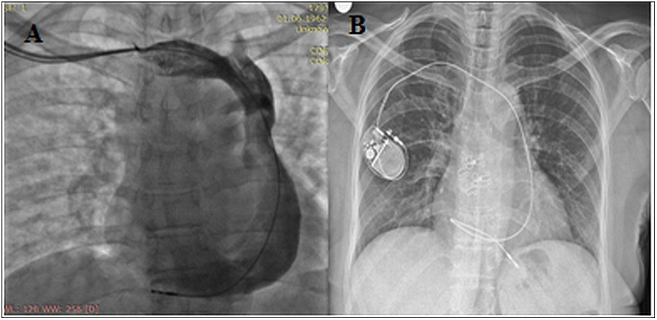
A 58-year-old female patient was admitted to our hospital with dizziness and near syncope. The diagnosis of Mobitz type 2 atrioventricular block was made on electrocardiogram. A dual-chamber pacemaker was inserted through the right side as the patient actively used her left arm. The right ventricular (RV) lead was advanced in an attempt to access the right superior vena cava (SVC), but resistance was encountered. Venography revealed persistent left superior vena cava (PLSVC) draining through the dilated coronary sinus (CS) into the right atrium with absent right SVC (Figure 1A). Then, a classic lead delivery system was used to reach the right atrium through CS and then through the tricuspid valve into the right ventricle. The RV lead was advanced via PLSVC through the CS and was actively fixed to the right ventricle apex with loop configuration. Following RV lead placement, an atrial active fixation lead was introduced, however we were unable to implant a pacemaker lead in a proper location. The parameters of the implanted VVIR pacemaker were ventricular sensing of 6 mV, pacing threshold of 0.4 V, and impedance of 630 ohms. Chest X-ray revealed satisfactory positioning of the ventricular lead (Figure 1B).

Figure 1.

A. Venogram from the right upper limb showing the absence of a right SVC with the persistent left superior vena cava draining into the dilated coronary sinus. B. Chest X-ray demonstrating the loop configuration of the ventricular lead of the VVIR pacemaker.

(0.09MB).

While implanting cardiac devices in patients with PLSVC and absent right SVC, it is difficult to pass through the tricuspid valve to place the ventricular lead. Although lead implantation is very challenging due to abnormal vena cava anatomy, different techniques can be used to facilitate the crossing tricuspid valve. Physicians should be aware of the use of active fixation leads, and conforming the loop configuration of the ventricular lead may overcome this technical challenge.

Conflict of interests

No potential conflict of interest was reported by the author(s).

Copyright © 2022. Sociedade Portuguesa de Cardiologia
Baixar PDF
Idiomas
Revista Portuguesa de Cardiologia
Opções de artigo
Ferramentas
en pt

Are you a health professional able to prescribe or dispense drugs?

Você é um profissional de saúde habilitado a prescrever ou dispensar medicamentos

Ao assinalar que é «Profissional de Saúde», declara conhecer e aceitar que a responsável pelo tratamento dos dados pessoais dos utilizadores da página de internet da Revista Portuguesa de Cardiologia (RPC), é esta entidade, com sede no Campo Grande, n.º 28, 13.º, 1700-093 Lisboa, com os telefones 217 970 685 e 217 817 630, fax 217 931 095 e com o endereço de correio eletrónico revista@spc.pt. Declaro para todos os fins, que assumo inteira responsabilidade pela veracidade e exatidão da afirmação aqui fornecida.