array:23 [
  "pii" => "S0870255119305463"
  "issn" => "08702551"
  "doi" => "10.1016/j.repc.2018.09.015"
  "estado" => "S300"
  "fechaPublicacion" => "2019-09-01"
  "aid" => "1447"
  "copyright" => "Sociedade Portuguesa de Cardiologia"
  "copyrightAnyo" => "2019"
  "documento" => "article"
  "crossmark" => 1
  "licencia" => "http://creativecommons.org/licenses/by-nc-nd/4.0/"
  "subdocumento" => "sco"
  "cita" => "Rev Port Cardiol. 2019;38:677-9"
  "abierto" => array:3 [
    "ES" => true
    "ES2" => true
    "LATM" => true
  ]
  "gratuito" => true
  "lecturas" => array:2 [
    "total" => 347
    "formatos" => array:3 [
      "EPUB" => 54
      "HTML" => 239
      "PDF" => 54
    ]
  ]
  "itemAnterior" => array:20 [
    "pii" => "S0870255119304822"
    "issn" => "08702551"
    "doi" => "10.1016/j.repc.2018.01.014"
    "estado" => "S300"
    "fechaPublicacion" => "2019-09-01"
    "aid" => "1424"
    "copyright" => "Sociedade Portuguesa de Cardiologia"
    "documento" => "simple-article"
    "crossmark" => 1
    "licencia" => "http://creativecommons.org/licenses/by-nc-nd/4.0/"
    "subdocumento" => "crp"
    "cita" => "Rev Port Cardiol. 2019;38:675.e1-675.e5"
    "abierto" => array:3 [
      "ES" => true
      "ES2" => true
      "LATM" => true
    ]
    "gratuito" => true
    "lecturas" => array:2 [
      "total" => 341
      "formatos" => array:3 [
        "EPUB" => 52
        "HTML" => 215
        "PDF" => 74
      ]
    ]
    "pt" => array:13 [
      "idiomaDefecto" => true
      "cabecera" => "<span class="elsevierStyleTextfn">Caso Cl&#237;nico</span>"
      "titulo" => "Electrocat&#233;ter como causa iatrog&#233;nica de insufici&#234;ncia card&#237;aca direita &#8211; relato de um caso"
      "tienePdf" => "pt"
      "tieneTextoCompleto" => "pt"
      "tieneResumen" => array:2 [
        0 => "pt"
        1 => "en"
      ]
      "paginas" => array:1 [
        0 => array:2 [
          "paginaInicial" => "675&#46;e1"
          "paginaFinal" => "675&#46;e5"
        ]
      ]
      "titulosAlternativos" => array:1 [
        "en" => array:1 [
          "titulo" => "Pacemaker lead as an iatrogenic cause of right heart failure&#58; Case report"
        ]
      ]
      "contieneResumen" => array:2 [
        "pt" => true
        "en" => true
      ]
      "contieneTextoCompleto" => array:1 [
        "pt" => true
      ]
      "contienePdf" => array:1 [
        "pt" => true
      ]
      "resumenGrafico" => array:2 [
        "original" => 0
        "multimedia" => array:7 [
          "identificador" => "fig0005"
          "etiqueta" => "Figura 1"
          "tipo" => "MULTIMEDIAFIGURA"
          "mostrarFloat" => true
          "mostrarDisplay" => false
          "figura" => array:1 [
            0 => array:4 [
              "imagen" => "gr1.jpeg"
              "Alto" => 2438
              "Ancho" => 2917
              "Tamanyo" => 659703
            ]
          ]
          "descripcion" => array:1 [
            "pt" => "<p id="spar0055" class="elsevierStyleSimplePara elsevierViewall">A&#46; Vista bidimensional &#40;esquerda&#41; e tridimensional &#40;direita&#41; apical de 4 c&#226;maras demonstrando interfer&#234;ncia do eletrocat&#233;ter no folheto septal com regurgita&#231;&#227;o tric&#250;spide&#59; B&#46; Vista tridimensional da perspetiva ventricular direita com n&#227;o coapta&#231;&#227;o dos folhetos valvulares e regurgita&#231;&#227;o grave com electrocat&#233;teres <span class="elsevierStyleItalic">in situ</span>&#59; C&#46; Vista tridimensional da perspetiva ventricular direita evidenciando dois eletrocat&#233;teres atrav&#233;s da v&#225;lvula tric&#250;spide com regurgita&#231;&#227;o moderada&#41;&#46;</p>"
          ]
        ]
      ]
      "autores" => array:1 [
        0 => array:2 [
          "autoresLista" => "Manuel Toscano, Z&#233;lia Neves, Carla Matias, Madalena Carvalho, Regina Ribeiras, Francisco Morgado, &#194;ngelo Nobre"
          "autores" => array:7 [
            0 => array:2 [
              "nombre" => "Manuel"
              "apellidos" => "Toscano"
            ]
            1 => array:2 [
              "nombre" => "Z&#233;lia"
              "apellidos" => "Neves"
            ]
            2 => array:2 [
              "nombre" => "Carla"
              "apellidos" => "Matias"
            ]
            3 => array:2 [
              "nombre" => "Madalena"
              "apellidos" => "Carvalho"
            ]
            4 => array:2 [
              "nombre" => "Regina"
              "apellidos" => "Ribeiras"
            ]
            5 => array:2 [
              "nombre" => "Francisco"
              "apellidos" => "Morgado"
            ]
            6 => array:2 [
              "nombre" => "&#194;ngelo"
              "apellidos" => "Nobre"
            ]
          ]
        ]
      ]
    ]
    "idiomaDefecto" => "pt"
    "Traduccion" => array:1 [
      "en" => array:9 [
        "pii" => "S2174204919302442"
        "doi" => "10.1016/j.repce.2018.01.012"
        "estado" => "S300"
        "subdocumento" => ""
        "abierto" => array:3 [
          "ES" => true
          "ES2" => true
          "LATM" => true
        ]
        "gratuito" => true
        "lecturas" => array:1 [
          "total" => 0
        ]
        "idiomaDefecto" => "en"
        "EPUB" => "https://multimedia.elsevier.es/PublicationsMultimediaV1/item/epub/S2174204919302442?idApp=UINPBA00004E"
      ]
    ]
    "EPUB" => "https://multimedia.elsevier.es/PublicationsMultimediaV1/item/epub/S0870255119304822?idApp=UINPBA00004E"
    "url" => "/08702551/0000003800000009/v2_202001221225/S0870255119304822/v2_202001221225/pt/main.assets"
  ]
  "en" => array:16 [
    "idiomaDefecto" => true
    "cabecera" => "<span class="elsevierStyleTextfn">Image in Cardiology</span>"
    "titulo" => "Functional assessment of coronary disease using workstation-based computed tomography-derived fractional flow reserve"
    "tieneTextoCompleto" => true
    "paginas" => array:1 [
      0 => array:2 [
        "paginaInicial" => "677"
        "paginaFinal" => "679"
      ]
    ]
    "autores" => array:1 [
      0 => array:4 [
        "autoresLista" => "Nuno Dias Ferreira, Ricardo Ladeiras-Lopes, Gustavo Pires de Morais, Vasco Gama, Pedro Braga"
        "autores" => array:5 [
          0 => array:4 [
            "nombre" => "Nuno"
            "apellidos" => "Dias Ferreira"
            "email" => array:1 [
              0 => "ferreira.nmd@gmail.com"
            ]
            "referencia" => array:1 [
              0 => array:2 [
                "etiqueta" => "<span class="elsevierStyleSup">&#42;</span>"
                "identificador" => "cor0005"
              ]
            ]
          ]
          1 => array:2 [
            "nombre" => "Ricardo"
            "apellidos" => "Ladeiras-Lopes"
          ]
          2 => array:2 [
            "nombre" => "Gustavo"
            "apellidos" => "Pires de Morais"
          ]
          3 => array:2 [
            "nombre" => "Vasco"
            "apellidos" => "Gama"
          ]
          4 => array:2 [
            "nombre" => "Pedro"
            "apellidos" => "Braga"
          ]
        ]
        "afiliaciones" => array:1 [
          0 => array:2 [
            "entidad" => "Servi&#231;o de Cardiologia&#44; Centro Hospitalar de Vila Nova de Gaia&#47;Espinho&#44; Vila Nova de Gaia&#44; Portugal"
            "identificador" => "aff0005"
          ]
        ]
        "correspondencia" => array:1 [
          0 => array:3 [
            "identificador" => "cor0005"
            "etiqueta" => "&#8270;"
            "correspondencia" => "Corresponding author&#46;"
          ]
        ]
      ]
    ]
    "titulosAlternativos" => array:1 [
      "pt" => array:1 [
        "titulo" => "Avalia&#231;&#227;o funcional de doen&#231;a coron&#225;ria utilizando FFR derivado de TC"
      ]
    ]
    "resumenGrafico" => array:2 [
      "original" => 0
      "multimedia" => array:7 [
        "identificador" => "fig0020"
        "etiqueta" => "Figure 4"
        "tipo" => "MULTIMEDIAFIGURA"
        "mostrarFloat" => true
        "mostrarDisplay" => false
        "figura" => array:1 [
          0 => array:4 [
            "imagen" => "gr4.jpeg"
            "Alto" => 706
            "Ancho" => 1255
            "Tamanyo" => 85455
          ]
        ]
        "descripcion" => array:1 [
          "en" => "<p id="spar0020" class="elsevierStyleSimplePara elsevierViewall">Images from invasive coronary angiography before &#40;A&#41; and after &#40;B&#41; percutaneous coronary intervention in the proximal left anterior descending artery&#44; with successful implantation of a drug-eluting stent&#46;</p>"
        ]
      ]
    ]
    "textoCompleto" => "<span class="elsevierStyleSections"><p id="par0005" class="elsevierStylePara elsevierViewall">A 55-year-old man presented to our outpatient clinic reporting a six-month history of exertional dyspnea and intermittent atypical chest pain&#46; His medical history included essential hypertension&#46; The baseline electrocardiogram and echocardiogram were normal&#46; The patient underwent treadmill exercise stress testing under the Bruce protocol&#46; During stage 3&#44; the patient developed 1&#46;5-mm horizontal ST depression in V3-V6 and the inferior leads&#46; The test was interrupted at 9 min due to fatigue&#44; with no chest pain&#46;</p><p id="par0010" class="elsevierStylePara elsevierViewall">Due to intermediate risk findings on the treadmill test&#44; coronary computed tomography angiography &#40;CCTA&#41; was requested for further assessment&#46; The coronary calcium score was 105&#46;2 Agatston units and an intermediate stenosis was identified in the proximal left anterior descending artery &#40;LAD&#41; due to non-calcified plaque &#40;<a class="elsevierStyleCrossRef" href="#fig0005">Figure 1</a>A and D&#44; arrow&#41;&#46; Because the physiologic significance of the lesion was unclear&#44; computed tomography-derived fractional flow reserve &#40;CT-FFR&#41; was determined using a workstation-based software prototype &#40;cFFR&#44; Siemens AG Healthcare&#44; Forchheim&#44; Germany&#41;&#46; The value of CT-FFR just beyond the proximal LAD stenosis was 0&#46;75 &#40;<a class="elsevierStyleCrossRef" href="#fig0010">Figure 2</a>&#41;&#46; The patient was referred for invasive coronary angiography &#40;ICA&#41; which confirmed a 75&#37; stenosis in the proximal LAD &#40;<a class="elsevierStyleCrossRef" href="#fig0015">Figure 3</a>B and Supplementary Video S1&#41;&#46; Invasive FFR assessed in the LAD showed a similar result &#40;0&#46;76&#41; to that observed on CCTA &#40;<a class="elsevierStyleCrossRef" href="#fig0015">Figures 3</a>A and 3C&#41;&#46; The patient successfully underwent percutaneous coronary intervention with a drug-eluting stent &#40;<a class="elsevierStyleCrossRef" href="#fig0020">Figure 4</a>&#41; and was asymptomatic after the procedure&#46; This case illustrates the value of CT-FFR as a gatekeeper prior to ICA&#44; to appropriately select patients and vessels for revascularization&#46;</p><elsevierMultimedia ident="fig0005"></elsevierMultimedia><elsevierMultimedia ident="fig0010"></elsevierMultimedia><elsevierMultimedia ident="fig0015"></elsevierMultimedia><elsevierMultimedia ident="fig0020"></elsevierMultimedia><span id="sec0005" class="elsevierStyleSection elsevierViewall"><span class="elsevierStyleSectionTitle" id="sect0005">Conflicts of interest</span><p id="par0015" class="elsevierStylePara elsevierViewall">The authors have no conflicts of interest to declare&#46;</p></span></span>"
    "textoCompletoSecciones" => array:1 [
      "secciones" => array:1 [
        0 => array:2 [
          "identificador" => "sec0005"
          "titulo" => "Conflicts of interest"
        ]
      ]
    ]
    "pdfFichero" => "main.pdf"
    "tienePdf" => true
    "fechaRecibido" => "2018-08-12"
    "fechaAceptado" => "2018-09-10"
    "apendice" => array:1 [
      0 => array:1 [
        "seccion" => array:1 [
          0 => array:4 [
            "apendice" => "<p id="par0025" class="elsevierStylePara elsevierViewall">The following are the supplementary material to this article&#58;<elsevierMultimedia ident="upi0005"></elsevierMultimedia></p>"
            "etiqueta" => "Appendix A"
            "titulo" => "Supplementary material"
            "identificador" => "sec0015"
          ]
        ]
      ]
    ]
    "multimedia" => array:5 [
      0 => array:7 [
        "identificador" => "fig0005"
        "etiqueta" => "Figure 1"
        "tipo" => "MULTIMEDIAFIGURA"
        "mostrarFloat" => true
        "mostrarDisplay" => false
        "figura" => array:1 [
          0 => array:4 [
            "imagen" => "gr1.jpeg"
            "Alto" => 1258
            "Ancho" => 1255
            "Tamanyo" => 174191
          ]
        ]
        "descripcion" => array:1 [
          "en" => "<p id="spar0005" class="elsevierStyleSimplePara elsevierViewall">Coronary computed tomography angiography images&#46; Curved multiplanar reformats of the left anterior descending artery &#40;LAD&#41; &#40;A&#41;&#44; left circumflex artery &#40;B&#41; and right coronary artery &#40;C&#41;&#59; three-dimensional volume rendering image of the heart &#40;D&#41;&#46; Note the presence of significant stenosis in the proximal LAD due to non-calcified plaque &#40;arrows&#41;&#46;</p>"
        ]
      ]
      1 => array:7 [
        "identificador" => "fig0010"
        "etiqueta" => "Figure 2"
        "tipo" => "MULTIMEDIAFIGURA"
        "mostrarFloat" => true
        "mostrarDisplay" => false
        "figura" => array:1 [
          0 => array:4 [
            "imagen" => "gr2.jpeg"
            "Alto" => 680
            "Ancho" => 905
            "Tamanyo" => 38286
          ]
        ]
        "descripcion" => array:1 [
          "en" => "<p id="spar0010" class="elsevierStyleSimplePara elsevierViewall">Computed tomography &#40;CT&#41;-derived fractional flow reserve &#40;FFR&#41; imaging showing an intermediate stenosis in the proximal left anterior descending artery&#46; The computed value of CT-derived FFR was 0&#46;75&#46;</p>"
        ]
      ]
      2 => array:7 [
        "identificador" => "fig0015"
        "etiqueta" => "Figure 3"
        "tipo" => "MULTIMEDIAFIGURA"
        "mostrarFloat" => true
        "mostrarDisplay" => false
        "figura" => array:1 [
          0 => array:4 [
            "imagen" => "gr3.jpeg"
            "Alto" => 1263
            "Ancho" => 1255
            "Tamanyo" => 147217
          ]
        ]
        "descripcion" => array:1 [
          "en" => "<p id="spar0015" class="elsevierStyleSimplePara elsevierViewall">Correlation between invasive coronary angiography and coronary computed tomography &#40;CT&#41; angiography&#46; &#40;A&#41; CT-derived fractional flow reserve &#40;FFR&#41; imaging showing an intermediate stenosis in the proximal left anterior descending artery &#40;LAD&#41; with a computed FFR of 0&#46;75&#59; &#40;B&#41; invasive coronary angiography confirming a 75&#37; stenosis in the proximal LAD&#59; &#40;C&#41; the measured value of invasive FFR was 0&#46;76&#44; which showed a good agreement with the CT-derived value &#40;A&#41;&#46;</p>"
        ]
      ]
      3 => array:7 [
        "identificador" => "fig0020"
        "etiqueta" => "Figure 4"
        "tipo" => "MULTIMEDIAFIGURA"
        "mostrarFloat" => true
        "mostrarDisplay" => false
        "figura" => array:1 [
          0 => array:4 [
            "imagen" => "gr4.jpeg"
            "Alto" => 706
            "Ancho" => 1255
            "Tamanyo" => 85455
          ]
        ]
        "descripcion" => array:1 [
          "en" => "<p id="spar0020" class="elsevierStyleSimplePara elsevierViewall">Images from invasive coronary angiography before &#40;A&#41; and after &#40;B&#41; percutaneous coronary intervention in the proximal left anterior descending artery&#44; with successful implantation of a drug-eluting stent&#46;</p>"
        ]
      ]
      4 => array:5 [
        "identificador" => "upi0005"
        "tipo" => "MULTIMEDIAECOMPONENTE"
        "mostrarFloat" => false
        "mostrarDisplay" => true
        "Ecomponente" => array:3 [
          "fichero" => "mmc1.mp4"
          "ficheroTamanyo" => 3420733
          "Video" => array:2 [
            "flv" => array:5 [
              "fichero" => "mmc1.flv"
              "poster" => "mmc1.jpg"
              "tiempo" => 0
              "alto" => 0
              "ancho" => 0
            ]
            "mp4" => array:5 [
              "fichero" => "mmc1.m4v"
              "poster" => "mmc1.jpg"
              "tiempo" => 0
              "alto" => 0
              "ancho" => 0
            ]
          ]
        ]
      ]
    ]
  ]
  "idiomaDefecto" => "en"
  "url" => "/08702551/0000003800000009/v2_202001221225/S0870255119305463/v2_202001221225/en/main.assets"
  "Apartado" => array:4 [
    "identificador" => "75834"
    "tipo" => "SECCION"
    "en" => array:2 [
      "titulo" => "Imagem em Cardiologia"
      "idiomaDefecto" => true
    ]
    "idiomaDefecto" => "en"
  ]
  "PDF" => "https://static.elsevier.es/multimedia/08702551/0000003800000009/v2_202001221225/S0870255119305463/v2_202001221225/en/main.pdf?idApp=UINPBA00004E&text.app=https://revportcardiol.org/"
  "EPUB" => "https://multimedia.elsevier.es/PublicationsMultimediaV1/item/epub/S0870255119305463?idApp=UINPBA00004E"
]
Partilhar
Informação da revista
Vol. 38. Núm. 9.
Páginas 677-679 (setembro 2019)
Partilhar
Partilhar
Baixar PDF
Mais opções do artigo
Vol. 38. Núm. 9.
Páginas 677-679 (setembro 2019)
Image in Cardiology
Open Access
Functional assessment of coronary disease using workstation-based computed tomography-derived fractional flow reserve
Avaliação funcional de doença coronária utilizando FFR derivado de TC
Visitas
3195
Nuno Dias Ferreira
Autor para correspondência
ferreira.nmd@gmail.com

Corresponding author.
, Ricardo Ladeiras-Lopes, Gustavo Pires de Morais, Vasco Gama, Pedro Braga
Serviço de Cardiologia, Centro Hospitalar de Vila Nova de Gaia/Espinho, Vila Nova de Gaia, Portugal
Este item recebeu

Under a Creative Commons license
Informação do artigo
Texto Completo
Baixar PDF
Estatísticas
Figuras (4)
Mostrar maisMostrar menos
Material adicional (1)
Texto Completo

A 55-year-old man presented to our outpatient clinic reporting a six-month history of exertional dyspnea and intermittent atypical chest pain. His medical history included essential hypertension. The baseline electrocardiogram and echocardiogram were normal. The patient underwent treadmill exercise stress testing under the Bruce protocol. During stage 3, the patient developed 1.5-mm horizontal ST depression in V3-V6 and the inferior leads. The test was interrupted at 9 min due to fatigue, with no chest pain.

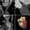
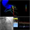
Due to intermediate risk findings on the treadmill test, coronary computed tomography angiography (CCTA) was requested for further assessment. The coronary calcium score was 105.2 Agatston units and an intermediate stenosis was identified in the proximal left anterior descending artery (LAD) due to non-calcified plaque (Figure 1A and D, arrow). Because the physiologic significance of the lesion was unclear, computed tomography-derived fractional flow reserve (CT-FFR) was determined using a workstation-based software prototype (cFFR, Siemens AG Healthcare, Forchheim, Germany). The value of CT-FFR just beyond the proximal LAD stenosis was 0.75 (Figure 2). The patient was referred for invasive coronary angiography (ICA) which confirmed a 75% stenosis in the proximal LAD (Figure 3B and Supplementary Video S1). Invasive FFR assessed in the LAD showed a similar result (0.76) to that observed on CCTA (Figures 3A and 3C). The patient successfully underwent percutaneous coronary intervention with a drug-eluting stent (Figure 4) and was asymptomatic after the procedure. This case illustrates the value of CT-FFR as a gatekeeper prior to ICA, to appropriately select patients and vessels for revascularization.

Figure 1.

Coronary computed tomography angiography images. Curved multiplanar reformats of the left anterior descending artery (LAD) (A), left circumflex artery (B) and right coronary artery (C); three-dimensional volume rendering image of the heart (D). Note the presence of significant stenosis in the proximal LAD due to non-calcified plaque (arrows).

(0.17MB).
Figure 2.

Computed tomography (CT)-derived fractional flow reserve (FFR) imaging showing an intermediate stenosis in the proximal left anterior descending artery. The computed value of CT-derived FFR was 0.75.

(0.04MB).
Figure 3.

Correlation between invasive coronary angiography and coronary computed tomography (CT) angiography. (A) CT-derived fractional flow reserve (FFR) imaging showing an intermediate stenosis in the proximal left anterior descending artery (LAD) with a computed FFR of 0.75; (B) invasive coronary angiography confirming a 75% stenosis in the proximal LAD; (C) the measured value of invasive FFR was 0.76, which showed a good agreement with the CT-derived value (A).

(0.14MB).
Figure 4.

Images from invasive coronary angiography before (A) and after (B) percutaneous coronary intervention in the proximal left anterior descending artery, with successful implantation of a drug-eluting stent.

(0.08MB).
Conflicts of interest

The authors have no conflicts of interest to declare.

Appendix A
Supplementary material

The following are the supplementary material to this article:

(3.26MB)

Copyright © 2019. Sociedade Portuguesa de Cardiologia
Baixar PDF
Idiomas
Revista Portuguesa de Cardiologia
Opções de artigo
Ferramentas
Material Suplementar
en pt

Are you a health professional able to prescribe or dispense drugs?

Você é um profissional de saúde habilitado a prescrever ou dispensar medicamentos

Ao assinalar que é «Profissional de Saúde», declara conhecer e aceitar que a responsável pelo tratamento dos dados pessoais dos utilizadores da página de internet da Revista Portuguesa de Cardiologia (RPC), é esta entidade, com sede no Campo Grande, n.º 28, 13.º, 1700-093 Lisboa, com os telefones 217 970 685 e 217 817 630, fax 217 931 095 e com o endereço de correio eletrónico revista@spc.pt. Declaro para todos os fins, que assumo inteira responsabilidade pela veracidade e exatidão da afirmação aqui fornecida.