


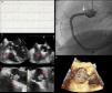
We report the case of a 65-year-old woman admitted to the emergency department with constricting chest pain of one hour's duration. The pre-hospital electrocardiogram (ECG) showed ST-segment elevation in the inferior leads (Figure 1A). On admission the patient was asymptomatic and the ECG alterations had resolved. Intermittent periods of ST-segment elevation were observed on telemetry while the patient was being prepared for urgent catheterization, which revealed interruption of contrast in the ostium of the right coronary artery, causing elevated intracoronary gradients during selective angiography (Figure 1B). This was initially thought to represent dissection and two stents were implanted, but an echocardiogram on the same day completely changed our view of the case: transesophageal echocardiography performed for a more accurate characterization showed akinesia of the inferior wall and a mobile structure, 1.4cm×0.9cm, pedunculated, between the right coronary sinus and the non-coronary sinus, which did not impede valve function, but intermittently partially obstructed the stent (Figure 1C–E). On magnetic resonance imaging, this structure revealed gadolinium enhancement. A few days later the mass was excised and histological study showed it to be a cardiac papillary fibroelastoma, which had been one of the possible diagnoses postulated during the investigation.

Diagnostic exams performed during the investigation of this case: (A) initial electrocardiogram showing ST-segment elevation in the inferior leads; (B) right coronary angiography showing interruption of contrast (arrow) in the ostium; (C and D) transesophageal echocardiogram of the tumor (dashed arrow) showing its location and relation to the stent (solid arrow) in the right coronary artery; (E) three-dimensional transesophageal echocardiogram of the tumor (dashed arrow).
Bedside echocardiography is an accessible tool that can completely change the diagnostic and therapeutic approach in some cases, such as the one presented.
Ethical disclosuresProtection of human and animal subjectsThe authors declare that no experiments were performed on humans or animals for this study.
Confidentiality of dataThe authors declare that no patient data appear in this article.
Right to privacy and informed consentThe authors declare that no patient data appear in this article.
Conflicts of interestThe authors have no conflicts of interest to declare.
Please cite this article as: Ferreira C, Baptista A, Leão S, et al. Fibroelastoma condicionando obstrução coronária intermitente e enfarte do miocárdio. Quando o ecocardiograma poderia ter feito a diferença?…. Rev Port Cardiol. 2014;33:395–396.
