Informação da revista
Vol. 40. Núm. 3.
Páginas 247-248 (Março 2021)
Partilhar
Partilhar
Baixar PDF
Mais opções do artigo
Vol. 40. Núm. 3.
Páginas 247-248 (Março 2021)
Image in Cardiology
Open Access
Embolization of a left atrial appendage closure device
Embolização de dispositivo de encerramento do apêndice auricular esquerdo
Visitas
2620
Alzira Nunesa,
Autor para correspondência
zira.nunes.2@gmail.com

Corresponding author.
, Diana Pissarrab, Marta Tavares Silvaa,c, Pedro Bernardo Almeidaa, João Carlos Silvaa, Maria Júlia Maciela,c
a Serviço de Cardiologia, Centro Hospitalar Universitário de São João, E.P.E., Porto, Portugal
b Serviço de Cirurgia Cardiotorácica, Centro Hospitalar Universitário de São João, E.P.E., Porto, Portugal
c Faculdade de Medicina da Universidade do Porto, Porto, Portugal
Este item recebeu

Under a Creative Commons license
Informação do artigo
Texto Completo
Baixar PDF
Estatísticas
Figuras (4)
Mostrar maisMostrar menos
Texto Completo

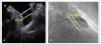
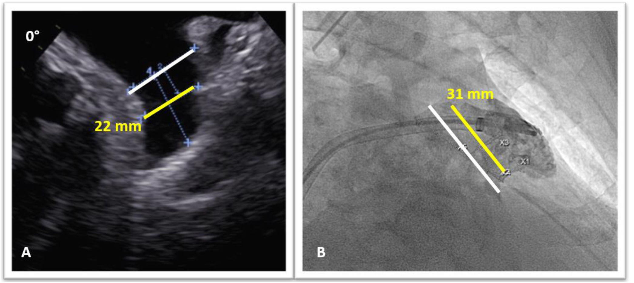
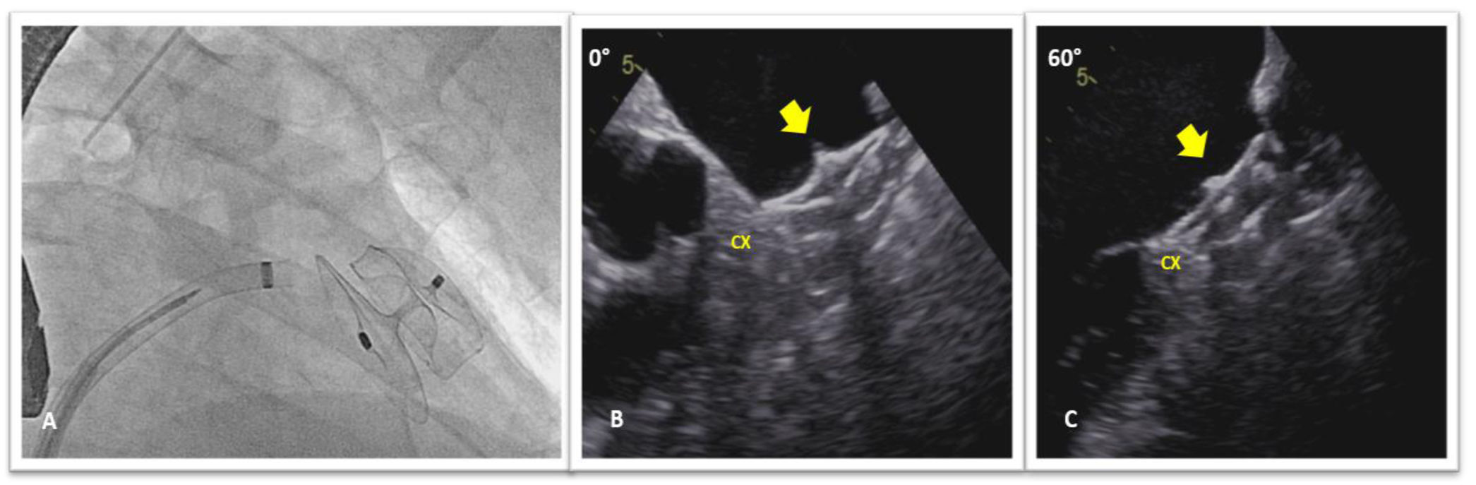
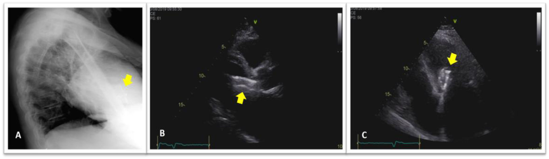
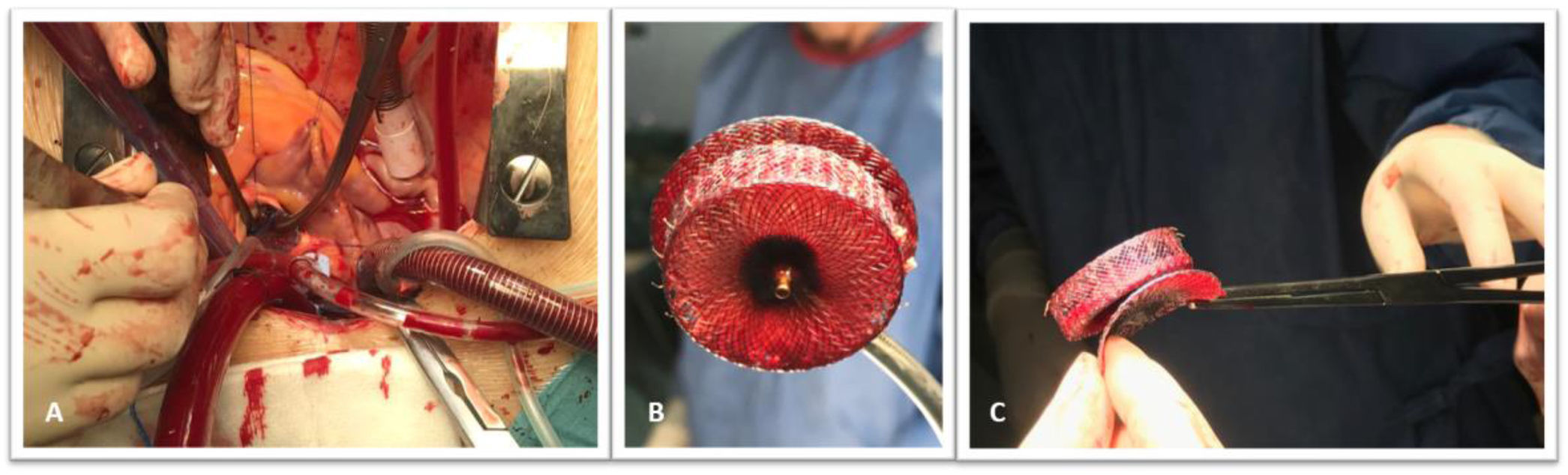
A 68-year old hypertensive man with diabetes and a history of persistent atrial fibrillation and an ischemic cerebral event, was referred for percutaneous left atrial appendage (LAA) closure. He was at high thromboembolic risk based on a CHA2DS2-VASc score of five and oral anticoagulants were contraindicated due to a firearm-related brain injury. A pre-procedure transesophageal echocardiogram (TOE) showed a LAA compatible with percutaneous closure (Figure 1A). It was performed under general anesthesia and with TOE guidance. After transseptal puncture, a windsock-shaped LAA with a landing zone of 31 mm was observed on fluoroscopy (Figure 1B). Based on TOE and fluoroscopy measurements, a 34-mm Amplatzer Amulet device was chosen and deployed in the LAA with a residual leak of 3 mm. Once in place, device stability was confirmed: tire shape of the lobe, concavity of the disk and good separation of the lobe from the disk (Figure 2-A); device lobe was aligned with appendage neck and more than two thirds of the device lobe was located distal to the circumflex artery (Figure 2B and C). The next day, he was asymptomatic and hemodynamically stable, however, a routine chest X-ray (Figure 3A) and transthoracic echocardiogram (Figure 3B and C) showed embolization of the LAA device into the left ventricular outflow tract, without impairment of mitral valve function. Because of the risk of damage to the aortic and mitral valves by the device unfolded anchoring barbs, urgent surgical device removal (Figure 4) and appendage amputation was the first line strategy and the procedure was uneventful.

Figure 1.

Transesophageal echocardiogram (A) and fluoroscopy (B) images showing the diameter of the left atrial appendage at the landing zone (yellow lines) 10 mm inside the left atrial appendage orifice (gray lines).

(0,18MB).
Figure 2.

Fluoroscopy (A) and transesophageal echocardiogram (B and C) images showing correct Amplatzer Amulet device implantation in the left atrial appendage (arrows). CX indicates circumflex artery.

(0,13MB).
Figure 3.

Lateral chest X-ray view (A) and two-dimensional transthoracic echocardiogram parasternal long-axis (B) and apical four-chamber (C) views revealing embolized device in the left ventricle outflow tract (arrows).

(0,07MB).
Figure 4.

Intraoperative image (A) shows the embolized left atrial appendage closure device being removed from the left ventricular outflow tract just below the aortic valve plane. Extraoperative images (B and C) showing the removed device.

(0,17MB).
Conflicts of interest

The authors have no conflicts of interest to declare.

Copyright © 2021. Sociedade Portuguesa de Cardiologia
Idiomas
Revista Portuguesa de Cardiologia
Opções de artigo
Ferramentas
en pt

Are you a health professional able to prescribe or dispense drugs?

Você é um profissional de saúde habilitado a prescrever ou dispensar medicamentos

Ao assinalar que é «Profissional de Saúde», declara conhecer e aceitar que a responsável pelo tratamento dos dados pessoais dos utilizadores da página de internet da Revista Portuguesa de Cardiologia (RPC), é esta entidade, com sede no Campo Grande, n.º 28, 13.º, 1700-093 Lisboa, com os telefones 217 970 685 e 217 817 630, fax 217 931 095 e com o endereço de correio eletrónico revista@spc.pt. Declaro para todos os fins, que assumo inteira responsabilidade pela veracidade e exatidão da afirmação aqui fornecida.