Informação da revista
Vol. 34. Núm. 10.
Páginas 629-630 (Outubro 2015)
Image in Cardiology
Open Access
Double aortic arch as cause of persistent stridor: Sequential imaging for preoperative assessment
Duplo arco aórtico na origem de estridor persistente: sequência de imagens para orientação cirúrgica
Visitas
9213
Bruno Sanchesa,
Autor para correspondência
brsan82@gmail.com

Corresponding author.
, Duarte Rebelob, Sara Britoc, Rui Anjosd, Luísa Loboe, Teresa Bandeiraf, Ana Saiandaf
a Serviço de Pediatria, Hospital Garcia de Orta, Almada, Portugal
b Departamento de Pediatria, Hospital de Santa Maria, Centro Hospitalar Lisboa Norte, Centro Académico de Medicina de Lisboa, Lisboa, Portugal
c Serviço de Pediatria, Centro Hospitalar de Leiria, Leiria, Portugal
d Serviço de Cardiologia Pediátrica, Hospital de Santa Cruz, Centro Hospitalar Lisboa Ocidental, Carnaxide, Portugal
e Serviço de Radiologia, Hospital de Santa Maria, Centro Hospitalar Lisboa Norte, Centro Académico de Medicina de Lisboa, Lisboa, Portugal
f Unidade de Pneumologia Pediátrica, Departamento de Pediatria, Hospital de Santa Maria, Centro Hospitalar Lisboa Norte, Centro Académico de Medicina de Lisboa, Lisboa, Portugal
Este item recebeu

Under a Creative Commons license
Informação do artigo
Texto Completo
Baixar PDF
Estatísticas
Figuras (4)
Mostrar maisMostrar menos
Texto Completo

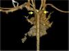
A boy aged 2.5 years presented biphasic stridor at three months of age, which worsened with crying, feeding and upper respiratory tract infections. Fiberoptic bronchoscopy (FOB) at five months diagnosed tracheomalacia, with 80% dynamic collapse of the lower two-thirds of the trachea. The child had normal growth and no difficulty feeding. FOB at 21 months due to persistent stridor showed marked dynamic collapse and extrinsic compression of the trachea. Thoracic computed tomography (angio-CT) angiography identified a vascular ring due to a double aortic arch (Figures 1–3), causing extrinsic compression of the trachea with marked reduction in its caliber (Figure 4). There were no other cardiac malformations on echocardiographic study. At 23 months the child underwent ligation and division of the more hypoplastic arch (left anterior) by thoracotomy. The postoperative course was good, and the child currently has mild stridor only.

Figure 1.

Thoracic computed tomography (angio-CT) angiography, axial view, showing two aortic arches (arrows) forming a double aortic arch, which surround the airway and reduce tracheal caliber. The right aortic arch is dominant.

Figure 2.

Thoracic angio-CT angiography, coronal maximum intensity projection reconstruction, showing two aortic arches (arrows) originating in the ascending aorta. The brachiocephalic trunk originates in the right aortic arch, while the left common carotid originates in the left aortic arch.

Figure 3.

Thoracic angio-CT angiography, three-dimensional reconstruction, posterior view, showing two aortic arches (arrows) that join in the proximal descending aorta. The right aortic arch is of larger caliber.

Figure 4.

Thoracic angio-CT angiography, three-dimensional reconstruction visualizing the airway. Note the tracheal compression (arrows) caused by the vascular ring.

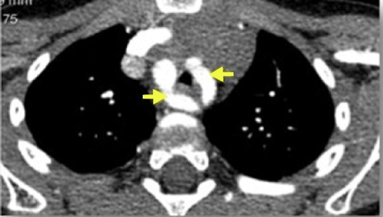
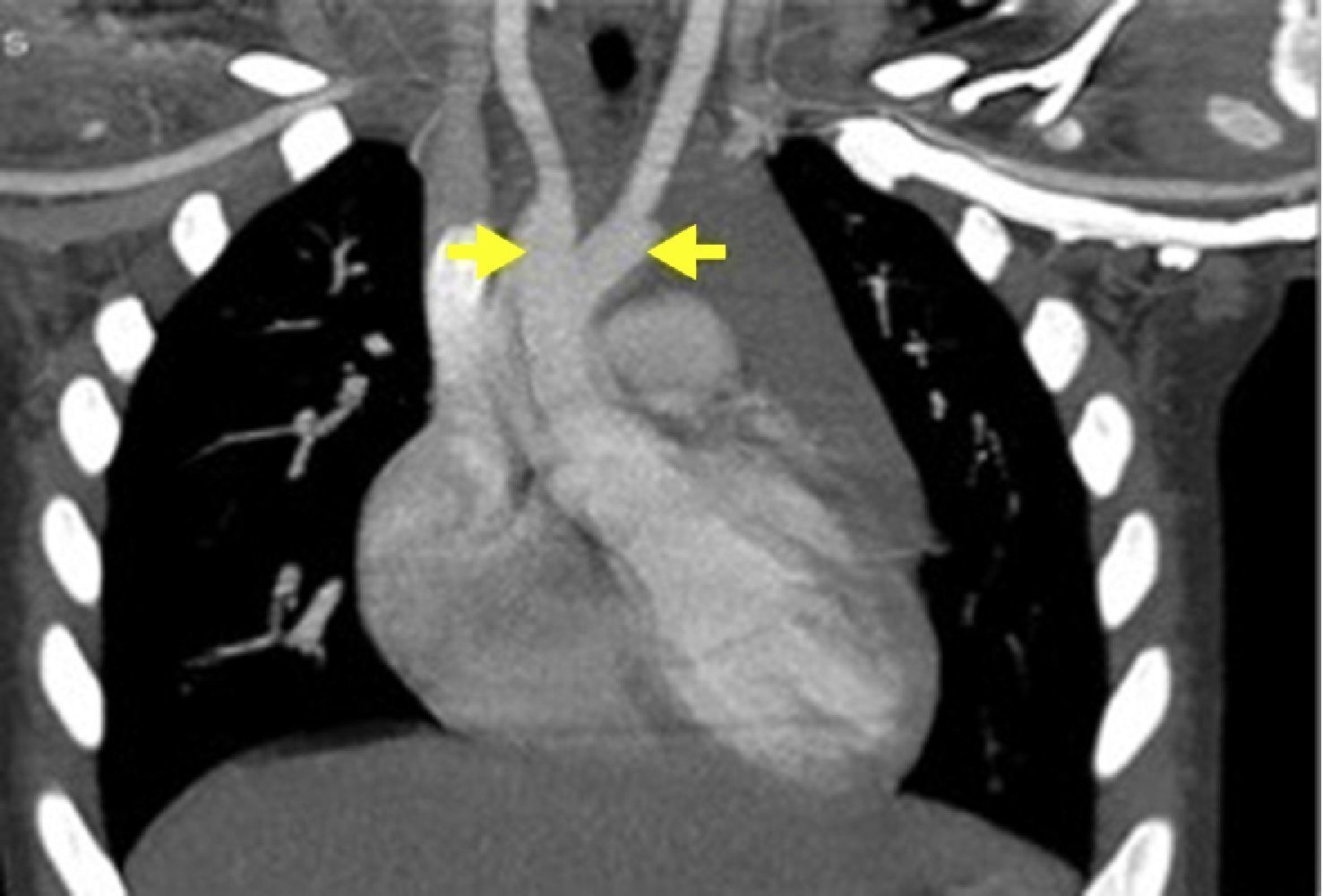
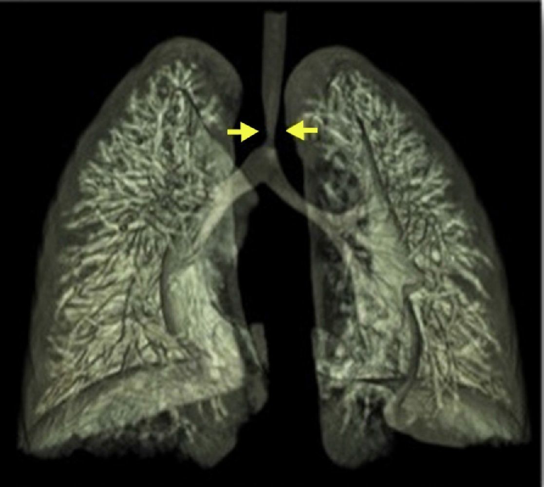
Vascular rings account for 1% of congenital heart defects and are the most common malformation causing airway compression. In this case, the clinical presentation dictated the sequence of imaging exams – FOB followed by angio-CT. This combination of imaging techniques is essential for preoperative assessment. angio-CT angiography provides better spatial and temporal resolution, rapid image acquisition requiring only sedation or brief anesthesia in uncooperative patients, and three-dimensional image reconstruction, and is thus the preferred imaging modality, especially with the lower radiation levels required with modern equipment. Assessment of tracheomalacia or other airway dysfunction still requires video bronchoscopy.

Ethical disclosuresProtection of human and animal subjects

The authors declare that no experiments were performed on humans or animals for this investigation.

Confidentiality of data

The authors declare that no patient data appears in this article.

Right to privacy and informed consent

The authors declare that no patient data appears in this article.

Conflicts of interest

The authors have no conflicts of interest to declare.

Please cite this article as: Sanches B, Rebelo D, Brito S, et al. Duplo arco aórtico na origem de estridor persistente: sequência de imagens para orientação cirúrgica. Rev Port Cardiol. 2015;34:629–630.

Copyright © 2015. Sociedade Portuguesa de Cardiologia
Baixar PDF
Idiomas
Revista Portuguesa de Cardiologia
Opções de artigo
Ferramentas
en pt

Are you a health professional able to prescribe or dispense drugs?

Você é um profissional de saúde habilitado a prescrever ou dispensar medicamentos

Ao assinalar que é «Profissional de Saúde», declara conhecer e aceitar que a responsável pelo tratamento dos dados pessoais dos utilizadores da página de internet da Revista Portuguesa de Cardiologia (RPC), é esta entidade, com sede no Campo Grande, n.º 28, 13.º, 1700-093 Lisboa, com os telefones 217 970 685 e 217 817 630, fax 217 931 095 e com o endereço de correio eletrónico revista@spc.pt. Declaro para todos os fins, que assumo inteira responsabilidade pela veracidade e exatidão da afirmação aqui fornecida.