
Congenital atrioventricular block (CAVB), classified as such when diagnosed in utero, at birth or during the first month of life, is a rare condition with an estimated incidence between 1/15 000 and 1/22 000 live births. It is now accepted that the pathophysiology of this condition is predominantly associated with an immunologically mediated response to the conduction system, which occurs due to transplacental passage of maternal autoantibodies from mothers diagnosed, in most cases, with systemic lupus erythematosus or Sjögren syndrome. Fetal echocardiography continues to be the diagnostic gold standard, however there are other techniques with good results and advantages. Regarding therapeutics, both pharmacological measures and cardiac stimulation techniques have been developed to increase the safety of procedures, decrease associated mortality and morbidity, and provide a better quality of life for patients, although there are disagreements in deciding the best therapeutic plan. This review aims to summarize and elucidate the best diagnostic approach as well as the best therapeutic strategies. A search was performed in the PubMed and Science Direct databases of articles published and accepted for publication. The following search terms were used: “Congenital atrioventricular block”, “Neonatal lupus”, “Pacemaker”, “Pathophysiology”, “Electrophysiology”, and “Prenatal diagnosis”. Articles in Portuguese and English were selected. No time constraints were used. Repeated articles were excluded from the two databases.
O bloqueio auriculoventricular congénito (BAVC), assim classificado quando diagnosticado in utero, ao nascimento ou durante o primeiro mês de vida, é uma patologia rara com uma incidência estimada que varia entre 1/15.000 e 1/22.000 nados vivos. É atualmente aceite que a fisiopatologia inerente a esta condição está predominantemente associada a uma resposta imunologicamente mediada contra o sistema de condução que ocorre devido à passagem transplacentária de autoanticorpos maternos de mães diagnosticadas, na maioria dos casos, com lúpus eritematoso sistémico (LES) ou síndrome de Sjögren (SS). A ecocardiografia fetal continua a ser o gold-standard diagnóstico, contudo existem outras técnicas com bons resultados e vantagens relativamente à primeira. No que concerne à terapêutica, tanto as estratégias farmacológicas quanto as técnicas de estimulação cardíaca têm evoluído no sentido de aumentar a segurança dos procedimentos, diminuir a mortalidade e morbilidade associadas e promover uma maior qualidade de vida dos doentes, continuando, apesar disso, a existir divergências no momento de decidir qual o melhor plano terapêutico. Deste modo, esta revisão tem como objetivo o resumo e a clarificação da melhor abordagem diagnóstica, bem como das melhores estratégias terapêuticas. Foi feita uma pesquisa nas bases de dados Pubmed e Science Direct de artigos publicados e aceites para publicação. Foram utilizadas as seguintes expressões de pesquisa: “Congenital atrioventricular block”, “Neonatal lupus”, “Pacemaker”, “Pathophysiology”, “Electrophysiology”, “Prenatal diagnosis”. Foram selecionados artigos escritos em português e inglês. Não foram utilizadas restrições temporais. Foram excluídos artigos repetidos nas duas bases de dados.
Congenital atrioventricular block (CAVB) is a rare condition with an estimated incidence that ranges between 1/15 000 and 1/22 000 live births.1–8
Atrioventricular (AV) block is a disturbance of cardiac conduction with abnormal propagation of the electrical impulse due to a structural or functional abnormality.7,9 It is said to be congenital when diagnosed in utero, at birth or during the first month of life.3,4
CAVB can occur in a structurally normal heart as isolated atrioventricular block, or in association with congenital heart defects, in which cases the prognosis is worse.3–6
The most prevalent form of CAVB is autoimmune, diagnosed in children of mothers diagnosed, in most cases, with systemic lupus erythematosus (SLE) or Sjögren syndrome (SS). However, even when circulating maternal antibody levels are elevated, only 2-5% of pregnancies result in fetuses with CAVB.3,4,10–12
Clinically, the condition can manifest with bradycardia, with heart rates of less than 100 bpm, pericardial effusion, ventricular dilatation, hyperechogenicity of the atrial walls due to fibrosis and reduced ventricular contractility, alterations visible on the echocardiogram revealing a proinflammatory state and probable concomitant myocarditis, endocardial fibroelastosis, ascites and fetal hydrops.13
There is no effective prophylactic treatment, although some drugs can improve the prognosis. Permanent pacemaker implantation is virtually imperative, since in most cases the block is complete and medically irreversible.
EtiologyOn the basis of its incidence and current knowledge of its pathophysiology, CAVB can be immune-modulated or autoimmune, and associated with congenital heart defects or idiopathic.
In 56-90% of cases it is autoimmune, defined as such when the heart has no anatomical malformation that could explain the condition and the mother has a diagnosis of autoimmune disease and/or is positive for autoantibodies.4,14,15
The second most frequent cause is congenital heart malformations, which are found in 14-42% of cases.4 Defects in cardiac embryogenesis including congenital malformations of the anterior endocardium and the AV node are associated with loss of function and/or anomalous location of these structures, weak propagation of the electrical signal, and a greater likelihood of CAVB.3–5
Transposition of the great vessels (TGV) and defects of the AV septum are the most common structural malformations.16 It is estimated that 5% of TGV cases diagnosed at birth are accompanied by complete CAVB.17
Idiopathic CAVB is associated with genetic abnormalities that have no apparent relationship with autoimmune disease and in a structurally normal heart.17
Genetic alterationsIn a retrospective multicenter study that included 141 children diagnosed with CAVB without structural heart disease or maternal antibodies, in which cardiac conduction disorders were demonstrated in both parents and children, the authors hypothesized that an inherited genetic alteration causes isolated, non-immune and idiopathic CAVB.18
Various mutations have been associated with increased risk for development of CAVB, in SCN5A, SCN1B, SCN10A, TRPM4, KCNK17, KCNJ2, HCN4, LMNA, ANKB, NKX2-5 and TBX5, among other genes.2,7,19,20
The gene that is generally agreed to be most closely associated with the pathogenesis of CAVB is SCN5A, which is responsible for phase 0 of the cardiac action potential. Alterations in the alpha subunit of the sodium channel NaV1.5 are associated with a 70% reduction in the density of these channels, and hence a decrease in the INa current generated.7
Mutations in the SCN5A gene are also the third most frequently associated with long QT syndrome, particularly type 3, caused by gain-of-function mutations in the sodium channel. Mutations in this gene are also associated with 20-30% of cases of Brugada syndrome, in this case with loss-of-function mutations in the sodium channel, classified as type 1 Brugada.16
The concomitant presence of manifestations of more than one of the above syndromes can lead to cardiac sodium channelopathy overlap syndrome, which is associated with higher risk of alterations in the conduction system.3
Ainsworth et al. identified a link between an allele of the HLA-C Asn80Lys polymorphism, which codes for the C2 epitope, a high-affinity ligand for the inhibitory natural killer (NK) cell receptor KIR2DL1, and CAVB. This epitope inhibits the action of NKs on macrophages and giant cells, leading to inflammation and damage to cardiac tissue.21
Maternal risk factorsAdvanced maternal age and the time of year of pregnancy, when there are increased levels of anti-Ro/SSA or anti-La/SSB antibodies, are believed to be the most important maternal risk factors.4,26
Other risk factors include maternal infections, particularly respiratory infections and those occurring between the 18th and 24th week of pregnancy, traumatic events during the same period including emotional stress due to the risk of fetal heart disease, low vitamin D levels and hypothyroidism. Conversely, higher maternal vitamin D levels are protective, associated with later need for pacemaker implantation.4,8,18,22–26
PathophysiologyPositivity for maternal anti-Ro/SSA or anti-La/SSB antibodies is found in 60-95% of cases,3,4,10–12 in most of which SLE or SS has been diagnosed.4,25,27,28
However, only 2-5% of pregnancies in these women result in CAVB,3,4,10–12,29 rising to 12-25% for subsequent pregnancies. The results of several studies suggest that the increased risk in subsequent pregnancies may be overestimated, since the selected groups consisted mainly of families with previous cases of neonatal lupus. However, the fact that women are older at the time of a subsequent birth may explain this increase.3,4
Damage to the conduction system usually occurs between the 16th and 24th weeks, a period during which maternal antibodies most often cross the placenta via the trophoblast FcγRn receptor.4,26,29,30
Maternal antibodies bind to L-type calcium channels in fetal cardiomyocytes, particularly those in the AV node, and reversibly inhibit these channels’ current. This results in inflammation, calcification and fibrosis of the conduction tissue, leading to irreversible damage, even in structurally normal hearts.3,4,14,29,31–36
In a case report analyzing an autopsy of cardiac tissue from a 19-week fetus with CAVB, Friedman et al. demonstrated extensive calcification and inflammatory infiltrate in the AV node and conduction tissue.37
Apoptosis and inflammation trigger macrophages to secrete TGF-β1 and TNF-α, inflammatory mediators that are permanently elevated in the conduction tissue of babies with heart disease on autopsy.8,36 The AV node and surrounding tissues are replaced by fibrotic tissue and areas of calcification that interrupt the electrical signal.3,32,33,38
The polymorphism of codon 25 in the TGFB1 gene has been associated with interindividual variability24 and increased risk for developing CAVB.
Another molecular mechanism is increased levels of interferon alpha (INF-α), which were found in 78% of cases in a recent study, and increase the expression of class II MHC on CD14+ monocytes. While the former increase synthesis of SIGLEC1, a proinflammatory protein, the latter increases trafficking of maternal IgG across the placenta.12
Analysis of umbilical cord blood showed that levels of C-reactive protein, N-terminal pro-B-type natriuretic peptide (NT-proBNP), matrix metalloproteinases (particularly type 2), plasminogen and urokinase plasminogen activator are increased in cases of neonatal lupus with severe cardiac damage. This is further evidence for the hypothesis that immune-mediated inflammation and fibrosis are associated with these alterations.8
Clinical manifestationsIn utero, the first manifestation is usually fetal bradycardia with a heart rate of less than 100 bpm.39 However, the phenotype is variable, depending on etiology, age at presentation and ventricular function.40
Differential diagnoses of fetal bradyarrhythmia include transient sinus bradycardia, complete CAVB and partial CAVB.13 The heart block may be intermittent in the neonatal period but usually becomes permanent.39,41
Fetal hydrops, ventricular response of <55 bpm, prematurity and complex congenital defects are markers of poor prognosis and are associated with higher mortality.1,3,5,6,28,42
Fetal hydrops is defined as the pathological accumulation of fluid in at least two fetal compartments, which may include the pleural or pericardial spaces, abdominal cavity, integument, or placenta. It can result from a combination of increased hydrostatic pressure, decreased oncotic pressure, and in some cases, lymphatic obstruction. Approximately 15-25% of fetuses with nonimmune hydrops have cardiac abnormalities.26
Patients with autoimmune CAVB may present with other concomitant cardiac abnormalities, endocardial fibroelastosis, dilated cardiomyopathy (DCM) and/or valve disease. The incidence of the latter is 1.6%.3,4,32
These alterations are among the manifestations of neonatal lupus syndrome (NLS), which is found in around 5% of newborns of mothers with SLE and positivity for anti-Ro (25-40%) and anti-La (10-15%) antibodies. The two most frequent forms of NLS are neonatal lupus erythematosus (NLE) and CAVB, although hematological and hepatic manifestations have also been documented.43 Maternal antibodies usually cease to be detectable between the 6th and 8th month of postnatal life followed by regression of dermal, hepatic and hematological manifestations, but this regeneration does not occur in cardiac tissue. Consequently, most cases of established CAVB are complete and irreversible.27
Data from the US Research Registry for Neonatal Lupus on neonatal outcomes in a group of pregnant women with SLE show that 49% of newborns had NLS, 30% with cutaneous NLE, 18% CAVB and 1% hematological/hepatic NLE.43
Comparison of late pediatric diagnoses with those made in utero or at birth shows that the former are less likely to have an autoimmune etiology4,28 and have lower mortality and better prognosis.3,4,8
Overall mortality associated with CAVB ranges between 9% and 25%, with 70% of deaths occurring in utero. Pacemaker implantation is required in the first year of life in 12-70% of babies.1,3–5,7,8,44
Most births (81%) are live, 38% of which are premature. Vaginal birth should be the aim, but due to the increased risk of preterm birth associated with maternal autoimmune disease, cesarean delivery is preferred in around 75% of cases.4,7,43 Cesarean delivery is indicated whenever there is evidence of fetal distress or risk to the life of mother or fetus; however, it is associated with higher rates of infection and bleeding, and should therefore only be performed for obstetric indications.43 The AV block diagnosed may be first or second degree, but in around 80% of cases it is third degree, complete, and irreversible.4,7
DiagnosisA thorough medical history, particularly of the mother, is essential to obtain an accurate and timely diagnosis. In high-risk pregnancies, fetal echocardiographic cardiac monitoring should begin in the 16th week and continue weekly until the 24th week, and thereafter fortnightly until birth.4,25,26
Indicators of high risk include the presence of maternal SSA/SSB antibodies, a family history of congenital heart disease, the presence of structural heart defects or rhythm disturbances on a routine obstetric echocardiogram, and evidence of fetal hydrops.26
Echocardiography remains the gold standard for diagnosis of CAVB, with a diagnostic rate of 90%, although this is dependent on the experience and skill of the operator.26 Echocardiography is also the gold standard for anatomical and functional study of the fetal heart, screening for abnormalities in anatomy, rhythm and rate by assessing atrial and ventricular rates, atrioventricular conduction, and the presence of a ventricular contraction after every atrial contraction.13,26
Other techniques for detecting arrhythmias and conduction defects have been proposed, but, although promising, there is little agreement concerning their usefulness in clinical practice. Cardiac magnetic resonance imaging is used to assess venous anatomy and associated extracardiac abnormalities, Doppler echocardiography to determine rhythm and the PR interval, fetal electrocardiography for fetal monitoring after rupture of membranes, and magnetocardiography for more precise assessment of conduction and rhythm in fetuses with known conduction disorders (class of recommendation IIa, level of evidence B/C).26,28,45
Around 75% of cases of CAVB are diagnosed between the 20th and 28th week of pregnancy. Presentation tends to be earlier in cases of autoimmune CAVB.3,4,6,39
In a study by Morel et al.46 of 187 neonates with CAVB over a median follow-up of seven years, 94.4% of cases were diagnosed in utero. Pacemakers were implanted in 80% of cases; 18.8% developed DCM, a median of 8.6 months after implantation. Ten-year survival was 23% for newborns diagnosed neonatally with DCM, 54% for those who developed late-onset DCM, and 98.6% for those without DCM. Fetal hydrops, in-utero DCM and maternal treatment with hydroxychloroquine (HCQ) were risk factors for neonatal DCM, while late-onset DCM was associated with in-utero mitral valve insufficiency and pacemaker implantation.
The same study reported high levels of IgG, IgM, CD43 T cells, and other markers of myocarditis, only in newborns with neonatal DCM.46
TreatmentPrenatalThere is little agreement on the treatment for CAVB, with various strategies having been proposed including steroids, beta-adrenergic receptor agonists, HCQ, plasmapheresis, and intravenous immunoglobulin (IVIG). The choice of therapy depends on the etiology of the AV block, ventricular function, and degree of heart failure.26,30,47,48
Beta-adrenergic agonists, preferably terbutaline, increase fetal heart rate and are indicated when the rate is below 55 bpm. However, side effects include anxiety, palpitations and headache and may not be tolerated by the mother.3,26,30,43,49
Plasmapheresis reduces the concentration of circulating maternal antibodies and hence damage to fetal cardiac tissue.
IVIG increases elimination and reduces placental transcytosis of maternal antibodies, and modulates inhibitory signaling on macrophages, reducing the inflammatory response and fibrosis. It has not been shown to prevent CAVB but it may be indicated for treatment of cardiomyopathy at a dose of 400 mg/kg/day.30,48,50
HCQ inhibits Toll-like receptors and thereby reduces plasma INF-α levels and the proinflammatory state in both mother and baby. It is currently indicated for treating exacerbations of the mother's autoimmune disease during pregnancy and for prevention of NLS.12,13,30,37,51–53 The Preventive Approach to Congenital Heart Block with Hydroxychloroquine (PATCH) prospective trial analyzed the effectiveness of this drug in preventing recurrence of cardiac manifestations of NLS in children of high-risk mothers.30
Fluorinated steroids, which are partially inactivated by placental 11beta-hydroxysteroid dehydrogenase and have satisfactory bioavailability in the fetus, have been used,5,54,55 mainly in immune-mediated CAVB (class IIb, level C).26 Some authors have proposed oral betamethasone or dexamethasone at doses of between 4 mg and 8 mg/day for six weeks.4,56,57 This regimen should be maintained until birth if AV block is reversed but should be discontinued when it is not, when there is first- or second-degree block, or if the fetus presents hydrops, myocarditis or ascites, even with complete block.57,58
These drugs reduce the need for pacemaker implantation, the degree of incomplete block, and the risk of myocarditis. They also inhibit the inflammatory cascade and suggest that there is a window of opportunity for treatment at the time when inflammation of the conduction system progresses to fibrosis. They have not been shown to have significant effects in cases of complete CAVB, which is irreversible,26,44,49,60,61 but if there is uncertainty as to the degree of block they can be used until this is confirmed.10,57,59,62
The side effects of fluorinated steroids need to be taken into consideration, bearing in mind the principle of “first, do no harm”. Adverse effects on the fetus can include miscarriage, oligohydramnios, delayed development, growth retardation, and adrenal insufficiency, while diabetes, hypertension and weight gain have been observed in mothers and must also be considered.6,44,47,57,63,64
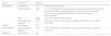
The main therapeutic options for medical therapy in utero are summarized in Table 1.
Treatment of congenital atrioventricular block in utero (adapted from 26).
| Cause | Treatment | CR/LE | |
|---|---|---|---|
| Autoimmune | Observation | I/A | Structurally normal heart |
| Dexamethazone | IIb/B | 1st- or 2nd-degree block with evidence of inflammation (pericardial effusion, ventricular dilatation, hyperechogenicity of the atrial walls or reduced biventricular contractility)Prophylactic, to reduce mortality and cardiomyopathy | |
| IVIG | IIb/B | Not recommended for prophylaxis | |
| Beta-adrenergic agonists | IIa/C | Heart rate <55 bpm, fetal hydrops, congenital malformations or cardiac dysfunction | |
| Cardiac malformations | Observation | I/A | |
| Beta-adrenergic agonists | IIa/C | Heart rate <55 bpm, fetal hydrops, congenital malformations or cardiac dysfunction | |
| Idiopathic | Observation | I/A |
bpm: beats per minute; CR: class of recommendation; IVIG: intravenous immunoglobulin; LE: level of evidence.
Percutaneous pacing techniques have been described, but results are unsatisfactory and the risk of fetal death is high. In most cases, therefore, the pregnancy runs its normal course and treatment only begins after birth.26,28,65
PostnatalAfter birth, fetal bradycardia (heart rate <70 bpm) can initially be controlled by drugs such as isoprenaline, atropine, epinephrine and/or dopamine, alone or in combination with transcutaneous pacing and/or temporary cardiac pacing, in order to prevent sudden death.3,27
Temporary cardiac pacing is also indicated in cases of cardiogenic shock or fetal hydrops. Temporary transesophageal pacing can also be used, but the risk of esophageal stenosis is high.42
Unfortunately CAVB is irreversible and hence a permanent pacemaker is necessary in 80% of cases,39 which improves long-term survival and reduces presyncope and syncope, even in asymptomatic cases.3,28,48
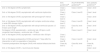
The latest indications for pacemaker implantation in children with CAVB are summarized in Table 2.
Indications for permanent pacemaker implantation in children with congenital atrioventricular block (adapted from 16,98,99).
| ESC guidelines98 | ACCF/AHA/HRS guidelines99 | ESC pediatric guidelines16 | |
|---|---|---|---|
| 2nd- or 3rd-degree CAVB, symptomatic | Class I, level C | Class I, level C | Class I, level C |
| 2nd- or 3rd-degree CAVB, asymptomatic with ventricular dysfunction | Class I, level C | Class I, level B | Class I, level C |
| 2nd- or 3rd-degree CAVB, asymptomatic with prolonged QT interval | Class I, level C | - | Class I, level C |
| 2nd- or 3rd-degree CAVB, asymptomatic with complex ventricular ectopy or wide QRS escape rhythm | Class I, level C | Class I, level B | Class I, level B |
| 2nd- or 3rd-degree CAVB, asymptomatic with abrupt ventricular pauses >2-3 basic cycle length | Class I, level C | Class IIa, level B | Class I, level B |
| 3rd-degree CAVB, asymptomatic + ventricular rate <55 bpm or with congenital heart disease + ventricular rate <70 bpm | - | Class I, level C | Class I, level C |
| 2nd- or 3rd-degree CAVB, asymptomatic + ventricular rate <50 bpm | Class I, level C | - | - |
| 2nd- or 3rd-degree postoperative CAVB, permanent 7 days after the intervention or with no expectation of resolution | Class I, level B | Class I, level B | Class I, level B |
| Transient postoperative 3rd-degree CAVB, with residual bifascicular block | Class IIa, level C | Class IIb, level C | Class IIb, level C |
| 2nd- or 3rd-degree CAVB + neuromuscular disease | - | - | Class I, level B |
bpm: beats per minute; CAVB: congenital atrioventricular block.
Pacemaker implantation is technically challenging, and various complications have been reported due to both the size of the patients and their rapid growth, and the presence of cardiac malformations.6,66 The task is further complicated by the small size of the vessels involved and the significant discrepancy between the size of the device and of the child's body.66,67
Opinions differ concerning the best approach, mode of stimulation, and location of the pulse generator.3
With regard to approach, the current options are epicardial and endocardial (transvenous) pacing. The most appropriate approach depends on body size, age, venous diameter, and presence of congenital cardiac defects.67 Access can be via lateral thoracotomy, sternotomy or a subxiphoid approach.
There is agreement that epicardial pacing is the first-line option in children weighing less than 15-20 kg and with structurally normal hearts3,66–68 and when venous access to the heart is not feasible, especially in a univentricular heart.16 However, periodic reviews are needed to reduce the rate of complications.69,70
Transvenous implantation of an endocardial pacemaker via tributaries of the superior vena cava or branches of the iliac veins is minimally invasive and has a low risk of malfunction, although the risk of systemic thrombosis is higher and the rate of venous occlusion is 25%.70–72
Intracardiac shunts increase the risk of systemic embolism and may need to be corrected before pacemaker implantation.16 According to Khairy et al., endocardial pacing in patients with intracardiac shunts is associated with less frequent lead replacement compared to epicardial pacing, but incurs a higher thromboembolic risk that is not reduced by anticoagulation.72
Complications of a transvenous approach include lead dislodgement, pocket hematoma or bleeding, pneumothorax, heart perforation, cardiac tamponade, and infection.16 The most common complication is infection, which has an incidence of 1-8% and is the most frequent indication for lead removal, which in turn results in heart perforation in 1-2% of cases and death in 0.1-0.4%.16,73
The small body size of the patients means that the chest wall is not a feasible location for the generator, and so the abdominal wall is generally used.3,66 In children weighing less than 2.5 kg, the generator can be placed in the pleural cavity, which affords better protection.74
A recent study by Costa et al. assessed the long-term results of epicardial pacemaker implantation with subxiphoid access. They concluded that the technique is viable and had excellent results and system longevity, reducing surgical trauma by placing the generator submuscularly in the preperitoneal space, reducing cardiac fibrosis, and diminishing the effect of the child's growth on the system by using a rectilinear trajectory.66
In epicardial pacing, ventricular lead placement improves mechanical synchrony and contraction efficiency.75 Apical pacing in the right ventricle is a common approach, but left ventricular apical or lateral wall pacing result in better left ventricular function and are thus currently recommended.76
Lead placement in the right ventricular lateral wall or outflow tract is associated with left ventricular dysfunction and dyssynchrony.75 Implantation in the posterior ventricle is also possible, but increases the risk of cardiac and coronary compression and sudden death.77–80
The type of pacemaker to be used depends on the patient's body size, level of activity and ventricular function.81–83
Some authors report that VVI pacing is more likely to lead to left ventricular dysfunction than DDD pacing.84 However, multicenter studies have shown little difference between the two modes in terms of mortality, cardiac function and quality of life.85–87
VVI pacemakers are often the first option in smaller children due to their smaller area. When the child reaches a certain weight, a second, atrial lead can be added; this change to a dual-chamber system improves quality of life and cardiac function, smaller ventricular dimensions and lower natriuretic peptide levels.81–83 However, the risk-benefit ratio of further invasive surgery and the need for future reviews must be taken into account.88,89
AV synchrony should be preserved, since dyssynchronous and non-physiological activation induces pacemaker-induced cardiomyopathy in 7% of patients with permanent pacing, leading to pathological myocardial remodeling and ventricular dilatation.92
Permanent His bundle pacing has fewer adverse effects on right ventricular function and cardiac function in general.90,91 Although technically demanding, especially in pediatric patients, it may become an alternative option in the near future.92–94
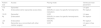
Recommendations for pacemaker implantation in pediatric patients with CAVB are summarized in Table 3.
Recommendations for pacemaker implantation in pediatric patients with congenital atrioventricular block (adapted from 16).
| Patient size | Access | Pacing mode | Ventricular lead placement |
|---|---|---|---|
| <10 kg | Epicardial | VVIR | LV apex |
| Endocardial (when epicardial access fails) | DDD(R) (in case of a specific hemodynamic indication) | RV septum | |
| 10-20 kg | Epicardial | VVIR | LV apex |
| Endocardial | DDD(R) (in case of a specific hemodynamic indication) | RV septum | |
| >20 kg | Endocardial | DDD(R) | RV septum |
| Epicardial (e.g. concomitant with other cardiac surgery) | VVIR | LV apex or free wall |
LV: left ventricular; RV: right ventricular.
The first case reported in Portugal of pediatric cardiac resynchronization in a case of cardiomyopathy induced by right ventricular pacing due to CAVB showed improved ejection fraction, reduced left ventricular end-diastolic diameter and significantly reduced mitral regurgitation, as well as improvement in New York Heart Association function class from III-IV to I.95
Fujioka et al. successfully applied a staged therapeutic approach to a premature low-birth-weight infant. Following diagnosis of CAVB and myocarditis in the 27th week of pregnancy, with a fetal atrial rate of 152 bpm and ventricular rate of 48 bpm, 4 mg/day of dexamethasone was administered. Cesarean delivery took place at 29 weeks and two days due to fetal distress, and temporary right ventricular pacing (120 bpm) was begun. A second lead was subsequently implanted in the ventricular apex and prednisolone (1 mg/kg/day for two weeks) and IGIV (1 g/day for three days) were administered. Permanent pacemaker implantation (VVI mode at 120 bpm) was performed on day 64.96
Future prospectsGene therapy for biological pacing, using an adenovirus as vector in cardiomyocytes, is a promising research field in constant development.3
Antagonism of molecular pathways, particularly of MMP-2, is based on discoveries that link these pathways with increased inflammation and fibrosis.8
Measurement of molecules including NT-proBNP can be used as a short-term diagnostic aid when no alterations are observed on imaging studies.8
A study of 38 patients showed that permanent His bundle pacing preserves left ventricular function and cardiac synchrony as compared with right ventricular septal pacing.92
Micropacemakers are an alternative under development to treat progressive CAVB associated with hydrops and may be applied in future projects.97
ConclusionsAlthough rare, CAVB is an important and treatable cause of pediatric mortality. Its etiology is predominantly autoimmune, mothers of affected fetuses usually being diagnosed with SLE or SS.
Various genetic polymorphisms and inflammatory markers are associated with the condition, which is caused by fibrosis and calcification of the AV node and conduction tissue.
Maternal risk is higher in cases of autoimmune disease, hypothyroidism or infection during pregnancy, which may explain why only 2-5% of pregnancies in which autoimmune disease is the only risk factor result in CAVB.
Fetal echocardiography remains the diagnostic gold standard.
Regarding therapy, effective antagonism of the inflammatory cascade is of value to prevent the genesis and evolution of AV block.
In most cases, monitoring of the pregnancy until birth is all that is recommended. Postnatally, pacemaker implantation is necessary in the majority of affected infants.
New techniques aimed at reducing morbidity and mortality have been proposed and continue to be studied. Some have been applied in clinical practice with satisfactory results, but most have only been analyzed in small samples or isolated cases, so there is still hesitancy and uncertainty concerning the use of these therapies.
Further studies are therefore required to optimize both diagnosis and treatment.
Conflicts of interestThe authors have no conflicts of interest to declare.
