


A 73-year-old man with a history of coronary artery disease and inferoposterior myocardial infarction was referred for cardiology consultation with symptoms of chronic heart failure in NYHA class III.
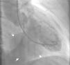
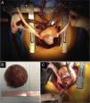
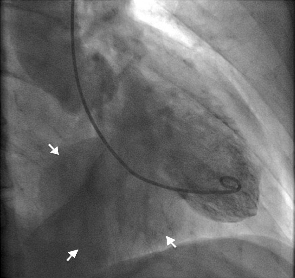
Transthoracic echocardiography revealed a thrombus partially filling a giant pseudoaneurysm/aneurysm of the inferoposterior wall, causing significant deformation of the ventricular geometry (Figure 1; Videos 1–4). Despite the size of the aneurysm, the subvalvular mitral apparatus was intact, with no regurgitation. Given the uncertainty surrounding the differential diagnosis, cardiac magnetic resonance imaging was performed, which confirmed a giant ventricular aneurysm (long axis 6.7 cm) containing a thrombus (Figure 2; Videos 5 and 6), and anticoagulant therapy was begun. Myocardial perfusion scintigraphy excluded ischemia and confirmed necrosis of the inferoposterior territory. Preoperative cardiac catheterization showed chronic occlusion of the proximal right coronary artery but no significant left coronary disease, while ventriculography documented a large ventricular aneurysm (Figure 3; Video 7). The patient underwent surgical repair of the aneurysm of the left ventricular inferior wall (Figure 4) by partial aneurysmectomy under extracorporeal circulation, with removal of a large wall thrombus and closure of the aneurysm neck with a 2 cm × 3 cm Dacron® patch (Figure 4). The left ventricle was closed using continuous sutures. The postoperative period was uneventful and the patient was discharged home on the fifth postoperative day.
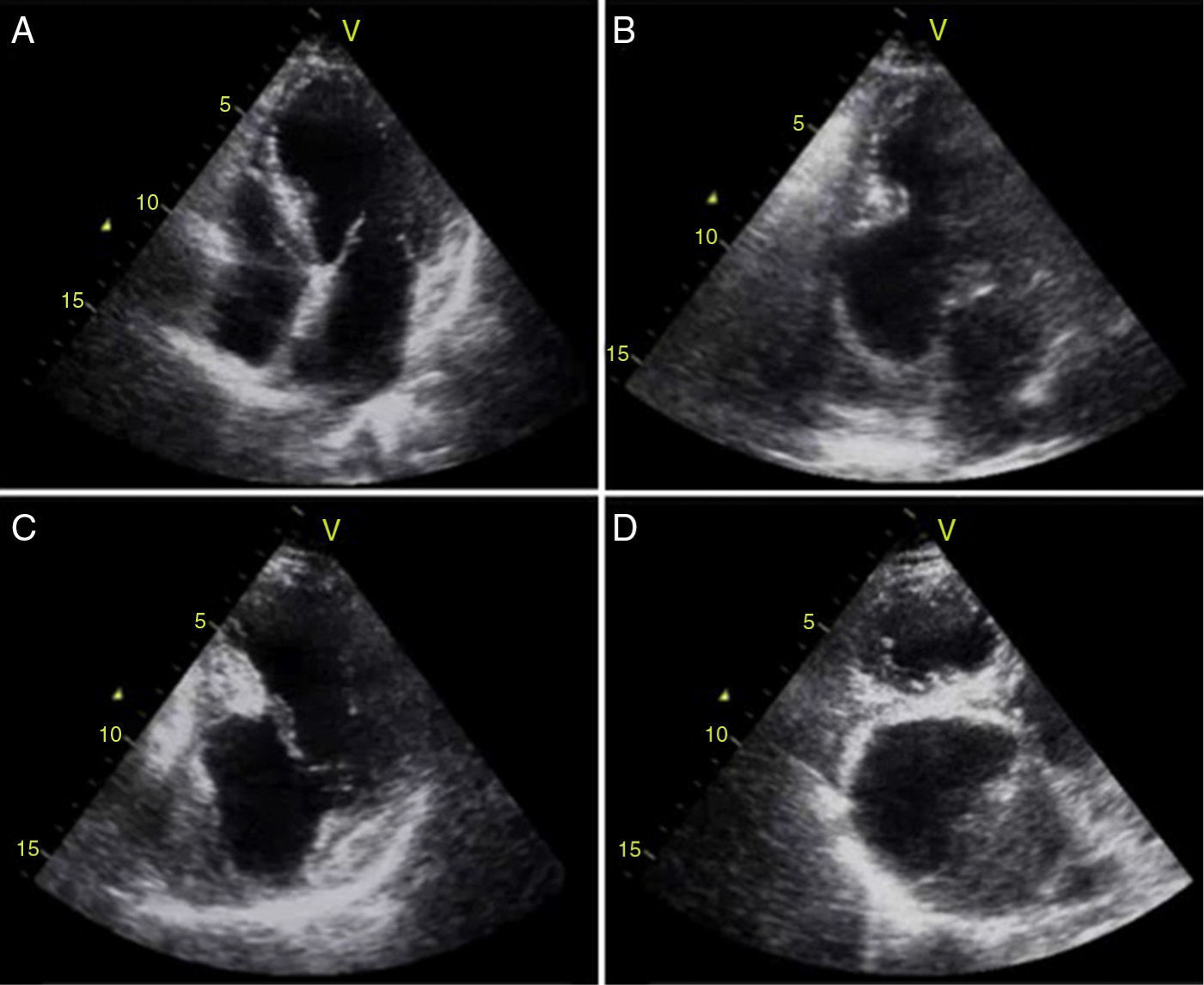
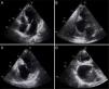
Two-dimensional transthoracic echocardiography: (A) apical 4-chamber view showing no alterations; (B) apical 2-chamber view revealing a giant ventricular inferior wall aneurysm containing a thrombus; (C) modified apical 2-chamber view showing intact subvalvular mitral apparatus; (D) modified apical 3-chamber view showing a large ventricular posterior wall aneurysm containing a thrombus.
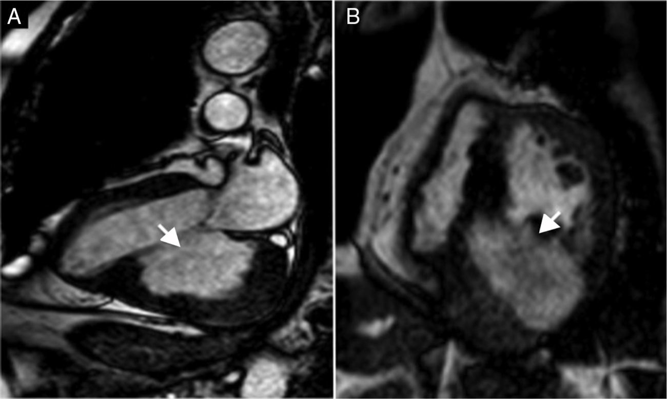
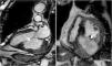
Cardiac cine steady-state free precession magnetic resonance imaging: (A) long-axis view showing a giant ventricular aneurysm (long axis 6.7 cm) of the posterior wall (arrow) containing a thrombus; (B) short-axis view above the level of the papillary muscles showing a large ventricular aneurysm of the inferoposterior wall (arrow), containing a thrombus.

The authors declare that no experiments were performed on humans or animals for this study.
Confidentiality of dataThe authors declare that they have followed the protocols of their work center on the publication of patient data.
Right to privacy and informed consentThe authors declare that no patient data appear in this article.
Conflicts of interestThe authors have no conflicts of interest to declare.
Please cite this article as: Lima da Silva G, Carvalheiro C, Cardoso P, et al. Imagiologia cardiovascular de um aneurisma ventricular esquerdo gigante. Rev Port Cardiol. 2015;34:501–503.
