


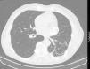
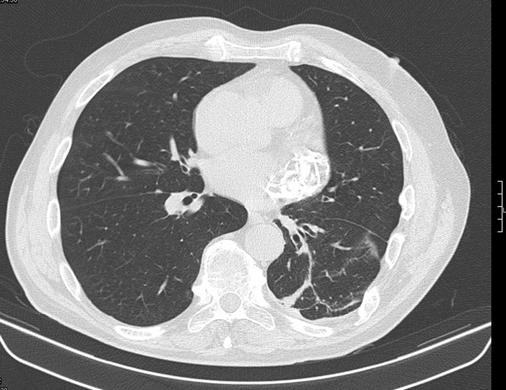
A 69-year-old man, with a history of hypertension, dyslipidemia, stroke in 1995 and left thoracic-abdominal trauma in 1999 resulting in hemopneumothorax after being hit by an ox, was referred for cardiology consultation following the incidental finding on thoracic-abdominal computed tomography (CT) of an extensive area (54 mm×36 mm×20 mm) of irregular, linear calcifications located posteriorly high in the pericardial sac (Figure 1). Transthoracic and transesophageal echocardiography and thoracic CT angiography were performed to clarify whether the calcification was intravascular or extravascular and to exclude a tumor.
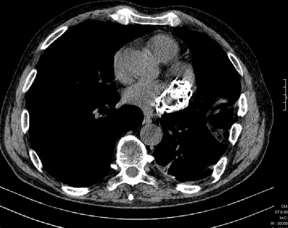
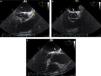
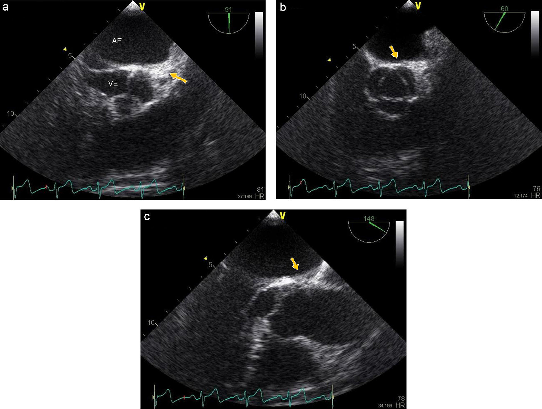
The echocardiogram showed a rounded mass with calcified walls attached to and slightly compressing the left atrium and the coronary sulcus of the lateral and anterior walls, with apparent fibrocalcification of the myocardium of the basal segment of the lateral wall, which was thinner, hyperechogenic and akinetic (Figure 2). CT angiography showed the lesion high in the pericardial sac in extensive contact with the pericardium, suggesting that its epicenter was within this structure. The lesion appeared to be of extravascular origin, possibly a consequence of the previous trauma (Figure 3).
The initial investigation focused on assessing the lesion, the other cardiac chambers, particularly the left atrial appendage, and the patient's hemodynamic status.
A brief review of the literature revealed descriptions of pericardial masses, cysts and calcifications (some due to trauma), but no case with similar characteristics to the one described above.
Ethical disclosuresProtection of human and animal subjectsThe authors declare that no experiments were performed on humans or animals for this study.
Confidentiality of dataThe authors declare that no patient data appear in this article.
Right to privacy and informed consentThe authors declare that no patient data appear in this article.
Conflicts of interestThe authors have no conflicts of interest to declare.
Please cite this article as: Lopes AJ, Ferreira Santos L, Gama P. Calcificação cardíaca: um achado acidental. Rev Port Cardiol. 2015;34:219–220.
