


A 64-year-old woman with severe mitral regurgitation (MR), pulmonary hypertension and patent foramen ovale (PFO) was referred for preoperative transthoracic echocardiography (TTE).
Besides her known conditions, the TTE showed a dilated coronary sinus (CS) and right ventricle that was not proportional to the MR. Transesophageal echocardiography (TEE) revealed prolapse of the mitral valve anterior leaflet (Figure 1A). The CS was markedly dilated, with no opacification after saline injection (Figure 1B). There was echo dropout in the left atrial wall and communication with the CS, raising the suspicion of unroofed CS (URCS) atrial septal defect (ASD). Three-dimensional echocardiography showed absence of the CS wall (Figure 1C) and the already known PFO (Figure 1D). Coronary computed tomography angiography (CCTA) confirmed the diagnosis of URCS (Figure 1E and F). Right catheterization showed a cardiac index of 11.47 l/min/m2, pulmonary artery pressure of 47 mmHg, and significant left-to-right shunt (Qp/Qs 2.2). The patient underwent mitral valve replacement and ASD closure. The PFO was left open. Postoperative course was uneventful and the patient was still asymptomatic at one year.

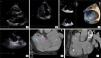
(A) Transthoracic echocardiography (TTE), parasternal long-axis view, showing the mitral valve (red arrow) and the enlarged coronary sinus (white arrow); (B) TTE, 4-chamber view, after injection of saline, without significant opacification; (C) three-dimensional echocardiography showing the coronary sinus (red dot) and left atrium (yellow dot) and revealing the absence of the coronary sinus wall; (D) transesophageal echocardiography showing the patent foramen ovale (green arrow); (E) coronary computed tomography angiography (CCTA) showing the right atrium (blue dot), left atrium (orange dot), the enlarged CS (green dot) with missing roof, communicating directly with the left atrium, and a communication between the right and left atria (purple arrow); (F) CCTA showing the right atrium (blue dot), right ventricle (white dot), left atrium (orange dot) and enlarged coronary sinus (green dot). The aorta (yellow arrow) and pulmonary artery (white arrow) can also be seen.
URCS is a rare type of interatrial communication (less than 1% of lesions related to interatrial shunting).1 Unlike a true ASD, the communication occurs through the ostium of the CS, resulting from the partial or complete absence of its roof.2,3 The diagnosis requires a high level of suspicion.3,4 Echocardiography remains the most important diagnostic modality.5 CCTA can provide further information.1
Conflicts of interestThe authors have no conflicts of interest to declare.
