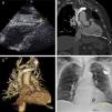
We present the case of a 90-year-old male patient transferred from another hospital with a diagnosis of pacemaker dysfunction. A permanent pacemaker had been placed one month previously for complete atrioventricular block. At a scheduled checkup with his cardiologist, he reported that he had started experiencing exertional dyspnea and pleuritic pain a few days after discharge. The electrocardiogram showed atrioventricular block with failure of pacemaker capture. The physical exam was unremarkable and he was hemodynamically stable. Suspecting lead malposition or fracture, a chest X-ray and transthoracic echocardiography (Figure 1A) were performed which showed the lead perforating the right ventricle (arrow) and mild cardiac effusion. Computed tomography (CT) (Figure 1B and C) was then performed in order to identify the entire course of the lead. After multidisciplinary discussion involving our surgical and electrophysiological team, it was decided to disconnect the old lead and leave it in place, as the wound was sealed, and to place a new passive fixation electrode (Figure 1D). The patient has been asymptomatic with a good clinical course since the procedure.
(A) Transthoracic echocardiography, subcostal view, showing the pacemaker lead perforating the right ventricle (arrow); (B) and (C) cardiac computed tomography showing the entire pacemaker lead and the perforation through the right ventricle; (D) chest X-ray at discharge showing the two leads (arrows). Black arrow: new lead, yellow arrow: old lead perforating the cardiac wall.
Lead perforation is a relatively rare complication after pacemaker implantation. Its presentation varies from life-threatening situations such as cardiac tamponade to mild and nonspecific symptoms. It should be suspected if the patient presents days after implantation with pleuritic pain, dyspnea, hiccup or device malfunction. The gold standard for the diagnosis is CT, and treatment consists of device extraction.1,2
In our case, given the patient's stability and characteristics, an unconventional approach was adopted, with a good outcome as far as we know.
Ethical disclosuresProtection of human and animal subjectsThe authors declare that no experiments were performed on humans or animals for this study.
Confidentiality of dataThe authors declare that they have followed the protocols of their work center on the publication of patient data.
Right to privacy and informed consentThe authors have obtained the written informed consent of the patients or subjects mentioned in the article. The corresponding author is in possession of this document.
Conflicts of interestThe authors have no conflicts of interest to declare.


