Informação da revista
Vol. 43. Núm. 3.
Páginas 143-145 (março 2024)
Partilhar
Partilhar
Baixar PDF
Mais opções do artigo
Vol. 43. Núm. 3.
Páginas 143-145 (março 2024)
Image in Cardiology
Acesso de texto completo
A galloping cardiac mass: An MRI perspective
Uma massa cardíaca galopante: uma perspetiva da ressonância magnética
Visitas
1314
Rui Files Flores
Autor para correspondência
ruifilesflores@gmail.com

Corresponding author.
, Fernando Guilherme Mané, Vítor Hugo Pereira, Catarina Vieira
Braga Hospital, Braga, Portugal
Este item recebeu
Informação do artigo
Texto Completo
Baixar PDF
Estatísticas
Figuras (1)
Material adicional (2)
Texto Completo

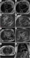
A 71-year-old woman was hospitalized for an unexplained fever. A positron emission tomography using PET-18FDG showed increased and diffuse uptake in the pericardium. An MRI revealed a small circumferential pericardial effusion and slight pericardial thickening over the right lateral side of the heart (Video 1, CINE sequences). The patient was discharged after the fever resolved. Some months later, she started having symptoms of exertional dyspnea and a dry cough. A CT raised concerns about a pericardial mass and multiple adenopathies. Reassessment by MRI showed a cardiac mass (84 mm×48 mm) with irregular borders in the right atrioventricular sulcus extending throughout the free wall of the right atrium (Video 2, CINE sequences). It was isointense on T1 (Panel, Image 1) and T2 (Panel, Image 2), hyperintense on T2 STIR (Panel, Image 3), and did not saturate in the fat saturation sequences (Panel, Image 4). It perfused on the first contrast passage (Video 2, perfusion sequences), with hypointense foci on early enhancement sequences (Panel, Image 5) and with a heterogeneous appearance on late enhancement sequences (Panel, Image 6). Exuberant adenopathy conglomerates were observed (Panel, Images 7 and 8, asterisk). A biopsy of an axillary lymph node revealed diffuse large B-cell lymphoma and chemotherapy was started.

Panel.

Panel exhibiting the cardiac mass characteristics on cardiac magnetic resonance imaging sequences. Image 1: T1 sequence. Image 2: T2 sequence. Image 3: T2 STIR sequence. Image 4: Fat saturation sequences. Image 5: Early enhancement sequence. Image 6: Late enhancement sequences. Images 7 and 8: Exuberant adenopathy conglomerates highlighted with an asterisk.

(0.5MB).
Conflicts of interest

The authors have no conflicts of interest to declare.

Appendix A
Supplementary data

The following are the supplementary data to this article:

(0.1MB)

CINE sequences of the magnetic resonance imaging performed at the first hospital admission showing a discrete thickening of the pericardium over its right lateral border, as well as a small pericardial effusion.

(0.05MB)

CINE sequences of the second MRI showing a large pericardial mass without a cleavage plane, with irregular contours, and with partial compression of the right heart chambers.

Copyright © 2023. Sociedade Portuguesa de Cardiologia
Baixar PDF
Idiomas
Revista Portuguesa de Cardiologia
Opções de artigo
Ferramentas
Material Suplementar
en pt

Are you a health professional able to prescribe or dispense drugs?

Você é um profissional de saúde habilitado a prescrever ou dispensar medicamentos

Ao assinalar que é «Profissional de Saúde», declara conhecer e aceitar que a responsável pelo tratamento dos dados pessoais dos utilizadores da página de internet da Revista Portuguesa de Cardiologia (RPC), é esta entidade, com sede no Campo Grande, n.º 28, 13.º, 1700-093 Lisboa, com os telefones 217 970 685 e 217 817 630, fax 217 931 095 e com o endereço de correio eletrónico revista@spc.pt. Declaro para todos os fins, que assumo inteira responsabilidade pela veracidade e exatidão da afirmação aqui fornecida.