


We present the case of a 71-year-old woman with a history of ischemic dilated cardiomyopathy, in NYHA class III, a cardiac resynchronization therapy device with defibrillator back-up having been implanted, and permanent atrial fibrillation, under oral anticoagulation therapy. She was admitted due to dyspnea, fatigue and angina on minimal exertion, pain and edema in the right leg and muscle weakness in both legs rendering her unable to walk, over the previous two weeks.
On physical examination she was cooperative, with blood pressure 100/40 mmHg, heart rate 70 bpm, good peripheral pulses, crackling rales in the lung bases, and flaccid paraparesis and signs of deep vein thrombosis in the right leg. Her d-dimers were elevated and the chest X-ray revealed cardiomegaly and fluid overload.
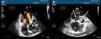
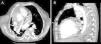
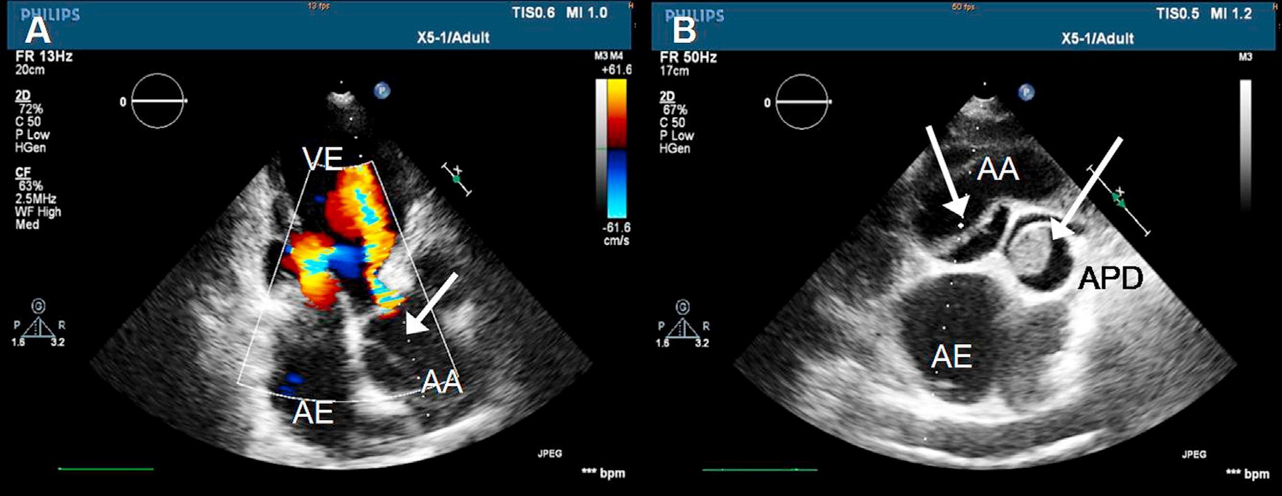
In view of the suspicion of pulmonary thromboembolism, she was referred for transthoracic echocardiography, which showed a dilated left ventricle and severe systolic dysfunction, moderate aortic regurgitation, dilated aortic root with an image suggestive of a double lumen, and a thrombus in the right pulmonary artery (Figure 1). Thoracic computed tomography angiography confirmed the diagnosis of pulmonary thromboembolism and type A aortic dissection with involvement of the descending thoracic aorta (Figure 2).
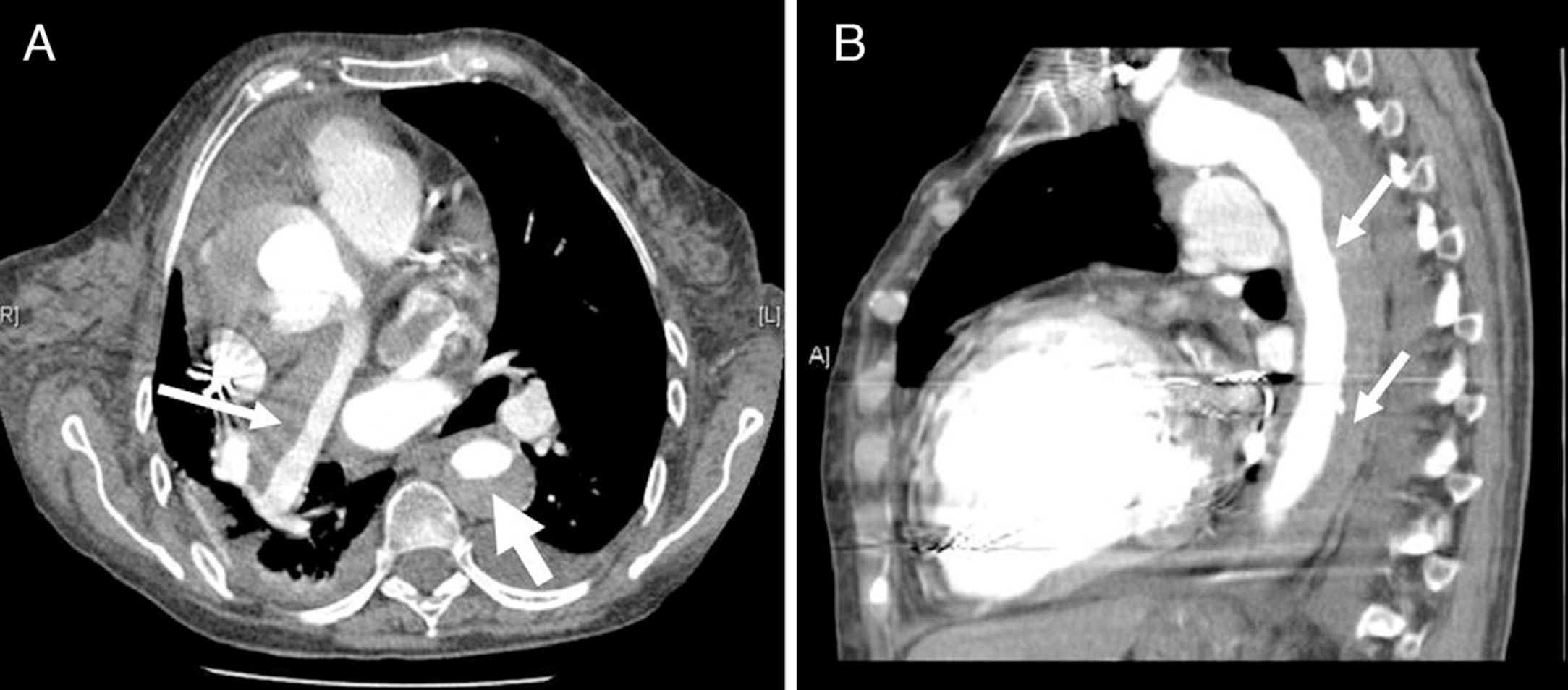
Thoracic computed tomography angiography with intravenous contrast: (A) transverse view showing an organized thrombus adhering to the wall of the right pulmonary artery (thin arrow) and filling of the true lumen of the aortic dissection (wide arrow); (B) sagittal view showing the dissection extending along the descending thoracic aorta (arrows).
Given the patient's high surgical risk a conservative approach was adopted, but the clinical course was unfavorable, leading to tetraparesis and renal failure, and she was discharged at her request to a continuing care home.
We highlight the role of transthoracic echocardiography, a readily available diagnostic exam, in the assessment of patients in the emergency department. This case also draws attention to the possibility of severe comorbidities and the desirability of using non-directed exams whenever possible.
Ethical disclosuresProtection of human and animal subjectsThe authors declare that no experiments were performed on humans or animals for this study.
Confidentiality of dataThe authors declare that they have followed the protocols of their work center on the publication of patient data and that all the patients included in the study received sufficient information and gave their written informed consent to participate in the study.
Right to privacy and informed consentThe authors declare that no patient data appear in this article.
Conflicts of interestThe authors have no conflicts of interest to declare.
Please cite this article as: Cruz I, et al. Um caso de tromboembolismo pulmonar e dissecção aórtica: importância do ecocardiograma. Rev Port Cardiol. 2013. http://dx.doi.org/10.1016/j.repc.2012.12.010.
