Journal Information
Vol. 31. Issue 3.
Pages 251-252 (March 2012)
Vol. 31. Issue 3.
Pages 251-252 (March 2012)
Image in cardiology
Open Access
Tubular image crossing the atrial septum
Imagem tubular “transversal ao septo inter-auricular”
Visits
5816
Ricardo Faria
Corresponding author
ricardofaria555@gmail.com

Corresponding author.
, Walter Santos, Ana Camacho, Nuno Marques, Rui Ferrinha, Vasco Marques, Ilídio de Jesus
Serviço de Cardiologia do Hospital de Faro, Faro, Portugal
This item has received

Under a Creative Commons license
Article information
Full Text
Bibliography
Download PDF
Statistics
Figures (5)
Show moreShow less
Full Text
Case report

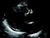
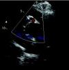
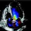
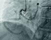
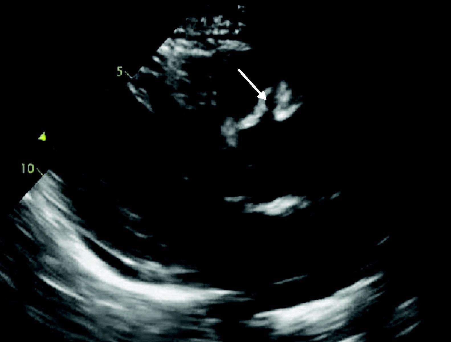
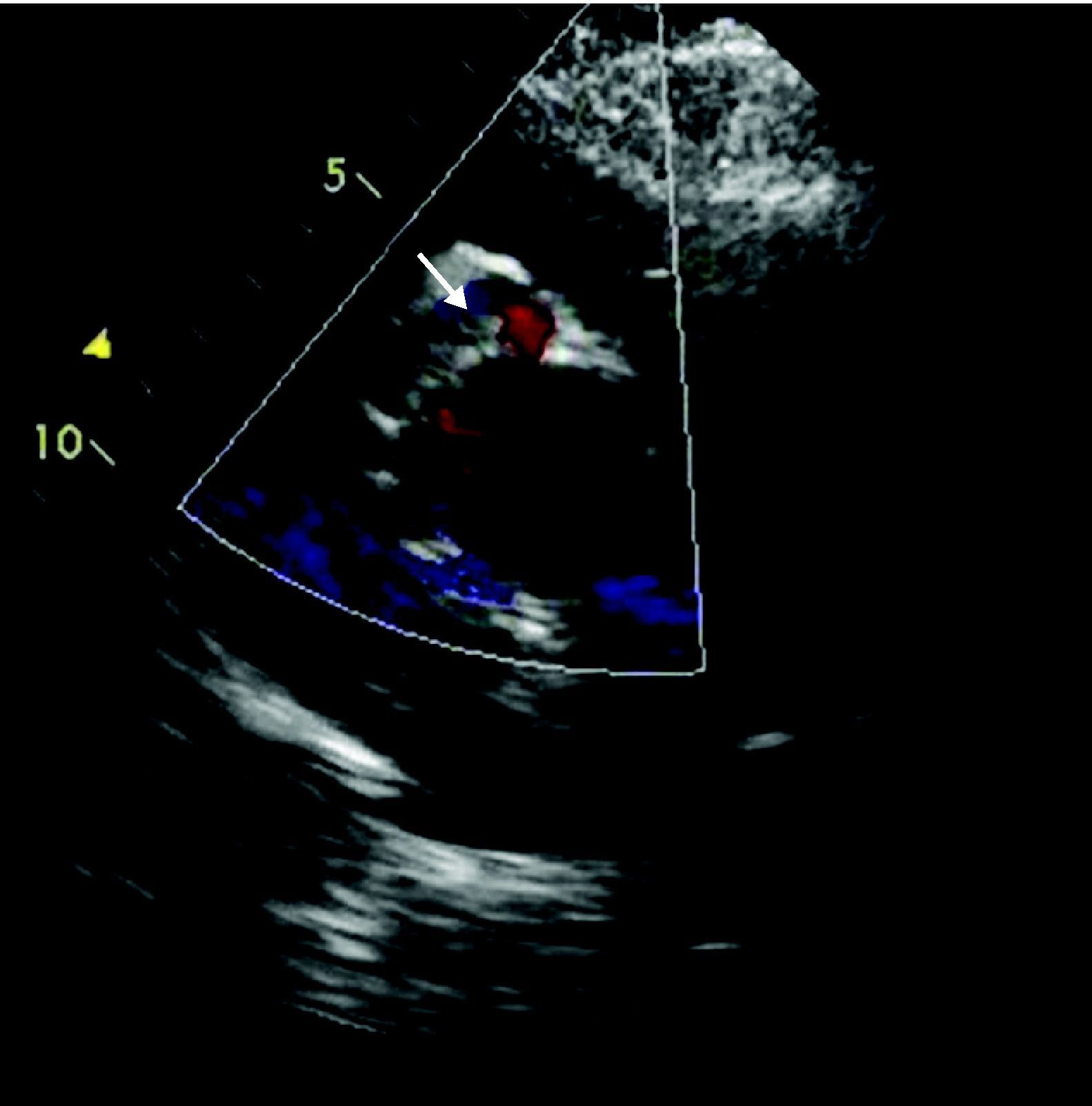
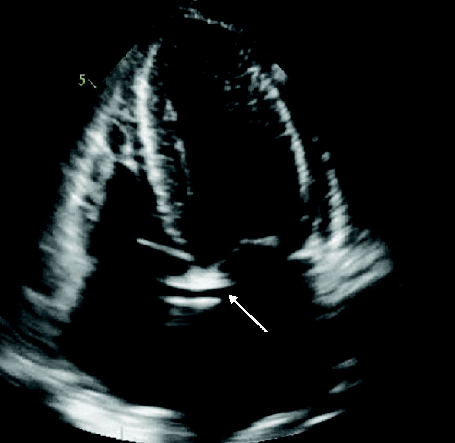
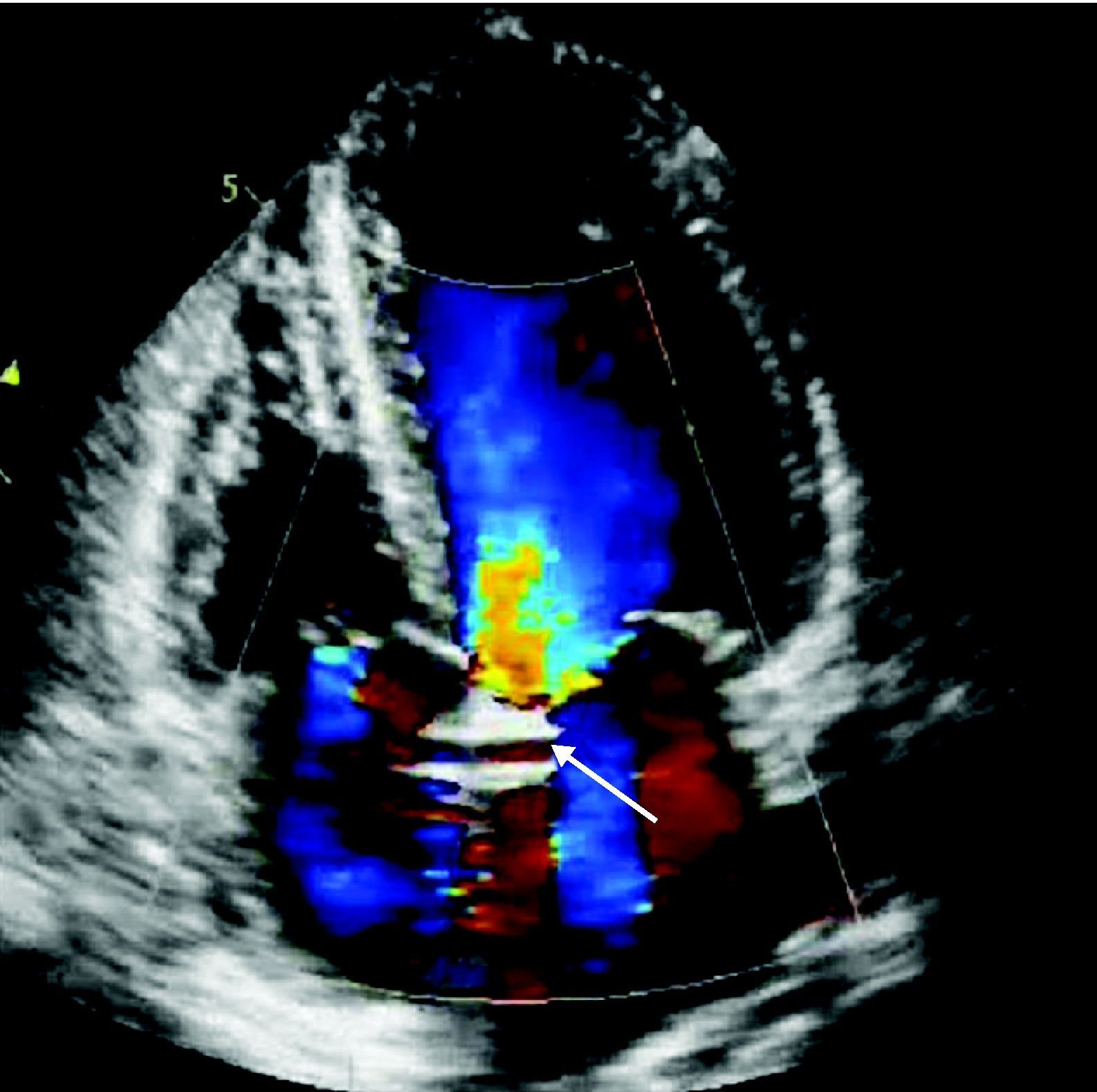
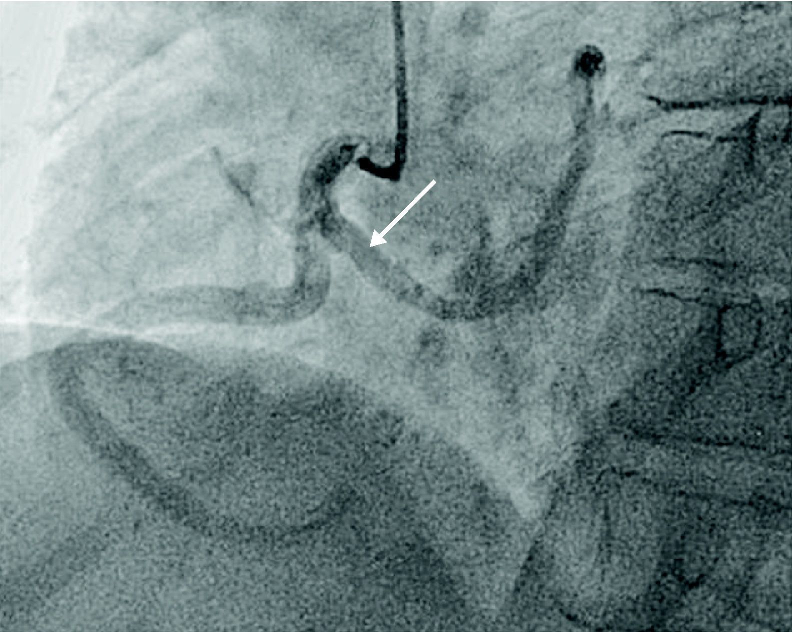
A 67-year-old woman with a history of scleroderma and hypertension but with no previous cardiac symptoms was hospitalized for assessment of systemic sclerosis. Physical examination revealed a grade II/VI systolic murmur on the apex; the electrocardiogram showed sinus rhythm and left anterior bundle branch block. Left parasternal long-axis echocardiography revealed an anomalous tubular image in continuity with the anterior wall of the ascending aorta (Fig. 1), while in parasternal short-axis view the same tubular structure was seen encircling the aorta (Fig. 2). In apical 4-chamber view, a tubular image, 2 mm in diameter and 42 mm in length, was observed crossing the atrial septum; its origin and end were not identified. Color Doppler echocardiography showed flow in its interior (Figs. 3 and 4), which raised the suspicion that it was a vascular structure, and cardiac catheterization was accordingly performed, which revealed anomalous origin of the left coronary artery (LCA) from the proximal segment of the right coronary artery, with an epicardial course anterior to the aorta (Fig. 5). The echocardiographic findings, most often associated with scleroderma, are pulmonary hypertension and pericardial involvement, including fibrinous pericarditis. There are no reports in the literature of an association between scleroderma and anomalous coronary artery origin, and hence this case may be simply a coincidence. Origin of the LCA from the right coronary ostium is found in only 0.15%1 of the population and is associated with sudden death.2

Figure 1.

Left parasternal long-axis echocardiogram showing a tubular image (arrow) in continuity with the anterior wall of the aorta.

(0.08MB).
Figure 2.

Left parasternal short-axis echocardiogram at the level of the aorta, showing a tubular structure (arrow) encircling the aorta.

(0.1MB).
Figure 3.

Apical 4-chamber echocardiogram showing a tubular image crossing the atrial septum.

(0.1MB).
Figure 4.

Apical 4-chamber echocardiogram with color Doppler revealing flow inside the tubular structure (arrow).

(0.17MB).
Figure 5.

Coronary angiography (left anterior oblique caudal) showing anomalous origin of the left coronary artery (arrow) coursing anterior to the aorta.

(0.24MB).
Conflicts of interest

The authors have no conflicts of interest to declare.

References
[1]
Coronary artery anomalies: a comprehensive approach, pp. 42
[2]
Y. von Kodolitsch, O. Franzen, G.K. Lund, et al.
Coronary artery anomalies Part II: Recent insights from clinical investigations.
Z Kardiol (Germany), 94 (2005), pp. 1-13

Please cite this article as: Faria R, et al. Imagem tubular «transversal ao septo inter-auricular». Rev Port Cardiol 2012. doi:10.1016/j.repc.2012.01.010.

Copyright © 2011. Sociedade Portuguesa de Cardiologia
Idiomas
Revista Portuguesa de Cardiologia (English edition)
Article options
Tools
en pt

Are you a health professional able to prescribe or dispense drugs?

Você é um profissional de saúde habilitado a prescrever ou dispensar medicamentos

By checking that you are a health professional, you are stating that you are aware and accept that the Portuguese Journal of Cardiology (RPC) is the Data Controller that processes the personal information of users of its website, with its registered office at Campo Grande, n.º 28, 13.º, 1700-093 Lisbon, telephone 217 970 685 and 217 817 630, fax 217 931 095, and email revista@spc.pt. I declare for all purposes that the information provided herein is accurate and correct.