


Heart rate variability (HRV) is a simple and noninvasive measure that estimates cardiac autonomic modulation, mainly the parasympathetic contribution. Increased sympathetic and/or decreased parasympathetic nervous activity is seen in post-myocardial infarction (MI) patients. Consequently, these patients present reduced HRV, which has been associated with increased risk of adverse events and mortality. Exercise training, recommended as a complementary therapy for patients with cardiovascular disease, has shown numerous beneficial effects. The main aim of the present manuscript was to provide a critical review of studies investigating the effects of exercise training on cardiac autonomic modulation, through HRV, in MI patients and the possible mechanisms involved. Despite conflicting evidence, exercise training appears to be a useful therapeutic intervention to improve the unbalanced autonomic function of MI patients. Finally, the mechanisms involved are not yet well understood, but nitric oxide bioavailability and angiotensin II levels seem to play an important role.
A variabilidade da frequência cardíaca é uma medida simples e não invasiva da modulação autonómica cardíaca, principalmente da contribuição da divisão parassimpática. O aumento da atividade simpática e/ou a diminuição da atividade parassimpática ocorre em doentes com enfarte agudo do miocárdio. Consequentemente, estes doentes apresentam reduzida variabilidade da frequência cardíaca, a qual tem sido associada com risco aumentado de eventos cardíacos adversos e de mortalidade. Existem evidências de numerosos benefícios do exercício físico regular, quando recomendado como terapêutica coadjuvante em sujeitos com doença cardiovascular. Assim sendo, o propósito do presente artigo é a realização de uma revisão crítica dos estudos investigando o efeito do exercício físico regular na variabilidade da frequência cardíaca e os potenciais mecanismos em doentes com história de enfarte agudo do miocárdio. O exercício físico regular parece ser um meio terapêutico para melhorar a desregulação da função do sistema nervoso autónomo em doentes com história de enfarte agudo do miocárdio, apesar de os resultados contraditórios entre os estudos. Finalmente, não estão claramente estabelecidos os possíveis mecanismos pelos quais o exercício melhora a variabilidade da frequência cardíaca, mas o aumento da biodisponibilidade do óxido nítrico e a redução dos níveis de angiotensina II induzidos pelo exercício parecem desempenhar um importante papel.
Autonomic Tone and Reflexes After Myocardial Infarction study
coronary artery bypass grafting
coronary artery disease
cardiovascular disease
high-frequency peak
heart rate
heart rate variability
low-frequency peak
ratio between low-frequency and high-frequency peak
myocardial infarction
intervals between consecutive normal heart beats or normal R wave peaks
nitric oxide
percentage of successive NN intervals differing by >50 ms
percutaneous transluminal coronary angioplasty
square root of the mean of the squared differences between successive NN intervals
standard deviation of average NN intervals calculated from 5-minute segments
standard deviation of all NN intervals
Cardiovascular disease (CVD) is the leading cause of death worldwide.1 According to the Global Atlas on Cardiovascular Disease Prevention and Control published by the World Health Organization,1 of the 57 million deaths that occurred globally in 2008, 17.3 million (30%) were due to CVD.1
A common element in CVD, including myocardial infarction (MI), is a detrimental change in cardiac autonomic modulation, i.e., increased sympathetic outflow and reduced parasympathetic activity.2,3 Cardiac autonomic modulation refers to the influence that these two branches of the autonomic nervous system have on the heart: they interact in a complex way to regulate the heart's chronotropism, dromotropism, and inotropism.4 Soon after MI, autonomic imbalance acts to preserve the proper functioning of the cardiovascular system and consequently of the whole body.5 Nevertheless, the maintenance of this imbalance can lead to harmful consequences in MI patients, such as decreased cardiomyocyte viability due to the cytotoxic effect of elevated catecholamine levels6,7 and adverse cardiac remodeling.5,8 Simultaneously, myocardial overload, due to the elevated heart rate (HR) and contractility induced by sympathetic overactivity, increases oxygen demand and reduces coronary perfusion time, impairing coronary blood flow in the weakened myocardium and thereby increasing the likelihood of ischemic insult and malignant ventricular arrhythmias and hence the risk of sudden cardiac death.9,10
A relatively easy noninvasive method that is reproducible and cost-effective for estimating cardiac autonomic modulation is evaluation of heart rate variability (HRV), which analyzes variations in the intervals between consecutive normal heart beats or normal R wave peaks (i.e., RR or NN intervals), measured in ms.4,11 Depressed HRV suggests poor functioning of cardiac autonomic modulation and therefore impaired ability of the heart to adapt to a range of physiological and environmental stimuli.12 Various HRV indices have been used to stratify the risk of mortality and cardiovascular events in patients after MI, and have shown promising results.13–15
Recommendations to join exercise training programs and to increase daily physical activity levels are included in the guidelines for secondary prevention through cardiac rehabilitation in patients with documented coronary artery disease (CAD).16,17 Exercise-based cardiac rehabilitation has been shown to be an effective therapy for reducing mortality and morbidity in these patients.18 There are many documented benefits arising from exercise training, such as improvements in aerobic fitness, exercise capacity, ventilatory threshold, modifiable cardiovascular disease risk factors, endothelial function, and vascular wall inflammation.19–21 The effect of exercise-based cardiac rehabilitation on the impaired autonomic function of CAD patients has also been studied,22–24 although there are conflicting results regarding the impact of exercise training on HRV.
This review aims to provide a critical appraisal of the literature linking exercise training and autonomic function in MI patients and a discussion of the possible mechanisms underlying the effects of exercise training on autonomic function.
Heart rate variability indices and prognostic implicationsIt is not the main purpose of the present review to describe the HRV indices employed in the literature; this has been done in detail by Malik et al.4 and Kleiger et al.11 However, it is important to mention that traditional methods, based on time and frequency domains, are by far the most commonly used. The main time domain indices include the standard deviation of all NN intervals (SDNN), the standard deviation of average NN intervals calculated from 5-minute segments (SDANN), the square root of the mean of the squared differences between successive NN intervals (RMSSD), and the percentage of successive NN intervals differing by >50 ms (pNN50).4,11 Frequency domain indices include low-frequency (LF) peak (0.04–0.15 Hz), high-frequency peak (0.15–0.4 Hz), expressed in absolute units of power (ms2) or in normalized units (nu), and the ratio between them (LF/HF).4,11 It is widely accepted that in general, time domain variables are related to each other and are affected to different degrees by parasympathetic blockade.25 Recent evidence supports the idea that they are markers of parasympathetic cardiac modulation.25 Nevertheless, HF peak is established as the foremost HRV marker of efferent cardiac vagal activity (i.e., vagal modulation of HR).26 There is no consensus regarding interpretation of the LF component, but it is considered a marker of both sympathetic and parasympathetic modulation.26 Lastly, the LF/HF ratio has been reported as a measure of sympathovagal balance.3
Various studies have been conducted to investigate the prognostic value of HRV after acute MI, employing: (i) different methods to analyze HRV data (e.g., traditional, geometric, and nonlinear), (ii) different periods of time after MI to evaluate baseline HRV (from days to weeks or months), (iii) different follow-up periods (from months to years), and (iv) different end-points (e.g., arrhythmic events, cardiac death, sudden death, and all-cause mortality).13–15 Kleiger et al.,13 in an elegant study, evaluated HRV with 24-hour Holter ECG monitoring in 808 patients recovering from MI (11±3 days after the acute event), and observed a relative risk of all-cause mortality of 2.7 for those with SDNN <50 ms compared to those with SDNN ≥50 ms, after a 31-month follow-up. In addition, after adjusting for measures such as ejection fraction, exercise capacity, post-infarction angina, ventricular arrhythmias, and drug treatment, SDNN was established as an independent predictor of mortality. La Rovere et al.,14 in the Autonomic Tone and Reflexes After Myocardial Infarction (ATRAMI) study, assessed 24-hour Holter HRV in a representative sample of recent (<28 days) post-MI patients (n=1284), of whom 63% received thrombolytic therapy. Over a mean follow-up of 21±8 months, it was observed that both one- and two-year cardiac mortality were higher among patients with SDNN <70 ms. In addition, Bigger et al.,15 evaluating LF and HF among other frequency domain indices, found that reduced levels of all measures were predictors of arrhythmic, all-cause, and cardiac mortality over a follow-up of two to four years. To summarize, these findings show that various HRV indices can provide valuable prognostic information on MI patients. However, for the purpose of dichotomous risk stratification in clinical practice, diverse HRV cut-off points have been reported.27 For instance, Kleiger et al.13 applied a cut-off of SDNN <50 ms, while in the ATRAMI study14 the cut-off was defined as SDNN <70 ms to differentiate MI patients with impaired HRV and consequently increased risk of developing the assessed end-points. Besides the conflicting results, the establishment of a single cut-off point did not allow normal HRV values to be distinguished from moderately and severely abnormal HRV values. This was the aim of Milicevic et al.,28 who analyzed various HRV indices in cardiac inpatient and outpatient groups. For the entire sample, the range of moderately depressed SDNN values was 59–92 ms, with values above these limits being considered normal, and below severely depressed. On the other hand, different values were expressed when the sample analysis was performed by gender and group. For outpatients, a moderately depressed SDNN was 75–105 ms for females and 65–100 ms for males. For inpatients attending a rehabilitation program, a moderately depressed SDNN was 47–74 ms for females and 53–83 ms for males.
There are numerous reasons for the considerable variability in the ranges of SDNN and other HRV indices: differences in infarct location and size, differences in age, gender, and experimental conditions, and difference in the statistical approach for data analysis.2,4 In view of this variety, dichotomized cut-off points or ranges to define normal, moderately abnormal, and severely abnormal HRV values have not been universally accepted, and further investigation and debate are thus warranted on this issue.
Effects of exercise training on heart rate variability in myocardial infarction patientsExercise-based cardiac rehabilitation has been shown to provide various health-related benefits,19 including improvements in exercise tolerance, endothelial function, cardiovascular risk factors, and vascular wall inflammation, which can collectively reduce morbidity and mortality in CAD patients.18,21,29,30 A meta-analysis of 47 studies randomizing 10794 CAD patients to exercise-based cardiac rehabilitation or usual care found significant reductions in total mortality (13%), cardiac mortality (26%), and hospital admissions (31%) in patients who exercised in a mean follow-up of 12 months or more.18 Furthermore, a significant reduction in sudden cardiac death (37%) was also observed in CAD patients participating in exercise training compared to usual medical care.29
As reduced HRV in CAD patients is associated with increased risk of all-cause mortality, cardiac mortality, and sudden death, one possible explanation for the reduction in morbidity and mortality in patients undergoing exercise-based cardiac rehabilitation could be exercise-induced improvement in cardiac autonomic modulation. However, no prospective study has been performed to test this hypothesis. Studies in healthy trained subjects undergoing aerobic exercise training31–33 have shown increased vagal and/or reduced sympathetic cardiac modulation, characterized by resting bradycardia and increased vagal-related HRV indices, compared to sedentary controls. Regarding the effects of exercise-based cardiac rehabilitation on HRV indices (Table 1), data from studies on post-MI patients have revealed heterogeneous results.22–24,34–42 However, not all of these studies were randomized controlled trials, which to some extent weakens their conclusions.
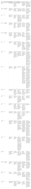
Studies of the effects of exercise training on heart rate variability in coronary artery disease patients.
| Study | Randomized/controlled | Population(sample size; age in years) | HRV evaluation | Rehabilitation program | HRV results | Comments |
| Laing et al.69 | No | CAD (n=17; 56±10) | Short-term ECG recording (5 min, supine resting) and during exercise recovery | Not reported | ←→ RMSSD, RMS (rest)↑ RMSSD, RMS (during exercise recovery) | No report of infarct locationDaily physical activity and diet not assessedOnly 6 patients completed the post rehabilitation testing |
| Sandercock et al.70 | Not randomized; control group | Post-MI, PTCA, CABGEG (n=38; 65.6±11.6)CG (n=23; 64.9±9.0) | Short-term ECG recording (5 min, supine resting) at ≈16 wks post-MI | EG: 8 wks; ≈1.5 h of aerobic exercises; ≈70% max HRCG: no specific exercise guidelines provided | EG: ↑ RR interval, SDNN, LF (ln), HF (ln) (vs. CG)←→ RMSSD, LF/HF | No report of infarct locationDaily physical activity assessed by exercise diary (CG only)Diet not assessedCG: HRV measures were more severely attenuated |
| Tsai et al.41 | RCT | After PTCA (post-MI)EG (n=42; 57.0±8.3)CG (n=42; 56.7±9.8) | Short-term ECG recording (5 min, supine resting) at ≈1 month after PTCA | EG: 8 wks; cycle ergometer at 60–85% HR reserve; 40 min/session; 1–3 sessions/wkandhome program: aerobic exercise (bicycling, walking, or jogging); similar intensityCG: usual daily activities without restriction | EG: ↑ HF (ln), mean of RR intervals (vs. CG)CG: ↓ mean of RR intervals, HF (ln) (vs. EG) | No report of infarct locationDaily physical activity assessed by questionnaire interviewDiet not assessed |
| Lucini et al.71 | Not randomized; control group | Post-MI, PTCA, CABGEG (n=29; 63±1.97)CG (n=11; 53±2.38) | Short-term ECG recording (10 min, supine resting, respiratory signal monitored) at 3–6 wks post-event | EG: 12 wks; 30–40 min of continuous upright aerobic and dynamic exercise; ≈70–85% max HRCG: usual daily activities without restriction | EG: ↑ RR interval variance (vs. CG)↑ RR interval (not different from CG)←→ LF (nu), HF (nu) | No report of infarct locationDaily physical activity and diet not assessedCG: younger, heavier, higher levels of total cholesterol, HDL and LDL than EGEG and CG: Encouraged to exercise 1–3 times per week outside of the rehabilitation program |
| Tygesen et al.72 | Randomized; no control group | Post-MI; CABG Normal-intensity group (n=33; 56.5±7.7)High-intensity group (n=29; 57.5±8.4) | Short-term ECG recording (10 min, supine resting, 15 respirations/min) and 24-h Holter recording at 1 month post-event | 12 wks; cycle ergometerNormal intensity training (2×/wk, 1 h)High intensity (3×/wk, 1 h + 3×/wk, 30 min, at home) | ↑ SDNN, SDANN (greater in the high intensity group)←→ RMSSD, HF, LF (both groups) | Daily physical activity and diet not assessedOne year later exercise capacity remained at the same level in the high intensity group and there was only an insignificant trend toward increased maximal workload in the normal intensity group |
| Duru et al.22 | RCT | Post-MIEG (n=12; 56±5)CG (n=13; 55±7) | Short-term ECG recording (15 min, supine resting) at ≈1 month post-MI | EG: 8 wks; 2× outdoor walking sessions (1 h) daily; 4 x 45-min/wk (cycling); 70% HR reserveCG: usual medical care and daily living activities | After rehabilitation:EG: ↑ pNN50←→ SDRR, HF (nu), LF (nu), LF/HFAfter 1 year:CG: ↓ SDRR, HF (nu)None of the HRV indices were significantly different between the two groups | Daily physical activity and diet not assessedHRV evaluated before and after 1 and 2 months of training and at 12 monthsCG: received usual clinical follow-up, and encouraged not to exercise beyond a level associated with normal activities of daily living |
| Iellamo et al.23 | RCT | CAD, Post-MI, CABGEG (n=45; 59.4±7.8)CG (n=41; 58.5±7.3) | Short-term ECG recording (10 min, supine resting) at 2 wks after CABG and at 2 months to 9 years post-MI (66% in the last year before the study) | EG: 2 wks; 2×/day; 30 min of cycle ergometer; 6×/wk; 85% max HRCG: walking and calisthenics with the same daily schedule as EG | EG: ↑ RR interval, SDRR (vs. CG)EG: ↑ RR interval, SDRR (both MI and non-MI patients) | Daily physical activity and diet not assessedAll patients resided at the rehabilitation center for 3 weeksEG: significant increase in peak VO2CG: small significant increase in peak VO2Half of the patients in each group were post-MI (stratified analysis showed no significant difference in baseline HRV between post-MI and non-MI patients)Considerable variability in time between MI and baseline evaluation |
| Pardo et al.73 | No | CAD, Post-MI, CABG, angina, angioplasty (n=20; 71±13) | 24-h Holter ECG recording at ≈40 days post-event | 12 wks; 3×/wk; 50–75% HR reserve | Entire sample:↑ HF (ln)←→ Mean NN, SDNN, SDANN index, SDNN index, RMSSD, pNN50, total power, LF (ln)After stratification by tertiles of MET and adjustment for baseline differences in resting systolic blood pressure and exercise duration:↑ HF (ln), SDNN in the highest MET increase tercile←→ Mean NN, SDANN index, SDNN index, RMSSD, pNN50, total power, LF (ln) | No report of infarct locationDaily physical activity assessed by questionnaireDiet not assessedDifferent exercise intensities used: 50–75% HR reserve, below the ischemic threshold (observed during exercise stress testing), and 20–30 bpm above resting HR (patients who had not undergone stress testing at program entry)68% of patients increased their kilocalorie expenditure by exercise training and daily physical activity over the 12-week periodExercise training significantly improved HRV in cardiac patients, particularly among those who achieved a threshold of >1.5 training METs increase over a 12-week period |
| Takeyama et al.37 | RCT | Post-MI, anginaEG (n=13; 58.8±6.3)CG (n=15; 61.7±8.7) | Short-term ECG recording (3 min, supine resting) and during aerobic exercise at 1 wk, 3 wks; 3, 6 and 12 months after CABG | EG: 2 wks; 2× daily; 30 min of cycle ergometer at anaerobic thresholdCG: 1st wk; 3× daily; 200 m-walking and 2nd wk, 500 m-walking | ↑ HF at rest in both groups (3, 6 and 12 months)EG: ↑ HF during exercise at 20 W vs. CG (3rd wk; 3rd, 6th and 12th months) | No report of infarct locationDaily physical activity and diet not assessedPatients using beta-blockers excluded |
| Stahle et al.40 | RCT | CAD, PTCA, post-MI, CABG, anginaEG (n=29; 71±4)CG (n=36; 72±5) | 24-h Holter ECG recording at 6 wks post-event | EG: 3 months; 3×/wk; 50 min; 3 × 4 min at >85% max HREG and CG: recommended to take a daily walk at a comfortable speed, and to increase the time, length and speed of the walk gradually | EG: ↑ SDNN, SDANN (daytime; vs. CG)←→ RMSSD, ULF, VLF, LF, HF (both groups) | No report of infarct locationDaily physical activity and diet not assessed |
| Oya et al.38 | RCT | Post-MIEG (n=16; 59±7)CG (n=12; 58±7) | Short-term ECG recording (3 min, supine resting) and during warm-up and aerobic exercise at 1 wk, 3 wks and 3 months post-MI | EG: 2 wks; 2× daily; 30 min of cycle ergometer at anaerobic thresholdCG: walking according to conventional rehabilitation protocol | EG: ↑ HF during exercise at 20 W vs. CG (3rd wk)Delta value of HF at 20 W from 1 to 3 weeks post-AMI was significantly higher in EG (30±6 ms2) vs. CG (10±8 ms2) | Daily physical activity and diet not assessed |
| Fujimoto et al.39 | RCT | First uncomplicated MIEG (n=20)CG (n=20)Total59±11 | 24-h Holter ECG recording at 2 wks post-MI | 2wks; 2× daily; 10 min of cycle ergometer at 80% of the anaerobic threshold + conventional cardiac rehabilitation program (daily living activities)Control group: conventional cardiac rehabilitation program (daily living activities) | EG: ↑ SDNN, SDANN, HF (vs. control group)↓ LF/HF (vs. control group)←→ mean NN, SDNN index | Daily physical activity and diet not assessed |
| Malfatto et al.24 | No | First uncomplicated MICR group (n=19; 52±2)BB + CR group (n=20; 53±2)BB group (n=14; 53±3) | Short-term ECG recording (15–20 min, supine resting) at 4 wks post-MI | 8 wks; 5×/wk; 1 h; cycle ergometer (80% max HR) and calisthenicsAfter 8 wk: regular physical exercise at home, at least 2–3×/wk;Compliance with the exercise schedule was assessed with a questionnaire during three-month follow-up phone calls, and at the end | After 3 months of MICR group: ←→ SDRR↑ MSSD, pNN50, HF (nu)↓ LF (nu), LF/HFBB + CR group: ↑ SDRR, MSSD, pNN50, HF (nu)↓ LF (nu), LF/HFBB group: ←→ SDRR, MSSD, pNN50, HF (nu), LF (nu), LF/HF (BB group)One year after MI:effects maintained | Daily physical activity and diet not assessedCR group: MSSD increased by 25% and pNN50 by 69%, LF/HF decreased by 40%BB + CR group: MSSD increased by 41%, pNN50 by 48%, and LF/HF decreased by 39%These results indicate that the effects of CR and BB on HRV are additive and not redundantOne year after MI only some patients were revaluated (CR group: n=16; BB + CR group: n=15, and BB group: n=9) |
| Leitch et al.36 | RCT | Uncomplicated MIEG (n=26; 56±1)CG (n=23; 59±1) | 24-h Holter ECG recording at 5–7 days post-MI | EG: 6 wks; supervised hospital-based exercise training program (leg ergometry and circuit training); ≈70% max HR; 3–4×/wk; 30 min (1st wk) to 60 min (3rd to last wk);+Unsupervised low-intensity home walking program (gradual increase in daily walking from 5 min, 2×/day [1st wk] to 30 min, 2×/day [6th wk])CG: only unsupervised low-intensity home walking program | EG: ↑ SDNN, SDANN, total power, LF (ln), HF (ln) (no significant differences between groups) | Significant differences between groups at baseline (CG: total power and LF were significantly reduced compared to EG)Daily physical activity and diet not assessedControl group not sedentary |
| Bryniarski et al.74 | No | First uncomplicated MIHypertensive group (n=34; 52±9.4)Normotensive group (n=30; 53±9.7) | 24-h Holter ECG recording at 14 days post-MI | 4 wks; 5×/wk; gymnastics (2×/day, 20 min), cycling (3×/wk, 30–45 min, 60–80% max HR), jogging (2×/wk, 1.5 km and 100 m) | ↑ SDNN, SDANN, SDNNi, RMSSD, pNN50 (both groups) | Daily physical activity and diet not assessedIncrease in exercise duration and working capacity (both groups)All medications known to influence autonomic function had been withdrawn >72 h before the study, except for short-acting nitrates for relief of anginal pain if necessary |
| Malfatto et al.42 | Not randomized; control group | First uncomplicated MIEG (n=22; 52±7)CG (n=8; 53±7) | Short-term ECG recording (15–20 min, supine resting) at 4 wks post-MI | EG: 8 wks; 5×/wk; 1 h; cycle ergometer (80% max HR) and calisthenicsAfter 8 wks: regular physical exercise at home, at least 2–3×/wk;Compliance with the exercise schedule was assessed with a questionnaire during three-month follow-up phone calls, and at the end | 3 months after MIEG: ↑ SDRR, MSSD, pNN50, HF (nu) (vs. CG)↓ LF (nu), LF/HF (vs. CG)↑ Mean RROne year after MI:effects maintained | Daily physical activity and diet not assessedOne year after MI, follow-up was completed by 18 patients (12 in EG and 6 in CG);One year after MI only some patients were revaluated (EG: n=12; CG: n=6) |
| Mazzuero et al.35 | RCT | Post-MIEG (n=22)CG (n=16)Total50±8 | 24-h Holter ECG recording at 4–6 wks post-MI | EG: 6 months; 3×/wk; 30 min; cycle ergometer; 80% peak HR;CG: free lifestyle of their choice | EG: ↑ Mean RR, SDRR←→ pNN50↑ LF/HFNone of the HRV indices were significant different between the EG and CG | Daily physical activity and diet not assessedCG: told to avoid any strenuous physical activityDrug wash-outSome patients had a very low ejection fraction (heart failure?)Ranges of HF and LF frequencies different from other studies |
| La Rovere et al.34 | RCT | First uncomplicated MITotal (n=22)EG 47±6CG 54±10 | Short-term ECG recording (15 min, supine resting; 15 min tilt test) and 24-h Holter ECG recording at 4 wks post-MI | EG: 4 wks, calisthenics and cycle ergometer; 75–95% anaerobic threshold | ←→ HF (nu), LF (nu), LF/HFEG (tilt test): ↑ LF; ↓ HF;↑ LF/HF (vs. CG) | Daily physical activity and diet not assessedDrug wash-outEG: improved response to the tilt test, suggesting improvement in the reflex activity of the autonomic nervous system |
Age: mean ± SD; BB: beta-blocker; CABG: coronary artery bypass grafting; CAD: coronary artery disease patients; CG: control group; CR: cardiac rehabilitation; ECG: electrocardiogram; EG: exercise training group; h: hours; HF: high-frequency power; HR: heart rate; LF: low-frequency power; LF/HF: ratio between low-frequency and high-frequency power; ln: natural logarithm; MET: metabolic equivalent; MI: myocardial infarction; NN50: number of successive NN intervals differing by >50 ms; nu: normalized units; pNN50: percentage of successive NN intervals differing by >50 ms; post-MI: patients post-myocardial infarction; PTCA: percutaneous transluminal coronary angioplasty; RCT: randomized controlled trial; RMS: root-mean-square residuals of the linear regression of R-R intervals; RMSSD: square root of the mean of the squared differences between successive NN intervals; SDANN: standard deviation of the average NN intervals calculated from 5-min segments; SDNN index: mean of the standard deviation of all normal RR intervals for all 5-min segments; SDNN or SDRR: standard deviation of all NN or RR intervals; TP: total power; VLF: very low-frequency power; wk: week; wks: weeks; ←→: no significant differences from baseline; ↑: significant increase from baseline (specified when the difference is also significant between groups); ↓: significant decrease from baseline (specified when the difference is also significant compared to a group).
In a randomized controlled study, Duru et al.22 assessed the time and frequency domain HRV (short-term, 30 min) of 25 post-MI patients, before and after an eight-week cycling/walking exercise program at roughly 70% of their HR reserve or on usual medical care only, and found no differences in HRV indices. Other studies showed similar results.34–36 La Rovere et al.34 also found no enhancement in frequency domain HRV indices at rest after a four-week exercise training program with calisthenics and cycle ergometer at 75–95% of anaerobic threshold. However, the exercise-training program significantly increased LF and decreased HF in response to the head-up tilt test. The fact that these responses were more marked in the trained group than in controls suggests that the reflex activity of the autonomic system may have been improved by a short-term exercise training program. A study comprising a very short (two weeks) exercise training program at an intensity corresponding to the anaerobic threshold also showed no significant effect on the HF component of HRV at rest.37 Nevertheless, during a constant load exercise test (20 W), a significant increase in HF was observed in the trained group.37 Similar results during a constant load exercise test (20 W) were reported by Oya et al.,38 who observed an HF delta value of 30±6 ms2.
Significant improvements in the HRV of the trained group compared to control groups were also reported in some randomized controlled trials.23,39–41 Forty patients, enrolled two weeks after their first uncomplicated MI, were randomly allocated to two-week exercise training on a cycle ergometer performed twice a day at an intensity of 80% of their anaerobic threshold or to the control group (n=20). Following exercise training, SDNN increased significantly from 104.9±16.3 to 121.7±26.8 ms, SDANN from 89.4±17.4 to 106.9±28.5 ms, and HF from 82.5±56.2 to 131.1±99.8 ms2, while the LF/HF ratio decreased from 3.9±2.2 to 2.6±1.3.39 Iellamo et al.23 applied a similar exercise program in 45 patients with and without previous MI. After two weeks of training, the resting RR interval was increased by a significant 7% and SDNN by 26% in the exercise group only (both MI and non-MI patients). Stahle et al.40 also reported higher HRV values (daytime SDNN and SDANN) for the exercise group after a three-month training period. Likewise, a more recent study was conducted on a heterogeneous sample of CAD patients, of whom at least half had previous MI.41 Eighty-four patients were randomly allocated to a control or exercise group and after eight weeks of exercise training, the exercise group showed a 10% increase in HF power and 5% in the mean of RR intervals, while in the control group these indices decreased. Furthermore, it was observed that the acute MI patients presented greater increases in HRV indices than those without acute MI.
Although not randomized, two studies by Malfatto et al., 24,42 deserve mention, especially regarding their design. In these studies, post-MI patients underwent supervised aerobic exercise five times a week for eight weeks. Afterwards, a non-supervised exercise schedule was prescribed to be followed for one year. Compliance with this schedule was assessed by phone calls. In the first study,42 SDNN, RMSSD, and pNN50 increased and LF/HF ratio decreased in the training group after exercise training by 25%, 69%, 120%, and 30%, respectively. In the second study,24 the same HRV indices improved in two of three patient groups, one treated with exercise-based cardiac rehabilitation only and another with the addition of beta-blocker therapy. The third group, treated only with beta-blockers, showed no change in HRV. Moreover, the higher vagal tone observed in the combined-therapy group compared to the exercise-only group suggests an additive effect with the two therapies. After one year, in both studies, the positive effects on HRV were maintained in the groups that underwent supervised exercise and had followed the non-supervised exercise schedule. The latter finding indicates a long-term beneficial effect in patients who complied with the home-based exercise schedule, which was later confirmed by the same research group in a different study.43 The beneficial effect on HRV of a home-based exercise program was suggested in a study by Leitch et al.,36 In this study, 49 patients with uncomplicated MI were randomly allocated to a six-week hospital-based exercise training program plus an unsupervised low-intensity home walking program (exercise group) or to a home walking program only (control group). The authors reported significant improvements in all HRV indices in both groups. However, the lack of a true control group makes it impossible to exclude the possibility that the natural recovery of HRV after the acute event could have contributed to the results.
There may be several reasons for the conflicting results reported by the above-mentioned studies, such as small sample sizes, dissimilar inclusion and exclusion criteria (e.g., patients over 70 years old), mixed groups of cardiac patients (acute MI vs. angina, coronary artery bypass grafting [CABG] vs. percutaneous transluminal coronary angioplasty [PTCA]), the baseline assessment being performed at different times after the acute event, lack of data on infarct size and location, and lack of control of potentially confounding variables such as dietary intake and daily physical activity performed by the study groups apart from the exercise training program.22,35,36,40,44 Other possible explanations include different methods and protocols used to assess HRV, such as the mathematical methods to calculate the power spectral density (autoregressive and fast Fourier transform), and the period used to record RR interval data.2,4,34,37,39,45
Furthermore, in short-term recordings of HRV, a critical element in the comparison of outcomes is data on breathing frequency,46 which was not reported in some studies.22,23,34,41 In addition, high baseline HRV values could limit the magnitude of changes induced by therapeutic exercise.36 Similarly, a plateau or saturation point has been reported, particularly in individuals with high vagal activity or with very low resting HR, beyond which there is no further increase in HRV or in indices related to cardiac vagal modulation (especially HF) with increases in cardiac vagal activity; indeed, there may even be a reduction in HRV.47,48 Thus, a simple statistical analysis including these individuals together with those with low cardiac vagal activity could have underestimated the beneficial effect of exercise. In these cases, a stratified analysis by baseline HRV values is recommended.
Regarding studies reporting beneficial effects of exercise on HRV, it is not possible to clearly ascertain which is the best combination of exercise training variables (duration of the program, mode of exercise, intensity, and frequency) to enhance HRV. Similar exercise training programs have provided different outcomes and, in addition, some studies do not provide complete information on training load variables.22,37–39,41 Conversely, studies with dissimilar training designs have shown significant improvements in HRV.23,41 On the other hand, four weeks of aerobic exercise training at 75–95% of anaerobic threshold did not increase HRV,34 whereas only two weeks of a similar training program were able to raise some HRV indices.39 Further studies are thus needed to examine the exercise-dose relationship required to improve HRV.
Mechanisms related to enhanced cardiac vagal modulation after exercise trainingUnderstanding of the mechanisms responsible for the therapeutic effect of exercise training on cardiac autonomic modulation and on HRV is limited. Nevertheless, studies on animals and humans have provided some indications, including decreases in catecholamine levels, beta-adrenergic receptor density and angiotensin II, and increased nitric oxide (NO) bioavailability.
The effects of exercise training on catecholamine levels have been studied in cardiovascular disease patients, particularly in heart failure patients.49,50 Coats et al. found a decrease in whole-body norepinephrine spillover following exercise training.49 In another study with heart failure patients enrolled in a six-month exercise-training program, resting plasma norepinephrine and epinephrine concentrations fell after training by 52% and 50%, respectively, while the control group showed no changes.50 Even without a significant decrease in catecholamine levels, exercise training can have a positive effect by reducing adrenergic receptor density; a study in animal models showed that a 10-week aerobic exercise program induced a significant reduction in beta-adrenergic receptor density, which was associated with bradycardia in trained animals.51
Another mechanism that could contribute to increased cardiac vagal modulation after exercise training is the increased cardiac acetylcholine content and choline acetyltransferase activity observed in the hearts of trained rats.52
A reduction in angiotensin II levels also appears to have an important influence on parasympathetic activity, as this peptide has been shown to increase sympathetic outflow and reduce vagal activity.53,54 A study using an animal model reported a significant decrease in angiotensin II levels in rats undergoing aerobic exercise training.55 Additionally, longitudinal studies have reported significant reductions in plasma renin with training,56,57 and cross-sectional studies have observed reduced plasma renin activity in runners compared to sedentary individuals.58,59 Nevertheless, a recent study in hypertensive patients indicated that aerobic exercise training improved cardiac autonomic function independently of angiotensin-converting enzyme inhibitor treatment.60 Thus, other potential mechanisms should be considered when analyzing the effects of exercise training on HRV, such as increased NO bioavailability.
Findings from animal and human research have shown that NO increases vagal activity at central and peripheral sites.61–64 The favorable effect of exercise training on NO bioavailability may be due to increased production and/or decreased degradation.19 Hambrecht et al. showed increased expression and phosphorylation of endothelial NO synthase in CAD patients after aerobic exercise training.65 There is also evidence of increased neuronal NO synthase activity after exercise training.66 In addition, overexpression of neuronal NO synthase in rats led to a significant attenuation of the increase in renal sympathetic nerve activity mediated by angiotensin II.54 Exercise training also reduces NO degradation by increasing antioxidant defenses, particularly by increasing the activity of superoxide dismutase and glutathione peroxidase.67,68 Exercise training thus increases NO bioavailability and this may mediate the effect of exercise on cardiac vagal activity.
ConclusionsCurrent knowledge shows that post-MI patients exhibit unbalanced cardiac autonomic function, with predominance of the sympathetic nervous system to the detriment of the parasympathetic system. This can be manifested by reduced HRV, which has been associated with increased risk of malignant ventricular arrhythmias, subsequent sudden death, and other cardiovascular events in these patients. However, cut-off or reference values for the different indices of HRV are not well established, which hampers their use in clinical practice for risk and prognostic stratification.
Despite the conflicting evidence, exercise training appears to be a useful therapeutic intervention to improve the unbalanced autonomic function of MI patients. Nevertheless, the small number of randomized controlled trials, the lack of uniformity in study designs, and the lack of control of potentially confounding variables should be taken into account. Furthermore, from studies observing an increase in cardiac vagal activity after aerobic exercise programs,23,39–41 it is not possible to establish the best combination of training load variables (length of the exercise program, exercise mode, intensity, frequency, and duration) to enhance HRV.
Finally, the possible mechanisms underlying the effects of exercise on cardiac autonomic modulation are not yet well understood, but some studies have shown that nitric oxide bioavailability and angiotensin II levels play an important role.
Ethical disclosuresProtection of human and animal subjectsThe authors declare that no experiments were performed on humans or animals for this investigation.
Confidentiality of dataThe authors declare that no patient data appear in this article.
Right to privacy and informed consentThe authors declare that no patient data appear in this article.
FundingThis work was funded by FEDER (European Regional Development Fund) through the Operational Competitiveness Programme (COMPETE), and by the Portuguese Foundation for Science and Technology (FCT) within the projects FCOMP-01-0124-FEDER-014706 (reference: FCT: PTDC/DES/113753/2009) and PEst-OE/SAU/UI0617/2011. FCT has supported the authors Nórton Luís Oliveira (grant no. SFRH/BD/48875/2008) and Fernando Ribeiro (grant no. SFRH/BPD/69965/2010).
Conflicts of interestsThe authors have no conflicts of interest to declare.
Please cite this article as: Oliveira NL, Ribeiro F, Alves AJ, Teixeira M, Miranda F, Oliveira J. Variabilidade da frequência cardíaca em pacientes após enfarte do miocárdio: efeitos do exercício físico regular 2012. http://dx.doi.org/.
