array:25 [
  "pii" => "S2174204915002263"
  "issn" => "21742049"
  "doi" => "10.1016/j.repce.2015.10.013"
  "estado" => "S300"
  "fechaPublicacion" => "2015-10-01"
  "aid" => "686"
  "copyright" => "Sociedade Portuguesa de Cardiologia"
  "copyrightAnyo" => "2015"
  "documento" => "article"
  "crossmark" => 1
  "licencia" => "http://creativecommons.org/licenses/by-nc-nd/4.0/"
  "subdocumento" => "sco"
  "cita" => "Rev Port Cardiol. 2015;34:627-8"
  "abierto" => array:3 [
    "ES" => true
    "ES2" => true
    "LATM" => true
  ]
  "gratuito" => true
  "lecturas" => array:2 [
    "total" => 2454
    "formatos" => array:3 [
      "EPUB" => 150
      "HTML" => 1880
      "PDF" => 424
    ]
  ]
  "Traduccion" => array:1 [
    "en" => array:20 [
      "pii" => "S0870255115002152"
      "issn" => "08702551"
      "doi" => "10.1016/j.repc.2015.05.002"
      "estado" => "S300"
      "fechaPublicacion" => "2015-10-01"
      "aid" => "686"
      "copyright" => "Sociedade Portuguesa de Cardiologia"
      "documento" => "article"
      "crossmark" => 1
      "licencia" => "http://creativecommons.org/licenses/by-nc-nd/4.0/"
      "subdocumento" => "sco"
      "cita" => "Rev Port Cardiol. 2015;34:627-8"
      "abierto" => array:3 [
        "ES" => true
        "ES2" => true
        "LATM" => true
      ]
      "gratuito" => true
      "lecturas" => array:2 [
        "total" => 2231
        "formatos" => array:3 [
          "EPUB" => 165
          "HTML" => 1565
          "PDF" => 501
        ]
      ]
      "en" => array:11 [
        "idiomaDefecto" => true
        "cabecera" => "<span class="elsevierStyleTextfn">Image in Cardiology</span>"
        "titulo" => "Close relationship between circumflex artery and left atrial appendage"
        "tienePdf" => "en"
        "tieneTextoCompleto" => "en"
        "paginas" => array:1 [
          0 => array:2 [
            "paginaInicial" => "627"
            "paginaFinal" => "628"
          ]
        ]
        "titulosAlternativos" => array:1 [
          "pt" => array:1 [
            "titulo" => "Art&#233;ria circunflexa e ap&#234;ndice arterial esquerdo&#58; rela&#231;&#227;o perigosa"
          ]
        ]
        "contieneTextoCompleto" => array:1 [
          "en" => true
        ]
        "contienePdf" => array:1 [
          "en" => true
        ]
        "resumenGrafico" => array:2 [
          "original" => 0
          "multimedia" => array:7 [
            "identificador" => "fig0010"
            "etiqueta" => "Figure 2"
            "tipo" => "MULTIMEDIAFIGURA"
            "mostrarFloat" => true
            "mostrarDisplay" => false
            "figura" => array:1 [
              0 => array:4 [
                "imagen" => "gr2.jpeg"
                "Alto" => 796
                "Ancho" => 975
                "Tamanyo" => 122564
              ]
            ]
            "descripcion" => array:1 [
              "en" => "<p id="spar0010" class="elsevierStyleSimplePara elsevierViewall">Thoracic computed tomography performed after an episode of alveolar hemorrhage also reveal the proximity of the circumflex artery to the left atrium and left atrial appendage &#40;arrow&#41;&#46;</p>"
            ]
          ]
        ]
        "autores" => array:1 [
          0 => array:2 [
            "autoresLista" => "Juan Caro-Cod&#243;n, Guillermo Galeote, Teresa L&#243;pez-Fern&#225;ndez, &#193;ngel S&#225;nchez-Recalde, Santiago Jim&#233;nez-Valero, Ra&#250;l Moreno"
            "autores" => array:6 [
              0 => array:2 [
                "nombre" => "Juan"
                "apellidos" => "Caro-Cod&#243;n"
              ]
              1 => array:2 [
                "nombre" => "Guillermo"
                "apellidos" => "Galeote"
              ]
              2 => array:2 [
                "nombre" => "Teresa"
                "apellidos" => "L&#243;pez-Fern&#225;ndez"
              ]
              3 => array:2 [
                "nombre" => "&#193;ngel"
                "apellidos" => "S&#225;nchez-Recalde"
              ]
              4 => array:2 [
                "nombre" => "Santiago"
                "apellidos" => "Jim&#233;nez-Valero"
              ]
              5 => array:2 [
                "nombre" => "Ra&#250;l"
                "apellidos" => "Moreno"
              ]
            ]
          ]
        ]
      ]
      "idiomaDefecto" => "en"
      "Traduccion" => array:1 [
        "en" => array:9 [
          "pii" => "S2174204915002263"
          "doi" => "10.1016/j.repce.2015.10.013"
          "estado" => "S300"
          "subdocumento" => ""
          "abierto" => array:3 [
            "ES" => true
            "ES2" => true
            "LATM" => true
          ]
          "gratuito" => true
          "lecturas" => array:1 [
            "total" => 0
          ]
          "idiomaDefecto" => "en"
          "EPUB" => "https://multimedia.elsevier.es/PublicationsMultimediaV1/item/epub/S2174204915002263?idApp=UINPBA00004E"
        ]
      ]
      "EPUB" => "https://multimedia.elsevier.es/PublicationsMultimediaV1/item/epub/S0870255115002152?idApp=UINPBA00004E"
      "url" => "/08702551/0000003400000010/v1_201510211302/S0870255115002152/v1_201510211302/en/main.assets"
    ]
  ]
  "itemSiguiente" => array:20 [
    "pii" => "S2174204915002032"
    "issn" => "21742049"
    "doi" => "10.1016/j.repce.2015.08.014"
    "estado" => "S300"
    "fechaPublicacion" => "2015-10-01"
    "aid" => "665"
    "copyright" => "Sociedade Portuguesa de Cardiologia"
    "documento" => "article"
    "crossmark" => 1
    "licencia" => "http://creativecommons.org/licenses/by-nc-nd/4.0/"
    "subdocumento" => "sco"
    "cita" => "Rev Port Cardiol. 2015;34:629-30"
    "abierto" => array:3 [
      "ES" => true
      "ES2" => true
      "LATM" => true
    ]
    "gratuito" => true
    "lecturas" => array:2 [
      "total" => 3654
      "formatos" => array:3 [
        "EPUB" => 153
        "HTML" => 3004
        "PDF" => 497
      ]
    ]
    "en" => array:11 [
      "idiomaDefecto" => true
      "cabecera" => "<span class="elsevierStyleTextfn">Image in Cardiology</span>"
      "titulo" => "Double aortic arch as cause of persistent stridor&#58; Sequential imaging for preoperative assessment"
      "tienePdf" => "en"
      "tieneTextoCompleto" => "en"
      "paginas" => array:1 [
        0 => array:2 [
          "paginaInicial" => "629"
          "paginaFinal" => "630"
        ]
      ]
      "titulosAlternativos" => array:1 [
        "pt" => array:1 [
          "titulo" => "Duplo arco a&#243;rtico na origem de estridor persistente&#58; sequ&#234;ncia de imagens para orienta&#231;&#227;o cir&#250;rgica"
        ]
      ]
      "contieneTextoCompleto" => array:1 [
        "en" => true
      ]
      "contienePdf" => array:1 [
        "en" => true
      ]
      "resumenGrafico" => array:2 [
        "original" => 0
        "multimedia" => array:7 [
          "identificador" => "fig0005"
          "etiqueta" => "Figure 1"
          "tipo" => "MULTIMEDIAFIGURA"
          "mostrarFloat" => true
          "mostrarDisplay" => false
          "figura" => array:1 [
            0 => array:4 [
              "imagen" => "gr1.jpeg"
              "Alto" => 833
              "Ancho" => 1468
              "Tamanyo" => 122651
            ]
          ]
          "descripcion" => array:1 [
            "en" => "<p id="spar0005" class="elsevierStyleSimplePara elsevierViewall">Thoracic computed tomography &#40;angio-CT&#41; angiography&#44; axial view&#44; showing two aortic arches &#40;arrows&#41; forming a double aortic arch&#44; which surround the airway and reduce tracheal caliber&#46; The right aortic arch is dominant&#46;</p>"
          ]
        ]
      ]
      "autores" => array:1 [
        0 => array:2 [
          "autoresLista" => "Bruno Sanches, Duarte Rebelo, Sara Brito, Rui Anjos, Lu&#237;sa Lobo, Teresa Bandeira, Ana Saianda"
          "autores" => array:7 [
            0 => array:2 [
              "nombre" => "Bruno"
              "apellidos" => "Sanches"
            ]
            1 => array:2 [
              "nombre" => "Duarte"
              "apellidos" => "Rebelo"
            ]
            2 => array:2 [
              "nombre" => "Sara"
              "apellidos" => "Brito"
            ]
            3 => array:2 [
              "nombre" => "Rui"
              "apellidos" => "Anjos"
            ]
            4 => array:2 [
              "nombre" => "Lu&#237;sa"
              "apellidos" => "Lobo"
            ]
            5 => array:2 [
              "nombre" => "Teresa"
              "apellidos" => "Bandeira"
            ]
            6 => array:2 [
              "nombre" => "Ana"
              "apellidos" => "Saianda"
            ]
          ]
        ]
      ]
    ]
    "idiomaDefecto" => "en"
    "Traduccion" => array:1 [
      "pt" => array:9 [
        "pii" => "S087025511500178X"
        "doi" => "10.1016/j.repc.2015.04.005"
        "estado" => "S300"
        "subdocumento" => ""
        "abierto" => array:3 [
          "ES" => true
          "ES2" => true
          "LATM" => true
        ]
        "gratuito" => true
        "lecturas" => array:1 [
          "total" => 0
        ]
        "idiomaDefecto" => "pt"
        "EPUB" => "https://multimedia.elsevier.es/PublicationsMultimediaV1/item/epub/S087025511500178X?idApp=UINPBA00004E"
      ]
    ]
    "EPUB" => "https://multimedia.elsevier.es/PublicationsMultimediaV1/item/epub/S2174204915002032?idApp=UINPBA00004E"
    "url" => "/21742049/0000003400000010/v2_201703210134/S2174204915002032/v2_201703210134/en/main.assets"
  ]
  "itemAnterior" => array:19 [
    "pii" => "S2174204915002251"
    "issn" => "21742049"
    "doi" => "10.1016/j.repce.2015.10.012"
    "estado" => "S300"
    "fechaPublicacion" => "2015-10-01"
    "aid" => "687"
    "documento" => "article"
    "crossmark" => 1
    "licencia" => "http://creativecommons.org/licenses/by-nc-nd/4.0/"
    "subdocumento" => "sco"
    "cita" => "Rev Port Cardiol. 2015;34:625-6"
    "abierto" => array:3 [
      "ES" => true
      "ES2" => true
      "LATM" => true
    ]
    "gratuito" => true
    "lecturas" => array:2 [
      "total" => 2317
      "formatos" => array:3 [
        "EPUB" => 151
        "HTML" => 1713
        "PDF" => 453
      ]
    ]
    "en" => array:11 [
      "idiomaDefecto" => true
      "cabecera" => "<span class="elsevierStyleTextfn">Image in Cardiology</span>"
      "titulo" => "Looking into and beyond the heart"
      "tienePdf" => "en"
      "tieneTextoCompleto" => "en"
      "paginas" => array:1 [
        0 => array:2 [
          "paginaInicial" => "625"
          "paginaFinal" => "626"
        ]
      ]
      "titulosAlternativos" => array:1 [
        "pt" => array:1 [
          "titulo" => "A olhar para e para al&#233;m do cora&#231;&#227;o"
        ]
      ]
      "contieneTextoCompleto" => array:1 [
        "en" => true
      ]
      "contienePdf" => array:1 [
        "en" => true
      ]
      "resumenGrafico" => array:2 [
        "original" => 0
        "multimedia" => array:7 [
          "identificador" => "fig0005"
          "etiqueta" => "Figure 1"
          "tipo" => "MULTIMEDIAFIGURA"
          "mostrarFloat" => true
          "mostrarDisplay" => false
          "figura" => array:1 [
            0 => array:4 [
              "imagen" => "gr1.jpeg"
              "Alto" => 1064
              "Ancho" => 2169
              "Tamanyo" => 180034
            ]
          ]
          "descripcion" => array:1 [
            "en" => "<p id="spar0005" class="elsevierStyleSimplePara elsevierViewall">Air-fluid level at the level of the cardiac silhouette &#40;A&#41; and behind the heart &#40;B&#41;&#46; Also seen are dual-chamber pacemaker generator and leads &#40;B&#44; arrows&#41; and 26<span class="elsevierStyleHsp" style=""></span>mm Edwards<span class="elsevierStyleSup">&#174;</span> biological aortic valve &#40;B&#44; arrowhead&#41;&#46;</p>"
          ]
        ]
      ]
      "autores" => array:1 [
        0 => array:2 [
          "autoresLista" => "Ana Isabel Azevedo, Ricardo Fontes-Carvalho, Adelaide Dias, Vasco Gama Ribeiro"
          "autores" => array:4 [
            0 => array:2 [
              "nombre" => "Ana Isabel"
              "apellidos" => "Azevedo"
            ]
            1 => array:2 [
              "nombre" => "Ricardo"
              "apellidos" => "Fontes-Carvalho"
            ]
            2 => array:2 [
              "nombre" => "Adelaide"
              "apellidos" => "Dias"
            ]
            3 => array:2 [
              "nombre" => "Vasco"
              "apellidos" => "Gama Ribeiro"
            ]
          ]
        ]
      ]
    ]
    "idiomaDefecto" => "en"
    "Traduccion" => array:1 [
      "en" => array:9 [
        "pii" => "S0870255115002164"
        "doi" => "10.1016/j.repc.2015.04.008"
        "estado" => "S300"
        "subdocumento" => ""
        "abierto" => array:3 [
          "ES" => true
          "ES2" => true
          "LATM" => true
        ]
        "gratuito" => true
        "lecturas" => array:1 [
          "total" => 0
        ]
        "idiomaDefecto" => "en"
        "EPUB" => "https://multimedia.elsevier.es/PublicationsMultimediaV1/item/epub/S0870255115002164?idApp=UINPBA00004E"
      ]
    ]
    "EPUB" => "https://multimedia.elsevier.es/PublicationsMultimediaV1/item/epub/S2174204915002251?idApp=UINPBA00004E"
    "url" => "/21742049/0000003400000010/v2_201703210134/S2174204915002251/v2_201703210134/en/main.assets"
  ]
  "en" => array:16 [
    "idiomaDefecto" => true
    "cabecera" => "<span class="elsevierStyleTextfn">Image in Cardiology</span>"
    "titulo" => "Close relationship between circumflex artery and left atrial appendage"
    "tieneTextoCompleto" => true
    "paginas" => array:1 [
      0 => array:2 [
        "paginaInicial" => "627"
        "paginaFinal" => "628"
      ]
    ]
    "autores" => array:1 [
      0 => array:4 [
        "autoresLista" => "Juan Caro-Cod&#243;n, Guillermo Galeote, Teresa L&#243;pez-Fern&#225;ndez, &#193;ngel S&#225;nchez-Recalde, Santiago Jim&#233;nez-Valero, Ra&#250;l Moreno"
        "autores" => array:6 [
          0 => array:4 [
            "nombre" => "Juan"
            "apellidos" => "Caro-Cod&#243;n"
            "email" => array:1 [
              0 => "juancarocd&#64;gmail&#46;com"
            ]
            "referencia" => array:1 [
              0 => array:2 [
                "etiqueta" => "<span class="elsevierStyleSup">&#42;</span>"
                "identificador" => "cor0005"
              ]
            ]
          ]
          1 => array:2 [
            "nombre" => "Guillermo"
            "apellidos" => "Galeote"
          ]
          2 => array:2 [
            "nombre" => "Teresa"
            "apellidos" => "L&#243;pez-Fern&#225;ndez"
          ]
          3 => array:2 [
            "nombre" => "&#193;ngel"
            "apellidos" => "S&#225;nchez-Recalde"
          ]
          4 => array:2 [
            "nombre" => "Santiago"
            "apellidos" => "Jim&#233;nez-Valero"
          ]
          5 => array:2 [
            "nombre" => "Ra&#250;l"
            "apellidos" => "Moreno"
          ]
        ]
        "afiliaciones" => array:1 [
          0 => array:2 [
            "entidad" => "Servi&#231;o de Cardiologia&#44; Hospital Universitario La Paz&#44; Madrid&#44; Spain"
            "identificador" => "aff0005"
          ]
        ]
        "correspondencia" => array:1 [
          0 => array:3 [
            "identificador" => "cor0005"
            "etiqueta" => "&#8270;"
            "correspondencia" => "Corresponding author&#46;"
          ]
        ]
      ]
    ]
    "titulosAlternativos" => array:1 [
      "pt" => array:1 [
        "titulo" => "Art&#233;ria circunflexa e ap&#234;ndice arterial esquerdo&#58; rela&#231;&#227;o perigosa"
      ]
    ]
    "resumenGrafico" => array:2 [
      "original" => 0
      "multimedia" => array:7 [
        "identificador" => "fig0010"
        "etiqueta" => "Figure 2"
        "tipo" => "MULTIMEDIAFIGURA"
        "mostrarFloat" => true
        "mostrarDisplay" => false
        "figura" => array:1 [
          0 => array:4 [
            "imagen" => "gr2.jpeg"
            "Alto" => 1327
            "Ancho" => 1626
            "Tamanyo" => 242555
          ]
        ]
        "descripcion" => array:1 [
          "en" => "<p id="spar0010" class="elsevierStyleSimplePara elsevierViewall">Thoracic computed tomography performed after an episode of alveolar hemorrhage also reveal the proximity of the circumflex artery to the left atrium and left atrial appendage &#40;arrow&#41;&#46;</p>"
        ]
      ]
    ]
    "textoCompleto" => "<span class="elsevierStyleSections"><p id="par0005" class="elsevierStylePara elsevierViewall">A 61-year-old man&#44; with a history of coronary heart disease and percutaneous coronary intervention&#44; with stents in the right coronary and circumflex arteries&#44; was referred to our center for left atrial appendage &#40;LAA&#41; closure&#46; He had atrial fibrillation with high thromboembolic risk &#40;CHA<span class="elsevierStyleInf">2</span>DS<span class="elsevierStyleInf">2</span>VASc&#61;4&#41; and oral anticoagulation was formally contraindicated because of recurrent severe bleeding&#46; Selective angiography of the LAA &#40;Video 1&#41; showed a close relationship between the LAA &#40;<a class="elsevierStyleCrossRef" href="#fig0005">Figure 1</a>&#44; asterisk&#41; and the circumflex artery &#40;<a class="elsevierStyleCrossRef" href="#fig0005">Figure 1</a>&#44; arrow&#41;&#44; well defined by the previously implanted stents&#46; A computed tomography scan performed after a previous episode of diffuse alveolar hemorrhage showed similar findings &#40;<a class="elsevierStyleCrossRef" href="#fig0010">Figure 2</a>&#44; arrow&#41;&#46; A 22-mm Amplatzer Amulet device was successfully placed&#44; with no residual leak&#46; Intraoperative monitoring by transesophageal echocardiography and post-procedural radioscopy confirmed the close relationship between the device and the circumflex artery &#40;<a class="elsevierStyleCrossRefs" href="#fig0015">Figures 3 and 4</a>&#44; Videos 2 and 3&#41;&#44; which is vital to keep in mind in order to prevent complications during the procedure&#46;</p><elsevierMultimedia ident="fig0005"></elsevierMultimedia><elsevierMultimedia ident="fig0010"></elsevierMultimedia><elsevierMultimedia ident="fig0015"></elsevierMultimedia><elsevierMultimedia ident="fig0020"></elsevierMultimedia><p id="par0010" class="elsevierStylePara elsevierViewall">LAA closure is a safe and effective intervention used to prevent stroke and systemic embolism in patients with atrial fibrillation and contraindications for oral anticoagulation&#46; Nevertheless&#44; there may be procedure-related complications such as device embolization and left atrial wall perforation&#46; The close relationship between the LAA and circumflex artery place this latter structure and its branches &#40;including the sinus node artery in 30&#37; of patients&#41; at risk during the procedure&#46; This condition must be considered if the patient develops atrial arrhythmias or signs of myocardial ischemia during the procedure&#46;</p><span id="sec0005" class="elsevierStyleSection elsevierViewall"><span class="elsevierStyleSectionTitle" id="sect0005">Ethical disclosures</span><span id="sec0010" class="elsevierStyleSection elsevierViewall"><span class="elsevierStyleSectionTitle" id="sect0010">Protection of human and animal subjects</span><p id="par0015" class="elsevierStylePara elsevierViewall">The authors declare that no experiments were performed on humans or animals for this study&#46;</p></span><span id="sec0015" class="elsevierStyleSection elsevierViewall"><span class="elsevierStyleSectionTitle" id="sect0015">Confidentiality of data</span><p id="par0020" class="elsevierStylePara elsevierViewall">The authors declare that no patient data appear in this article&#46;</p></span><span id="sec0020" class="elsevierStyleSection elsevierViewall"><span class="elsevierStyleSectionTitle" id="sect0020">Right to privacy and informed consent</span><p id="par0025" class="elsevierStylePara elsevierViewall">The authors declare that no patient data appear in this article&#46;</p></span></span><span id="sec0035" class="elsevierStyleSection elsevierViewall"><span class="elsevierStyleSectionTitle" id="sect0025">Conflicts of interest</span><p id="par0040" class="elsevierStylePara elsevierViewall">The authors have no conflicts of interest to declare&#46;</p></span></span>"
    "textoCompletoSecciones" => array:1 [
      "secciones" => array:2 [
        0 => array:3 [
          "identificador" => "sec0005"
          "titulo" => "Ethical disclosures"
          "secciones" => array:3 [
            0 => array:2 [
              "identificador" => "sec0010"
              "titulo" => "Protection of human and animal subjects"
            ]
            1 => array:2 [
              "identificador" => "sec0015"
              "titulo" => "Confidentiality of data"
            ]
            2 => array:2 [
              "identificador" => "sec0020"
              "titulo" => "Right to privacy and informed consent"
            ]
          ]
        ]
        1 => array:2 [
          "identificador" => "sec0035"
          "titulo" => "Conflicts of interest"
        ]
      ]
    ]
    "pdfFichero" => "main.pdf"
    "tienePdf" => true
    "fechaRecibido" => "2015-03-11"
    "fechaAceptado" => "2015-05-02"
    "apendice" => array:1 [
      0 => array:1 [
        "seccion" => array:1 [
          0 => array:4 [
            "apendice" => "<p id="par0035" class="elsevierStylePara elsevierViewall"><elsevierMultimedia ident="upi0005"></elsevierMultimedia><elsevierMultimedia ident="upi0010"></elsevierMultimedia><elsevierMultimedia ident="upi0015"></elsevierMultimedia></p>"
            "etiqueta" => "Appendix A"
            "titulo" => "Supplementary material"
            "identificador" => "sec0030"
          ]
        ]
      ]
    ]
    "multimedia" => array:7 [
      0 => array:7 [
        "identificador" => "fig0005"
        "etiqueta" => "Figure 1"
        "tipo" => "MULTIMEDIAFIGURA"
        "mostrarFloat" => true
        "mostrarDisplay" => false
        "figura" => array:1 [
          0 => array:4 [
            "imagen" => "gr1.jpeg"
            "Alto" => 1632
            "Ancho" => 1625
            "Tamanyo" => 347198
          ]
        ]
        "descripcion" => array:1 [
          "en" => "<p id="spar0005" class="elsevierStyleSimplePara elsevierViewall">Selective angiography of the left atrial appendage using a pigtail catheter shows the relationship between this structure &#40;asterisk&#41; and the stents previously implanted in the mid-circumflex artery &#40;arrow&#41;&#46;</p>"
        ]
      ]
      1 => array:7 [
        "identificador" => "fig0010"
        "etiqueta" => "Figure 2"
        "tipo" => "MULTIMEDIAFIGURA"
        "mostrarFloat" => true
        "mostrarDisplay" => false
        "figura" => array:1 [
          0 => array:4 [
            "imagen" => "gr2.jpeg"
            "Alto" => 1327
            "Ancho" => 1626
            "Tamanyo" => 242555
          ]
        ]
        "descripcion" => array:1 [
          "en" => "<p id="spar0010" class="elsevierStyleSimplePara elsevierViewall">Thoracic computed tomography performed after an episode of alveolar hemorrhage also reveal the proximity of the circumflex artery to the left atrium and left atrial appendage &#40;arrow&#41;&#46;</p>"
        ]
      ]
      2 => array:7 [
        "identificador" => "fig0015"
        "etiqueta" => "Figure 3"
        "tipo" => "MULTIMEDIAFIGURA"
        "mostrarFloat" => true
        "mostrarDisplay" => false
        "figura" => array:1 [
          0 => array:4 [
            "imagen" => "gr3.jpeg"
            "Alto" => 738
            "Ancho" => 3333
            "Tamanyo" => 185774
          ]
        ]
        "descripcion" => array:1 [
          "en" => "<p id="spar0015" class="elsevierStyleSimplePara elsevierViewall">Intraoperative monitoring of deployment of the Amplatzer device by transesophageal echocardiography &#40;A&#8211;D&#41;&#46; Stents previously placed in the circumflex artery &#40;yellow arrows&#41; can be seen in the vicinity of the left atrial appendage&#44; immediately next to the device landing zone&#46;</p>"
        ]
      ]
      3 => array:7 [
        "identificador" => "fig0020"
        "etiqueta" => "Figure 4"
        "tipo" => "MULTIMEDIAFIGURA"
        "mostrarFloat" => true
        "mostrarDisplay" => false
        "figura" => array:1 [
          0 => array:4 [
            "imagen" => "gr4.jpeg"
            "Alto" => 1458
            "Ancho" => 1459
            "Tamanyo" => 298778
          ]
        ]
        "descripcion" => array:1 [
          "en" => "<p id="spar0020" class="elsevierStyleSimplePara elsevierViewall">After the Amplatzer was completely deployed&#44; radioscopy confirmed the relationship between the distal lobe of the device and the coronary artery&#46; Although in this patient this latter structure was easily delineated by the previously implanted stents&#44; it is vital to consider this anatomical relationship during the occlusion procedure&#46;</p>"
        ]
      ]
      4 => array:5 [
        "identificador" => "upi0005"
        "tipo" => "MULTIMEDIAECOMPONENTE"
        "mostrarFloat" => false
        "mostrarDisplay" => true
        "Ecomponente" => array:3 [
          "fichero" => "mmc1.mp4"
          "ficheroTamanyo" => 1303633
          "Video" => array:2 [
            "mp4" => array:2 [
              "fichero" => "mmc1.m4v"
              "poster" => "mmc1.jpg"
            ]
            "flv" => array:5 [
              "fichero" => "mmc1.flv"
              "poster" => "mmc1.jpg"
              "tiempo" => 0
              "alto" => 0
              "ancho" => 0
            ]
          ]
        ]
      ]
      5 => array:5 [
        "identificador" => "upi0010"
        "tipo" => "MULTIMEDIAECOMPONENTE"
        "mostrarFloat" => false
        "mostrarDisplay" => true
        "Ecomponente" => array:3 [
          "fichero" => "mmc2.mp4"
          "ficheroTamanyo" => 2619037
          "Video" => array:2 [
            "mp4" => array:2 [
              "fichero" => "mmc2.m4v"
              "poster" => "mmc2.jpg"
            ]
            "flv" => array:5 [
              "fichero" => "mmc2.flv"
              "poster" => "mmc2.jpg"
              "tiempo" => 0
              "alto" => 0
              "ancho" => 0
            ]
          ]
        ]
      ]
      6 => array:5 [
        "identificador" => "upi0015"
        "tipo" => "MULTIMEDIAECOMPONENTE"
        "mostrarFloat" => false
        "mostrarDisplay" => true
        "Ecomponente" => array:3 [
          "fichero" => "mmc3.mp4"
          "ficheroTamanyo" => 1425004
          "Video" => array:2 [
            "mp4" => array:2 [
              "fichero" => "mmc3.m4v"
              "poster" => "mmc3.jpg"
            ]
            "flv" => array:5 [
              "fichero" => "mmc3.flv"
              "poster" => "mmc3.jpg"
              "tiempo" => 0
              "alto" => 0
              "ancho" => 0
            ]
          ]
        ]
      ]
    ]
  ]
  "idiomaDefecto" => "en"
  "url" => "/21742049/0000003400000010/v2_201703210134/S2174204915002263/v2_201703210134/en/main.assets"
  "Apartado" => array:4 [
    "identificador" => "9915"
    "tipo" => "SECCION"
    "en" => array:2 [
      "titulo" => "Images in cardiology"
      "idiomaDefecto" => true
    ]
    "idiomaDefecto" => "en"
  ]
  "PDF" => "https://static.elsevier.es/multimedia/21742049/0000003400000010/v2_201703210134/S2174204915002263/v2_201703210134/en/main.pdf?idApp=UINPBA00004E&text.app=https://revportcardiol.org/"
  "EPUB" => "https://multimedia.elsevier.es/PublicationsMultimediaV1/item/epub/S2174204915002263?idApp=UINPBA00004E"
]
Share
Journal Information
Vol. 34. Issue 10.
Pages 627-628 (October 2015)
Share
Share
Download PDF
More article options
Vol. 34. Issue 10.
Pages 627-628 (October 2015)
Image in Cardiology
Open Access
Close relationship between circumflex artery and left atrial appendage
Artéria circunflexa e apêndice arterial esquerdo: relação perigosa
Visits
5580
Juan Caro-Codón
Corresponding author
juancarocd@gmail.com

Corresponding author.
, Guillermo Galeote, Teresa López-Fernández, Ángel Sánchez-Recalde, Santiago Jiménez-Valero, Raúl Moreno
Serviço de Cardiologia, Hospital Universitario La Paz, Madrid, Spain
This item has received

Under a Creative Commons license
Article information
Full Text
Download PDF
Statistics
Figures (4)
Show moreShow less
Additional material (3)
Full Text

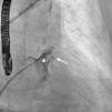
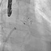
A 61-year-old man, with a history of coronary heart disease and percutaneous coronary intervention, with stents in the right coronary and circumflex arteries, was referred to our center for left atrial appendage (LAA) closure. He had atrial fibrillation with high thromboembolic risk (CHA2DS2VASc=4) and oral anticoagulation was formally contraindicated because of recurrent severe bleeding. Selective angiography of the LAA (Video 1) showed a close relationship between the LAA (Figure 1, asterisk) and the circumflex artery (Figure 1, arrow), well defined by the previously implanted stents. A computed tomography scan performed after a previous episode of diffuse alveolar hemorrhage showed similar findings (Figure 2, arrow). A 22-mm Amplatzer Amulet device was successfully placed, with no residual leak. Intraoperative monitoring by transesophageal echocardiography and post-procedural radioscopy confirmed the close relationship between the device and the circumflex artery (Figures 3 and 4, Videos 2 and 3), which is vital to keep in mind in order to prevent complications during the procedure.

Figure 1.

Selective angiography of the left atrial appendage using a pigtail catheter shows the relationship between this structure (asterisk) and the stents previously implanted in the mid-circumflex artery (arrow).

(0.33MB).
Figure 2.

Thoracic computed tomography performed after an episode of alveolar hemorrhage also reveal the proximity of the circumflex artery to the left atrium and left atrial appendage (arrow).

(0.23MB).
Figure 3.

Intraoperative monitoring of deployment of the Amplatzer device by transesophageal echocardiography (A–D). Stents previously placed in the circumflex artery (yellow arrows) can be seen in the vicinity of the left atrial appendage, immediately next to the device landing zone.

(0.18MB).
Figure 4.

After the Amplatzer was completely deployed, radioscopy confirmed the relationship between the distal lobe of the device and the coronary artery. Although in this patient this latter structure was easily delineated by the previously implanted stents, it is vital to consider this anatomical relationship during the occlusion procedure.

(0.28MB).

LAA closure is a safe and effective intervention used to prevent stroke and systemic embolism in patients with atrial fibrillation and contraindications for oral anticoagulation. Nevertheless, there may be procedure-related complications such as device embolization and left atrial wall perforation. The close relationship between the LAA and circumflex artery place this latter structure and its branches (including the sinus node artery in 30% of patients) at risk during the procedure. This condition must be considered if the patient develops atrial arrhythmias or signs of myocardial ischemia during the procedure.

Ethical disclosuresProtection of human and animal subjects

The authors declare that no experiments were performed on humans or animals for this study.

Confidentiality of data

The authors declare that no patient data appear in this article.

Right to privacy and informed consent

The authors declare that no patient data appear in this article.

Conflicts of interest

The authors have no conflicts of interest to declare.

Appendix A
Supplementary material

(1.24MB)
(2.5MB)
(1.36MB)

Copyright © 2015. Sociedade Portuguesa de Cardiologia
Download PDF
Idiomas
Revista Portuguesa de Cardiologia (English edition)
Article options
Tools
Supplemental materials
en pt

Are you a health professional able to prescribe or dispense drugs?

Você é um profissional de saúde habilitado a prescrever ou dispensar medicamentos

By checking that you are a health professional, you are stating that you are aware and accept that the Portuguese Journal of Cardiology (RPC) is the Data Controller that processes the personal information of users of its website, with its registered office at Campo Grande, n.º 28, 13.º, 1700-093 Lisbon, telephone 217 970 685 and 217 817 630, fax 217 931 095, and email revista@spc.pt. I declare for all purposes that the information provided herein is accurate and correct.