Journal Information
Vol. 34. Issue 7 - 8.
Pages 507-508 (July - August 2015)
Share
Share
Download PDF
More article options
Vol. 34. Issue 7 - 8.
Pages 507-508 (July - August 2015)
Image in Cardiology
Open Access
Cardiac papillary fibroelastoma
Fibloelastoma papilar cardíaco
Visits
4427
Sang-Wan Ryua, Min Sun Beomb, Sung Ryong Kimb, Gwan Sic Kimb,
Corresponding author
lovingmylife@naver.com

Corresponding author.
a Department of Thoracic and Cardiovascular Surgery, Chosun University Hospital, Chosun College of Medicine, Gwangju, Republic of Korea
b Department of Thoracic and Cardiovascular Surgery, Saint Carollo Hospital, Suncheon, Republic of Korea
This item has received

Under a Creative Commons license
Article information
Full Text
Download PDF
Statistics
Figures (3)
Show moreShow less
Full Text

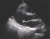
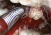
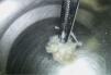
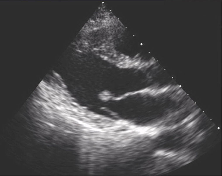
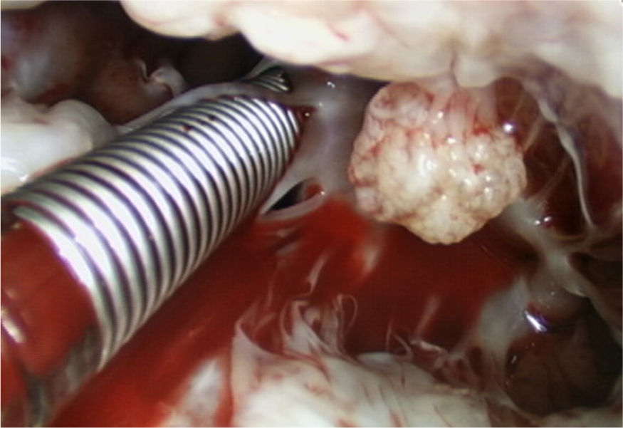
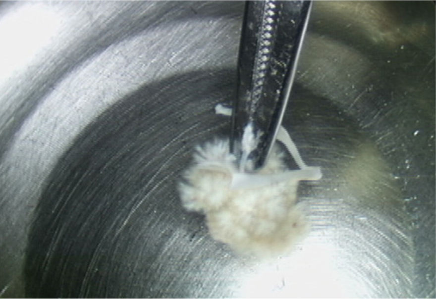
A 41-year-old man presented to the cardiovascular department with paroxysmal twinges in the left chest for three days. The initial chest radiograph, 12-lead electrocardiogram and laboratory findings were unremarkable. Transthoracic echocardiography revealed a 10.6 mm × 8.3 mm mobile mass attached to the anterior mitral leaflet (Figure 1). Because of the potential systemic embolic risk, the patient was referred for complete surgical removal. On operative inspection, the lesion was between the anterior mitral leaflet and primary chordae (Figure 2). A mitral valve repair (28 mm ring annuloplasty, triangular resection of the anterior leaflet and new chordae formation) with complete excision of the mass was undertaken. The mass had a sea-anemone appearance with frond-like projections after immersion in saline solution (Figure 3). The histopathologic examination confirmed a papillary fibroelastoma. The patient's postoperative course was uneventful. Six months after the operation, the patient's clinical findings were normal, without evidence of recurrence by transthoracic echocardiography.

Figure 1.

Transthoracic echocardiography revealing a 10.6 mm × 8.3 mm mobile mass attached to the anterior mitral leaflet.

(0.06MB).
Figure 2.

On operative inspection, the lesion was between the anterior mitral leaflet and primary chordae.

(0.08MB).
Figure 3.

The mass had a sea-anemone appearance with frond-like projections after immersion in saline solution.

(0.08MB).
Ethical disclosuresProtection of human and animal subjects

The authors declare that no experiments were performed on humans or animals for this study.

Confidentiality of data

The authors declare that no patient data appear in this article.

Right to privacy and informed consent

The authors declare that no patient data appear in this article.

Conflicts of interest

The authors have no conflicts of interest to declare.

Copyright © 2015. Sociedade Portuguesa de Cardiologia
Idiomas
Revista Portuguesa de Cardiologia (English edition)
Article options
Tools
en pt

Are you a health professional able to prescribe or dispense drugs?

Você é um profissional de saúde habilitado a prescrever ou dispensar medicamentos

By checking that you are a health professional, you are stating that you are aware and accept that the Portuguese Journal of Cardiology (RPC) is the Data Controller that processes the personal information of users of its website, with its registered office at Campo Grande, n.º 28, 13.º, 1700-093 Lisbon, telephone 217 970 685 and 217 817 630, fax 217 931 095, and email revista@spc.pt. I declare for all purposes that the information provided herein is accurate and correct.