Journal Information
Vol. 31. Issue 10.
Pages 683-684 (October 2012)
Share
Share
Download PDF
More article options
Visits
9038
Vol. 31. Issue 10.
Pages 683-684 (October 2012)
Image in Cardiology
Open Access
Anomalous insertion of the papillary muscle in a patient with sickle cell disease: A normal variant with no left ventricular outflow obstruction
Inserção anómala do músculo papilar num doente com drepanocitose: Uma variante do normal sem obstrução do tracto de saída do ventriculo esquerdo
Visits
9038
Inês Zimbarra Cabritaa,
Corresponding author
ines.cabrita05@imperial.ac.uk

Corresponding author.
, Julia Grapsaa, David Dawsona, Fausto J. Pintob,c, J. Simon R. Gibbsa, Petros Nihoyannopoulosb
a National Heart and Lung Institute, Hammersmith Campus, Imperial College London, United Kingdom
b Instituto Cardiovascular, Lisboa, Portugal
c Hospital de Santa Maria, Centro Académico de Medicina de Lisboa, Serviço de Cardiologia I, CCUL, Lisboa, Portugal
This item has received

Under a Creative Commons license
Article information
Full Text
Bibliography
Download PDF
Statistics
Figures (2)
Additional material (2)
Full Text
Introduction

Abnormalities of the papillary muscle (PM) are a rare disorder that has been described in patients with hypertrophic cardiomyopathy1–3 or cardiac amyloidosis causing significant left ventricular outflow tract (LVOT) obstruction. These anomalies are often challenging to diagnose with 2D echocardiography, and real-time 3D echocardiography (RT3D) facilitates the diagnosis by providing accurate imaging and delineation of the spatial relationship between the cardiac structures.

Case report

A 43-year-old black African woman, asymptomatic at presentation (blood pressure 105/71mmHg, heart rate 82bpm, body surface area 1.52m2), diagnosed with sickle cell disease, was investigated for pulmonary hypertension in our department. Echocardiography was requested for the assessment of cardiac function and estimation of pulmonary artery pressures.

Full 2D and 3D transthoracic echocardiography (TTE) was performed and 3D images were acquired and analyzed using Philips Qlab offline analysis software. The echocardiogram revealed a dilated left ventricle (LV) with normal wall thickness and good global systolic function.

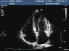
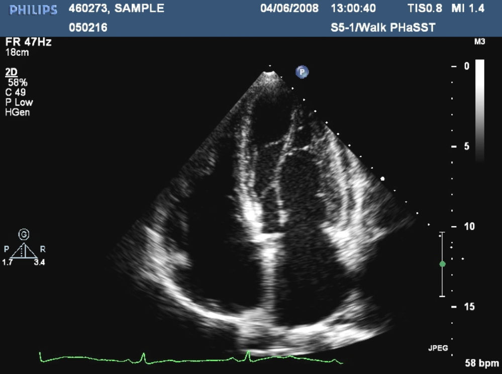
In parasternal long-axis 2D view, the anterolateral PM appeared in an unusual position, and in short-axis view and apical 4 chambers view (Figure 1) it was clearly inserted lower than normal. A complete Doppler assessment revealed no high velocity or turbulent flow pattern and no significant LVOT or intracavitary gradient was identified.

Figure 1.

Apical 4-chamber view of the anomalous insertion of the papillary muscles in 2D transthoracic echocardiography.

(0.08MB).

3D echocardiography clearly showed the lower insertion of the PM and the hypertrophied chordae (Figure 2). A follow-up echocardiography study was recommended to assess left ventricular hypertrophy but the patient died a few months later due to a subarachnoid hemorrhage. No post-mortem was performed.

Figure 2.

Real-time 3D echocardiography demonstrating the low insertion of the anterolateral papillary muscle in apical 4-chamber view.

(0.09MB).
Discussion

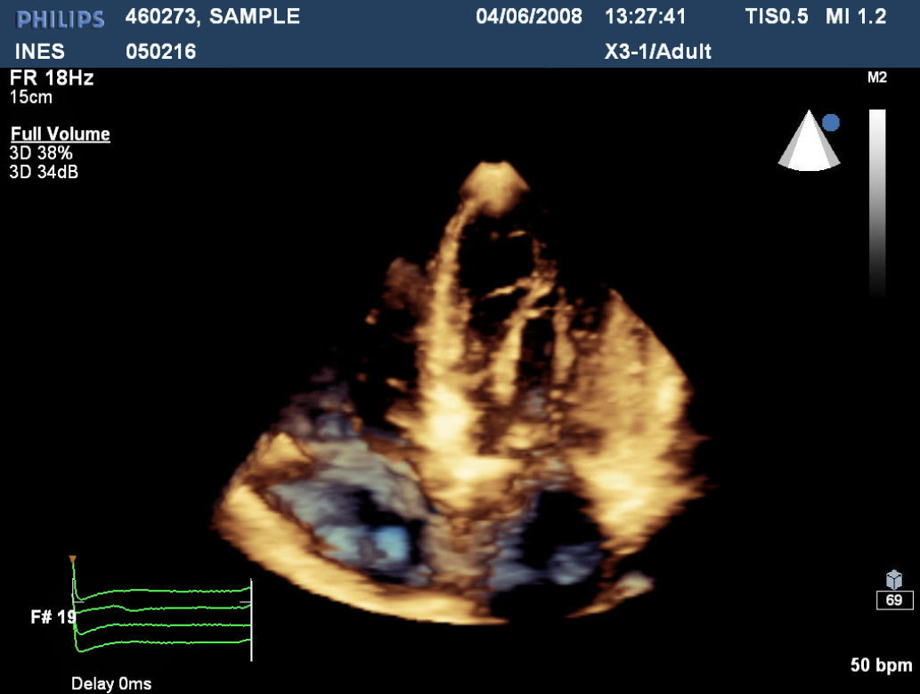
Anomalous insertion of PMs not causing LVOT obstruction is a rare abnormality.1–4 RT3D is being increasingly used in the clinical setting to accurately identify cardiac structures and anatomy.5,6 Its role is mainly in providing additional spatial information to 2D TTE in the identification of complex anatomic structures. The normal variant of low insertion of the anterolateral PM without LVOT obstruction may be observed in healthy individuals but an association with sickle cell disease cannot be excluded; further investigations are needed to confirm this association. To our knowledge, this is the first case in sickle cell disease of an anomalous insertion of PMs not causing LVOT obstruction reported in the literature.

Ethical disclosuresConfidentiality of data

The authors declare that they have followed the protocols of their work center on the publication of patient data and that all the patients included in the study have received sufficient information and have given their informed consent in writing to participate in the study.

Right to privacy and informed consent

The authors have obtained the informed consent of the patients and/or subjects mentioned in the article. The author for correspondence is in possession of this document.

Protection of human and animal subjects

The authors declare that no experiments were performed on humans or animals for this investigation.

Conflicts of interest

The authors have no conflicts of interest to declare.

Appendix A
Supplementary material

The following are the supplementary data to this article:

(2.45MB)
(2.36MB)

References
[1]
B.A. Austin, D.H. Kwon, N.G. Smedira, et al.
Abnormally thickened papillary muscle resulting in dynamic left ventricular outflow tract obstruction: an unusual presentation of hypertrophic cardiomyopathy.
J Am Soc Echocardiogr, 22 (2009), pp. e5-e6
[2]
T. Nomura, Y. Harada, Y. Suzaki, et al.
Left ventricular outflow tract obstruction due to anomalous insertion of papillary muscle.
Circ J, 68 (2004), pp. 1219-1222
[3]
H.G. Klues, B.J. Maron, A.L. Dollar, et al.
Diversity of structural mitral valve alterations in hypertrophic cardiomyopathy.
Circulation, 85 (1992), pp. 1651-1660
[4]
N. Watanabe, N. Wada, K. Yoshida.
Uncommon anomalous papillary muscle/chordae tendineae incidentally found in patient with transient ischaemic attack.
Heart, 91 (2005), pp. p1074
[5]
H.S. Yang, K.S. Lee, H.P. Chaliki, et al.
Anomalous insertion of the papillary muscle causing left ventricular outflow obstruction: visualization by real-time three-dimensional echocardiography.
Eur J Echocardiogr, 9 (2008), pp. 855-860
[6]
J. Hung, R. Lang, F. Flachskampf, et al.
3D echocardiography: a review of the current status and future directions.
J Am Soc Echocardiogr, 20 (2007), pp. 213-233
Copyright © 2012. Sociedade Portuguesa de Cardiologia
Idiomas
Revista Portuguesa de Cardiologia (English edition)
Article options
Tools
Supplemental materials
en pt

Are you a health professional able to prescribe or dispense drugs?

Você é um profissional de saúde habilitado a prescrever ou dispensar medicamentos

By checking that you are a health professional, you are stating that you are aware and accept that the Portuguese Journal of Cardiology (RPC) is the Data Controller that processes the personal information of users of its website, with its registered office at Campo Grande, n.º 28, 13.º, 1700-093 Lisbon, telephone 217 970 685 and 217 817 630, fax 217 931 095, and email revista@spc.pt. I declare for all purposes that the information provided herein is accurate and correct.