array:23 [
  "pii" => "S2174204912001596"
  "issn" => "21742049"
  "doi" => "10.1016/j.repce.2012.09.004"
  "estado" => "S300"
  "fechaPublicacion" => "2012-10-01"
  "aid" => "144"
  "copyright" => "Sociedade Portuguesa de Cardiologia"
  "copyrightAnyo" => "2012"
  "documento" => "article"
  "crossmark" => 0
  "licencia" => "http://creativecommons.org/licenses/by-nc-nd/4.0/"
  "subdocumento" => "sco"
  "cita" => "Rev Port Cardiol. 2012;31:685-6"
  "abierto" => array:3 [
    "ES" => true
    "ES2" => true
    "LATM" => true
  ]
  "gratuito" => true
  "lecturas" => array:2 [
    "total" => 4019
    "formatos" => array:3 [
      "EPUB" => 113
      "HTML" => 3243
      "PDF" => 663
    ]
  ]
  "itemAnterior" => array:19 [
    "pii" => "S2174204912001584"
    "issn" => "21742049"
    "doi" => "10.1016/j.repce.2012.09.003"
    "estado" => "S300"
    "fechaPublicacion" => "2012-10-01"
    "aid" => "143"
    "copyright" => "Sociedade Portuguesa de Cardiologia"
    "documento" => "article"
    "crossmark" => 0
    "licencia" => "http://creativecommons.org/licenses/by-nc-nd/4.0/"
    "subdocumento" => "sco"
    "cita" => "Rev Port Cardiol. 2012;31:683-4"
    "abierto" => array:3 [
      "ES" => true
      "ES2" => true
      "LATM" => true
    ]
    "gratuito" => true
    "lecturas" => array:2 [
      "total" => 5187
      "formatos" => array:3 [
        "EPUB" => 144
        "HTML" => 4361
        "PDF" => 682
      ]
    ]
    "en" => array:11 [
      "idiomaDefecto" => true
      "cabecera" => "<span class="elsevierStyleTextfn">Image in Cardiology</span>"
      "titulo" => "Anomalous insertion of the papillary muscle in a patient with sickle cell disease&#58; A normal variant with no left ventricular outflow obstruction"
      "tienePdf" => "en"
      "tieneTextoCompleto" => "en"
      "paginas" => array:1 [
        0 => array:2 [
          "paginaInicial" => "683"
          "paginaFinal" => "684"
        ]
      ]
      "titulosAlternativos" => array:1 [
        "pt" => array:1 [
          "titulo" => "Inser&#231;&#227;o an&#243;mala do m&#250;sculo papilar num doente com drepanocitose&#58; Uma variante do normal sem obstru&#231;&#227;o do tracto de sa&#237;da do ventriculo esquerdo"
        ]
      ]
      "contieneTextoCompleto" => array:1 [
        "en" => true
      ]
      "contienePdf" => array:1 [
        "en" => true
      ]
      "resumenGrafico" => array:2 [
        "original" => 0
        "multimedia" => array:7 [
          "identificador" => "fig0005"
          "etiqueta" => "Figure 1"
          "tipo" => "MULTIMEDIAFIGURA"
          "mostrarFloat" => true
          "mostrarDisplay" => false
          "figura" => array:1 [
            0 => array:4 [
              "imagen" => "gr1.jpeg"
              "Alto" => 744
              "Ancho" => 999
              "Tamanyo" => 87149
            ]
          ]
          "descripcion" => array:1 [
            "en" => "<p id="spar0005" class="elsevierStyleSimplePara elsevierViewall">Apical 4-chamber view of the anomalous insertion of the papillary muscles in 2D transthoracic echocardiography&#46;</p>"
          ]
        ]
      ]
      "autores" => array:1 [
        0 => array:2 [
          "autoresLista" => "In&#234;s Zimbarra Cabrita, Julia Grapsa, David Dawson, Fausto J&#46; Pinto, J&#46; Simon R&#46; Gibbs, Petros Nihoyannopoulos"
          "autores" => array:6 [
            0 => array:2 [
              "nombre" => "In&#234;s"
              "apellidos" => "Zimbarra Cabrita"
            ]
            1 => array:2 [
              "nombre" => "Julia"
              "apellidos" => "Grapsa"
            ]
            2 => array:2 [
              "nombre" => "David"
              "apellidos" => "Dawson"
            ]
            3 => array:2 [
              "nombre" => "Fausto J&#46;"
              "apellidos" => "Pinto"
            ]
            4 => array:2 [
              "nombre" => "J&#46; Simon R&#46;"
              "apellidos" => "Gibbs"
            ]
            5 => array:2 [
              "nombre" => "Petros"
              "apellidos" => "Nihoyannopoulos"
            ]
          ]
        ]
      ]
    ]
    "idiomaDefecto" => "en"
    "EPUB" => "https://multimedia.elsevier.es/PublicationsMultimediaV1/item/epub/S2174204912001584?idApp=UINPBA00004E"
    "url" => "/21742049/0000003100000010/v1_201305151709/S2174204912001584/v1_201305151709/en/main.assets"
  ]
  "en" => array:16 [
    "idiomaDefecto" => true
    "cabecera" => "<span class="elsevierStyleTextfn">Image in cardiology</span>"
    "titulo" => "Embolization of a Biostar device in the pulmonary artery"
    "tieneTextoCompleto" => true
    "paginas" => array:1 [
      0 => array:2 [
        "paginaInicial" => "685"
        "paginaFinal" => "686"
      ]
    ]
    "autores" => array:1 [
      0 => array:4 [
        "autoresLista" => "Paolo G&#46; Pino, Amedeo Pergolini, Giordano Zampi"
        "autores" => array:3 [
          0 => array:2 [
            "nombre" => "Paolo G&#46;"
            "apellidos" => "Pino"
          ]
          1 => array:2 [
            "nombre" => "Amedeo"
            "apellidos" => "Pergolini"
          ]
          2 => array:4 [
            "nombre" => "Giordano"
            "apellidos" => "Zampi"
            "email" => array:1 [
              0 => "giordano&#46;zampi&#64;alice&#46;it"
            ]
            "referencia" => array:1 [
              0 => array:2 [
                "etiqueta" => "<span class="elsevierStyleSup">¿</span>"
                "identificador" => "cor0005"
              ]
            ]
          ]
        ]
        "afiliaciones" => array:1 [
          0 => array:1 [
            "entidad" => "Department of Cardiovascular Science&#44; &#8220;S&#46; Camillo-Forlanini&#8221; Hospital&#44; Rome&#44; Italy"
          ]
        ]
        "correspondencia" => array:1 [
          0 => array:3 [
            "identificador" => "cor0005"
            "etiqueta" => "&#8270;"
            "correspondencia" => "Corresponding author&#46;"
          ]
        ]
      ]
    ]
    "titulosAlternativos" => array:1 [
      "pt" => array:1 [
        "titulo" => "Emboliza&#231;&#227;o duma pr&#243;tese Biostar na art&#233;ria pulmonar"
      ]
    ]
    "resumenGrafico" => array:2 [
      "original" => 0
      "multimedia" => array:7 [
        "identificador" => "fig0005"
        "etiqueta" => "Figure 1"
        "tipo" => "MULTIMEDIAFIGURA"
        "mostrarFloat" => true
        "mostrarDisplay" => false
        "figura" => array:1 [
          0 => array:4 [
            "imagen" => "gr1.jpeg"
            "Alto" => 2479
            "Ancho" => 2167
            "Tamanyo" => 478075
          ]
        ]
        "descripcion" => array:1 [
          "en" => "<p id="spar0005" class="elsevierStyleSimplePara elsevierViewall">&#40;A&#41; Transthoracic echocardiography&#59; &#40;B&#41; contrast echocardiography&#59; &#40;C&#41; 3D real-time transesophageal echocardiography&#59; &#40;D&#41; 3D off-line reconstruction of the PFO&#59; &#40;E and F&#41; cardioscopy&#46;</p>"
        ]
      ]
    ]
    "textoCompleto" => "<span class="elsevierStyleSections"><p id="par0005" class="elsevierStylePara elsevierViewall">An asymptomatic 50-year-old Caucasian woman was referred to our echocardiography laboratory to monitor a Biostar septal occluder device inserted six months before in order to close a patent foramen ovale&#46;</p><p id="par0010" class="elsevierStylePara elsevierViewall">On transthoracic echocardiography no residual shunt was observed but the device was not identifiable &#40;<a class="elsevierStyleCrossRef" href="#fig0005">Figure 1</a>A&#41;&#59; after injection of agitated saline solution in a vein of the right arm and the Valsalva maneuver&#44; a remarkable paradoxical right-to-left shunt appeared &#40;<a class="elsevierStyleCrossRef" href="#fig0005">Figure 1</a>B&#44; <a class="elsevierStyleCrossRef" href="#sec0035">video clip 1</a>&#41;&#46; The estimated systolic artery pressure was 25<span class="elsevierStyleHsp" style=""></span>mmHg&#46;</p><elsevierMultimedia ident="fig0005"></elsevierMultimedia><p id="par0015" class="elsevierStylePara elsevierViewall">To better understand the pathophysiology of the shunt 3D real-time transesophageal echocardiography was performed&#46; The septal occluder was not found on the interatrial septum &#40;<a class="elsevierStyleCrossRef" href="#fig0005">Figure 1</a>C&#41;&#46; A small interatrial defect was identified close to the foramen ovale &#40;<a class="elsevierStyleCrossRef" href="#fig0005">Figure 1</a>D&#44; <a class="elsevierStyleCrossRef" href="#sec0035">video clip 2</a>&#41; with a trivial left-to-right shunt &#40;Qp&#47;Qs 1&#46;1&#41; in baseline conditions&#59; the agitated saline contrast test after the Valsalva maneuver was markedly positive&#46; Given the suspicion of distal embolization of the device&#44; cardioscopy was performed&#58; the Biostar was found in the left pulmonary artery &#40;<a class="elsevierStyleCrossRef" href="#fig0005">Figure 1</a>E and F&#44; <a class="elsevierStyleCrossRef" href="#sec0035">video clips 3 and 4</a>&#41;&#44; without hemodynamic effects &#40;mean pulmonary artery pressure 18<span class="elsevierStyleHsp" style=""></span>mmHg&#41;&#46;</p><p id="par0020" class="elsevierStylePara elsevierViewall">In accordance with the clinical and instrumental findings&#44; the patient was enrolled for long-term follow-up&#44; with regular clinical and echocardiographic monitoring in order to prevent complications&#46;</p><span id="sec0005" class="elsevierStyleSection elsevierViewall"><span class="elsevierStyleSectionTitle">Ethical disclosures</span><span id="sec0010" class="elsevierStyleSection elsevierViewall"><span class="elsevierStyleSectionTitle">Right to privacy and informed consent</span><p id="par0025" class="elsevierStylePara elsevierViewall">The authors declare that no patient data appear in this article&#46;</p></span><span id="sec0015" class="elsevierStyleSection elsevierViewall"><span class="elsevierStyleSectionTitle">Confidentiality of data</span><p id="par0030" class="elsevierStylePara elsevierViewall">The authors declare that no patient data appear in this article&#46;</p></span><span id="sec0020" class="elsevierStyleSection elsevierViewall"><span class="elsevierStyleSectionTitle">Protection of human and animal subjects</span><p id="par0035" class="elsevierStylePara elsevierViewall">The authors declare that no experiments were performed on humans or animals for this investigation&#46;</p></span></span><span id="sec0025" class="elsevierStyleSection elsevierViewall"><span class="elsevierStyleSectionTitle">Conflicts of interest</span><p id="par0040" class="elsevierStylePara elsevierViewall">The authors have no conflicts of interest to declare&#46;</p></span></span>"
    "textoCompletoSecciones" => array:1 [
      "secciones" => array:2 [
        0 => array:3 [
          "identificador" => "sec0005"
          "titulo" => "Ethical disclosures"
          "secciones" => array:3 [
            0 => array:2 [
              "identificador" => "sec0010"
              "titulo" => "Right to privacy and informed consent"
            ]
            1 => array:2 [
              "identificador" => "sec0015"
              "titulo" => "Confidentiality of data"
            ]
            2 => array:2 [
              "identificador" => "sec0020"
              "titulo" => "Protection of human and animal subjects"
            ]
          ]
        ]
        1 => array:2 [
          "identificador" => "sec0025"
          "titulo" => "Conflicts of interest"
        ]
      ]
    ]
    "pdfFichero" => "main.pdf"
    "tienePdf" => true
    "fechaRecibido" => "2012-03-31"
    "fechaAceptado" => "2012-04-11"
    "apendice" => array:1 [
      0 => array:1 [
        "seccion" => array:1 [
          0 => array:4 [
            "apendice" => "<p id="par0050" class="elsevierStylePara elsevierViewall">The following is the supplementary data to this article&#58;<elsevierMultimedia ident="upi0005"></elsevierMultimedia><elsevierMultimedia ident="upi0010"></elsevierMultimedia><elsevierMultimedia ident="upi0015"></elsevierMultimedia><elsevierMultimedia ident="upi0020"></elsevierMultimedia></p>"
            "etiqueta" => "Appendix A"
            "titulo" => "Supplementary material"
            "identificador" => "sec0035"
          ]
        ]
      ]
    ]
    "multimedia" => array:5 [
      0 => array:7 [
        "identificador" => "fig0005"
        "etiqueta" => "Figure 1"
        "tipo" => "MULTIMEDIAFIGURA"
        "mostrarFloat" => true
        "mostrarDisplay" => false
        "figura" => array:1 [
          0 => array:4 [
            "imagen" => "gr1.jpeg"
            "Alto" => 2479
            "Ancho" => 2167
            "Tamanyo" => 478075
          ]
        ]
        "descripcion" => array:1 [
          "en" => "<p id="spar0005" class="elsevierStyleSimplePara elsevierViewall">&#40;A&#41; Transthoracic echocardiography&#59; &#40;B&#41; contrast echocardiography&#59; &#40;C&#41; 3D real-time transesophageal echocardiography&#59; &#40;D&#41; 3D off-line reconstruction of the PFO&#59; &#40;E and F&#41; cardioscopy&#46;</p>"
        ]
      ]
      1 => array:5 [
        "identificador" => "upi0005"
        "tipo" => "MULTIMEDIAECOMPONENTE"
        "mostrarFloat" => false
        "mostrarDisplay" => true
        "Ecomponente" => array:3 [
          "fichero" => "mmc1.mov"
          "ficheroTamanyo" => 2365147
          "Video" => array:2 [
            "flv" => array:5 [
              "fichero" => "mmc1.flv"
              "poster" => "mmc1.jpg"
              "tiempo" => 0
              "alto" => 0
              "ancho" => 0
            ]
            "mp4" => array:2 [
              "fichero" => "mmc1.m4v"
              "poster" => "mmc1.jpg"
            ]
          ]
        ]
      ]
      2 => array:5 [
        "identificador" => "upi0010"
        "tipo" => "MULTIMEDIAECOMPONENTE"
        "mostrarFloat" => false
        "mostrarDisplay" => true
        "Ecomponente" => array:3 [
          "fichero" => "mmc2.mov"
          "ficheroTamanyo" => 359294
          "Video" => array:2 [
            "flv" => array:5 [
              "fichero" => "mmc2.flv"
              "poster" => "mmc2.jpg"
              "tiempo" => 0
              "alto" => 0
              "ancho" => 0
            ]
            "mp4" => array:2 [
              "fichero" => "mmc2.m4v"
              "poster" => "mmc2.jpg"
            ]
          ]
        ]
      ]
      3 => array:5 [
        "identificador" => "upi0015"
        "tipo" => "MULTIMEDIAECOMPONENTE"
        "mostrarFloat" => false
        "mostrarDisplay" => true
        "Ecomponente" => array:3 [
          "fichero" => "mmc3.mov"
          "ficheroTamanyo" => 298085
          "Video" => array:2 [
            "flv" => array:5 [
              "fichero" => "mmc3.flv"
              "poster" => "mmc3.jpg"
              "tiempo" => 0
              "alto" => 0
              "ancho" => 0
            ]
            "mp4" => array:2 [
              "fichero" => "mmc3.m4v"
              "poster" => "mmc3.jpg"
            ]
          ]
        ]
      ]
      4 => array:5 [
        "identificador" => "upi0020"
        "tipo" => "MULTIMEDIAECOMPONENTE"
        "mostrarFloat" => false
        "mostrarDisplay" => true
        "Ecomponente" => array:3 [
          "fichero" => "mmc4.mov"
          "ficheroTamanyo" => 290280
          "Video" => array:2 [
            "flv" => array:5 [
              "fichero" => "mmc4.flv"
              "poster" => "mmc4.jpg"
              "tiempo" => 0
              "alto" => 0
              "ancho" => 0
            ]
            "mp4" => array:2 [
              "fichero" => "mmc4.m4v"
              "poster" => "mmc4.jpg"
            ]
          ]
        ]
      ]
    ]
  ]
  "idiomaDefecto" => "en"
  "url" => "/21742049/0000003100000010/v1_201305151709/S2174204912001596/v1_201305151709/en/main.assets"
  "Apartado" => array:4 [
    "identificador" => "9915"
    "tipo" => "SECCION"
    "en" => array:2 [
      "titulo" => "Images in cardiology"
      "idiomaDefecto" => true
    ]
    "idiomaDefecto" => "en"
  ]
  "PDF" => "https://static.elsevier.es/multimedia/21742049/0000003100000010/v1_201305151709/S2174204912001596/v1_201305151709/en/main.pdf?idApp=UINPBA00004E&text.app=https://revportcardiol.org/"
  "EPUB" => "https://multimedia.elsevier.es/PublicationsMultimediaV1/item/epub/S2174204912001596?idApp=UINPBA00004E"
]
Share
Journal Information
Vol. 31. Issue 10.
Pages 685-686 (October 2012)
Share
Share
Download PDF
More article options
Vol. 31. Issue 10.
Pages 685-686 (October 2012)
Image in cardiology
Open Access
Embolization of a Biostar device in the pulmonary artery
Embolização duma prótese Biostar na artéria pulmonar
Visits
6773
Paolo G. Pino, Amedeo Pergolini, Giordano Zampi
Corresponding author
giordano.zampi@alice.it

Corresponding author.
Department of Cardiovascular Science, “S. Camillo-Forlanini” Hospital, Rome, Italy
This item has received

Under a Creative Commons license
Article information
Full Text
Download PDF
Statistics
Figures (1)
Additional material (4)
Full Text

An asymptomatic 50-year-old Caucasian woman was referred to our echocardiography laboratory to monitor a Biostar septal occluder device inserted six months before in order to close a patent foramen ovale.

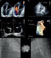
On transthoracic echocardiography no residual shunt was observed but the device was not identifiable (Figure 1A); after injection of agitated saline solution in a vein of the right arm and the Valsalva maneuver, a remarkable paradoxical right-to-left shunt appeared (Figure 1B, video clip 1). The estimated systolic artery pressure was 25mmHg.

Figure 1.

(A) Transthoracic echocardiography; (B) contrast echocardiography; (C) 3D real-time transesophageal echocardiography; (D) 3D off-line reconstruction of the PFO; (E and F) cardioscopy.

(0.46MB).

To better understand the pathophysiology of the shunt 3D real-time transesophageal echocardiography was performed. The septal occluder was not found on the interatrial septum (Figure 1C). A small interatrial defect was identified close to the foramen ovale (Figure 1D, video clip 2) with a trivial left-to-right shunt (Qp/Qs 1.1) in baseline conditions; the agitated saline contrast test after the Valsalva maneuver was markedly positive. Given the suspicion of distal embolization of the device, cardioscopy was performed: the Biostar was found in the left pulmonary artery (Figure 1E and F, video clips 3 and 4), without hemodynamic effects (mean pulmonary artery pressure 18mmHg).

In accordance with the clinical and instrumental findings, the patient was enrolled for long-term follow-up, with regular clinical and echocardiographic monitoring in order to prevent complications.

Ethical disclosuresRight to privacy and informed consent

The authors declare that no patient data appear in this article.

Confidentiality of data

The authors declare that no patient data appear in this article.

Protection of human and animal subjects

The authors declare that no experiments were performed on humans or animals for this investigation.

Conflicts of interest

The authors have no conflicts of interest to declare.

Appendix A
Supplementary material

The following is the supplementary data to this article:

(2.26MB)
(0.34MB)
(0.28MB)
(0.28MB)

Copyright © 2012. Sociedade Portuguesa de Cardiologia
Download PDF
Idiomas
Revista Portuguesa de Cardiologia (English edition)
Article options
Tools
Supplemental materials
en pt

Are you a health professional able to prescribe or dispense drugs?

Você é um profissional de saúde habilitado a prescrever ou dispensar medicamentos

By checking that you are a health professional, you are stating that you are aware and accept that the Portuguese Journal of Cardiology (RPC) is the Data Controller that processes the personal information of users of its website, with its registered office at Campo Grande, n.º 28, 13.º, 1700-093 Lisbon, telephone 217 970 685 and 217 817 630, fax 217 931 095, and email revista@spc.pt. I declare for all purposes that the information provided herein is accurate and correct.