array:25 [
  "pii" => "S2174204914002177"
  "issn" => "21742049"
  "doi" => "10.1016/j.repce.2014.02.023"
  "estado" => "S300"
  "fechaPublicacion" => "2014-09-01"
  "aid" => "506"
  "copyright" => "Sociedade Portuguesa de Cardiologia"
  "copyrightAnyo" => "2014"
  "documento" => "article"
  "crossmark" => 0
  "licencia" => "http://creativecommons.org/licenses/by-nc-nd/4.0/"
  "subdocumento" => "sco"
  "cita" => "Rev Port Cardiol. 2014;33:571-2"
  "abierto" => array:3 [
    "ES" => true
    "ES2" => true
    "LATM" => true
  ]
  "gratuito" => true
  "lecturas" => array:2 [
    "total" => 2699
    "formatos" => array:3 [
      "EPUB" => 160
      "HTML" => 2073
      "PDF" => 466
    ]
  ]
  "Traduccion" => array:1 [
    "en" => array:20 [
      "pii" => "S0870255114001887"
      "issn" => "08702551"
      "doi" => "10.1016/j.repc.2014.02.023"
      "estado" => "S300"
      "fechaPublicacion" => "2014-09-01"
      "aid" => "506"
      "copyright" => "Sociedade Portuguesa de Cardiologia"
      "documento" => "article"
      "crossmark" => 0
      "licencia" => "http://creativecommons.org/licenses/by-nc-nd/4.0/"
      "subdocumento" => "sco"
      "cita" => "Rev Port Cardiol. 2014;33:571-2"
      "abierto" => array:3 [
        "ES" => true
        "ES2" => true
        "LATM" => true
      ]
      "gratuito" => true
      "lecturas" => array:2 [
        "total" => 3983
        "formatos" => array:3 [
          "EPUB" => 160
          "HTML" => 3161
          "PDF" => 662
        ]
      ]
      "en" => array:11 [
        "idiomaDefecto" => true
        "cabecera" => "<span class="elsevierStyleTextfn">Image in Cardiology</span>"
        "titulo" => "A rare case of congenital aneurysm of the right atrium"
        "tienePdf" => "en"
        "tieneTextoCompleto" => "en"
        "paginas" => array:1 [
          0 => array:2 [
            "paginaInicial" => "571"
            "paginaFinal" => "572"
          ]
        ]
        "titulosAlternativos" => array:1 [
          "pt" => array:1 [
            "titulo" => "Um caso raro de aneurisma cong&#233;nito da aur&#237;cula direita"
          ]
        ]
        "contieneTextoCompleto" => array:1 [
          "en" => true
        ]
        "contienePdf" => array:1 [
          "en" => true
        ]
        "resumenGrafico" => array:2 [
          "original" => 0
          "multimedia" => array:7 [
            "identificador" => "fig0020"
            "etiqueta" => "Figure 4"
            "tipo" => "MULTIMEDIAFIGURA"
            "mostrarFloat" => true
            "mostrarDisplay" => false
            "figura" => array:1 [
              0 => array:4 [
                "imagen" => "gr4.jpeg"
                "Alto" => 877
                "Ancho" => 1658
                "Tamanyo" => 157319
              ]
            ]
            "descripcion" => array:1 [
              "en" => "<p id="spar0020" class="elsevierStyleSimplePara elsevierViewall">Intraoperative photograph &#40;surgeon&#39;s view&#41; revealing the right atrial aneurysm &#40;RAA&#41;&#46;</p>"
            ]
          ]
        ]
        "autores" => array:1 [
          0 => array:2 [
            "autoresLista" => "Alexandra da Silva Castro, Nuno Ferreira, Ricardo Ferraz, Nuno Bettencourt, Jo&#227;o Carlos Mota, Vasco Gama Ribeiro"
            "autores" => array:6 [
              0 => array:2 [
                "nombre" => "Alexandra"
                "apellidos" => "da Silva Castro"
              ]
              1 => array:2 [
                "nombre" => "Nuno"
                "apellidos" => "Ferreira"
              ]
              2 => array:2 [
                "nombre" => "Ricardo"
                "apellidos" => "Ferraz"
              ]
              3 => array:2 [
                "nombre" => "Nuno"
                "apellidos" => "Bettencourt"
              ]
              4 => array:2 [
                "nombre" => "Jo&#227;o Carlos"
                "apellidos" => "Mota"
              ]
              5 => array:2 [
                "nombre" => "Vasco"
                "apellidos" => "Gama Ribeiro"
              ]
            ]
          ]
        ]
      ]
      "idiomaDefecto" => "en"
      "Traduccion" => array:1 [
        "en" => array:9 [
          "pii" => "S2174204914002177"
          "doi" => "10.1016/j.repce.2014.02.023"
          "estado" => "S300"
          "subdocumento" => ""
          "abierto" => array:3 [
            "ES" => true
            "ES2" => true
            "LATM" => true
          ]
          "gratuito" => true
          "lecturas" => array:1 [
            "total" => 0
          ]
          "idiomaDefecto" => "en"
          "EPUB" => "https://multimedia.elsevier.es/PublicationsMultimediaV1/item/epub/S2174204914002177?idApp=UINPBA00004E"
        ]
      ]
      "EPUB" => "https://multimedia.elsevier.es/PublicationsMultimediaV1/item/epub/S0870255114001887?idApp=UINPBA00004E"
      "url" => "/08702551/0000003300000009/v1_201410010215/S0870255114001887/v1_201410010215/en/main.assets"
    ]
  ]
  "itemSiguiente" => array:20 [
    "pii" => "S217420491400172X"
    "issn" => "21742049"
    "doi" => "10.1016/j.repce.2014.03.005"
    "estado" => "S300"
    "fechaPublicacion" => "2014-09-01"
    "aid" => "482"
    "copyright" => "Sociedade Portuguesa de Cardiologia"
    "documento" => "article"
    "crossmark" => 0
    "licencia" => "http://creativecommons.org/licenses/by-nc-nd/4.0/"
    "subdocumento" => "sco"
    "cita" => "Rev Port Cardiol. 2014;33:573-4"
    "abierto" => array:3 [
      "ES" => true
      "ES2" => true
      "LATM" => true
    ]
    "gratuito" => true
    "lecturas" => array:2 [
      "total" => 3197
      "formatos" => array:3 [
        "EPUB" => 153
        "HTML" => 2405
        "PDF" => 639
      ]
    ]
    "en" => array:11 [
      "idiomaDefecto" => true
      "cabecera" => "<span class="elsevierStyleTextfn">Image in Cardiology</span>"
      "titulo" => "The lost needle of a seamstress"
      "tienePdf" => "en"
      "tieneTextoCompleto" => "en"
      "paginas" => array:1 [
        0 => array:2 [
          "paginaInicial" => "573"
          "paginaFinal" => "574"
        ]
      ]
      "titulosAlternativos" => array:1 [
        "pt" => array:1 [
          "titulo" => "A agulha perdida de uma costureira"
        ]
      ]
      "contieneTextoCompleto" => array:1 [
        "en" => true
      ]
      "contienePdf" => array:1 [
        "en" => true
      ]
      "resumenGrafico" => array:2 [
        "original" => 0
        "multimedia" => array:7 [
          "identificador" => "fig0005"
          "etiqueta" => "Figure 1"
          "tipo" => "MULTIMEDIAFIGURA"
          "mostrarFloat" => true
          "mostrarDisplay" => false
          "figura" => array:1 [
            0 => array:4 [
              "imagen" => "gr1.jpeg"
              "Alto" => 1174
              "Ancho" => 1800
              "Tamanyo" => 290816
            ]
          ]
          "descripcion" => array:1 [
            "en" => "<p id="spar0005" class="elsevierStyleSimplePara elsevierViewall">&#40;A&#41; Right coronary artery in left anterior oblique projection at 45&#176;&#44; caudal 10&#176;&#44; showing 60&#37; proximal stenosis and 70&#37; stenosis in the posterolateral branch&#59; &#40;B&#41; left coronary artery in right anterior oblique projection at 30&#176;&#44; caudal 10&#176;&#44; showing no significant lesions but revealing a sewing needle in the left hemithorax&#59; &#40;C&#41; anteroposterior view&#44; 0&#176; 0&#176;&#44; showing magnified image of the needle&#59; &#40;D&#41; thoracic computed tomography &#40;CT&#41;&#44; sagittal plane&#44; showing the needle in subpleural position&#44; posterior to the apical segment of the left inferior lobe&#59; &#40;E&#41; thoracic CT&#44; pulmonary window&#44; coronal plane&#44; showing the needle&#44; 40 mm in length&#44; in cranial-caudal orientation&#59; &#40;F&#41; myocardial scintigraphy&#58; after exercise &#40;above&#41; and at rest &#40;below&#41;&#44; documenting a lateral perfusion defect with significant reversibility&#46;</p>"
          ]
        ]
      ]
      "autores" => array:1 [
        0 => array:2 [
          "autoresLista" => "Ana Rita Godinho, Mariana Vasconcelos, In&#234;s Rangel, Domingos Magalh&#227;es, Maria J&#250;lia Maciel"
          "autores" => array:5 [
            0 => array:2 [
              "nombre" => "Ana Rita"
              "apellidos" => "Godinho"
            ]
            1 => array:2 [
              "nombre" => "Mariana"
              "apellidos" => "Vasconcelos"
            ]
            2 => array:2 [
              "nombre" => "In&#234;s"
              "apellidos" => "Rangel"
            ]
            3 => array:2 [
              "nombre" => "Domingos"
              "apellidos" => "Magalh&#227;es"
            ]
            4 => array:2 [
              "nombre" => "Maria J&#250;lia"
              "apellidos" => "Maciel"
            ]
          ]
        ]
      ]
    ]
    "idiomaDefecto" => "en"
    "Traduccion" => array:1 [
      "pt" => array:9 [
        "pii" => "S0870255114001498"
        "doi" => "10.1016/j.repc.2014.03.004"
        "estado" => "S300"
        "subdocumento" => ""
        "abierto" => array:3 [
          "ES" => true
          "ES2" => true
          "LATM" => true
        ]
        "gratuito" => true
        "lecturas" => array:1 [
          "total" => 0
        ]
        "idiomaDefecto" => "pt"
        "EPUB" => "https://multimedia.elsevier.es/PublicationsMultimediaV1/item/epub/S0870255114001498?idApp=UINPBA00004E"
      ]
    ]
    "EPUB" => "https://multimedia.elsevier.es/PublicationsMultimediaV1/item/epub/S217420491400172X?idApp=UINPBA00004E"
    "url" => "/21742049/0000003300000009/v1_201410030041/S217420491400172X/v1_201410030041/en/main.assets"
  ]
  "itemAnterior" => array:20 [
    "pii" => "S2174204914002165"
    "issn" => "21742049"
    "doi" => "10.1016/j.repce.2014.02.022"
    "estado" => "S300"
    "fechaPublicacion" => "2014-09-01"
    "aid" => "505"
    "copyright" => "Sociedade Portuguesa de Cardiologia"
    "documento" => "article"
    "crossmark" => 0
    "licencia" => "http://creativecommons.org/licenses/by-nc-nd/4.0/"
    "subdocumento" => "sco"
    "cita" => "Rev Port Cardiol. 2014;33:569-70"
    "abierto" => array:3 [
      "ES" => true
      "ES2" => true
      "LATM" => true
    ]
    "gratuito" => true
    "lecturas" => array:2 [
      "total" => 2753
      "formatos" => array:3 [
        "EPUB" => 165
        "HTML" => 2057
        "PDF" => 531
      ]
    ]
    "en" => array:11 [
      "idiomaDefecto" => true
      "cabecera" => "<span class="elsevierStyleTextfn">Image in Cardiology</span>"
      "titulo" => "Cardiac metastasis from colorectal cancer&#58; To be or not to be&#8230;"
      "tienePdf" => "en"
      "tieneTextoCompleto" => "en"
      "paginas" => array:1 [
        0 => array:2 [
          "paginaInicial" => "569"
          "paginaFinal" => "570"
        ]
      ]
      "titulosAlternativos" => array:1 [
        "pt" => array:1 [
          "titulo" => "Met&#225;stase card&#237;aca do cancro colorretal&#58; ser ou n&#227;o ser&#8230;"
        ]
      ]
      "contieneTextoCompleto" => array:1 [
        "en" => true
      ]
      "contienePdf" => array:1 [
        "en" => true
      ]
      "resumenGrafico" => array:2 [
        "original" => 0
        "multimedia" => array:7 [
          "identificador" => "fig0005"
          "etiqueta" => "Figure 1"
          "tipo" => "MULTIMEDIAFIGURA"
          "mostrarFloat" => true
          "mostrarDisplay" => false
          "figura" => array:1 [
            0 => array:4 [
              "imagen" => "gr1.jpeg"
              "Alto" => 1979
              "Ancho" => 2500
              "Tamanyo" => 300239
            ]
          ]
          "descripcion" => array:1 [
            "en" => "<p id="spar0005" class="elsevierStyleSimplePara elsevierViewall">&#40;a&#41; Transthoracic echocardiography&#44; apical 4-chamber view&#44; showing a homogenous ovoid mass&#59; &#40;b&#41; transthoracic echocardiography&#44; subcostal view&#44; showing the mass extending from the inferior vena cava to the right atrium&#59; &#40;c&#41; transesophageal echocardiography&#44; off-axis bicaval view&#44; showing invasion of the inferior vena cava&#59; &#40;d&#41; transesophageal echocardiography&#44; bicaval view&#44; showing the thin attachment of the mass to the right atrial inferoposterior wall&#46;</p>"
          ]
        ]
      ]
      "autores" => array:1 [
        0 => array:2 [
          "autoresLista" => "Daniele Pontillo, Giordano Zampi, Amedeo Pergolini, Vincenzo Polizzi, Denitza Tinti, Paolo Spadaccia"
          "autores" => array:6 [
            0 => array:2 [
              "nombre" => "Daniele"
              "apellidos" => "Pontillo"
            ]
            1 => array:2 [
              "nombre" => "Giordano"
              "apellidos" => "Zampi"
            ]
            2 => array:2 [
              "nombre" => "Amedeo"
              "apellidos" => "Pergolini"
            ]
            3 => array:2 [
              "nombre" => "Vincenzo"
              "apellidos" => "Polizzi"
            ]
            4 => array:2 [
              "nombre" => "Denitza"
              "apellidos" => "Tinti"
            ]
            5 => array:2 [
              "nombre" => "Paolo"
              "apellidos" => "Spadaccia"
            ]
          ]
        ]
      ]
    ]
    "idiomaDefecto" => "en"
    "Traduccion" => array:1 [
      "en" => array:9 [
        "pii" => "S0870255114001875"
        "doi" => "10.1016/j.repc.2014.02.022"
        "estado" => "S300"
        "subdocumento" => ""
        "abierto" => array:3 [
          "ES" => true
          "ES2" => true
          "LATM" => true
        ]
        "gratuito" => true
        "lecturas" => array:1 [
          "total" => 0
        ]
        "idiomaDefecto" => "en"
        "EPUB" => "https://multimedia.elsevier.es/PublicationsMultimediaV1/item/epub/S0870255114001875?idApp=UINPBA00004E"
      ]
    ]
    "EPUB" => "https://multimedia.elsevier.es/PublicationsMultimediaV1/item/epub/S2174204914002165?idApp=UINPBA00004E"
    "url" => "/21742049/0000003300000009/v1_201410030041/S2174204914002165/v1_201410030041/en/main.assets"
  ]
  "en" => array:16 [
    "idiomaDefecto" => true
    "cabecera" => "<span class="elsevierStyleTextfn">Image in Cardiology</span>"
    "titulo" => "A rare case of congenital aneurysm of the right atrium"
    "tieneTextoCompleto" => true
    "paginas" => array:1 [
      0 => array:2 [
        "paginaInicial" => "571"
        "paginaFinal" => "572"
      ]
    ]
    "autores" => array:1 [
      0 => array:4 [
        "autoresLista" => "Alexandra da Silva Castro, Nuno Ferreira, Ricardo Ferraz, Nuno Bettencourt, Jo&#227;o Carlos Mota, Vasco Gama Ribeiro"
        "autores" => array:6 [
          0 => array:4 [
            "nombre" => "Alexandra"
            "apellidos" => "da Silva Castro"
            "email" => array:1 [
              0 => "alexecas&#64;hotmail&#46;com"
            ]
            "referencia" => array:2 [
              0 => array:2 [
                "etiqueta" => "<span class="elsevierStyleSup">a</span>"
                "identificador" => "aff0005"
              ]
              1 => array:2 [
                "etiqueta" => "<span class="elsevierStyleSup">&#42;</span>"
                "identificador" => "cor0005"
              ]
            ]
          ]
          1 => array:3 [
            "nombre" => "Nuno"
            "apellidos" => "Ferreira"
            "referencia" => array:1 [
              0 => array:2 [
                "etiqueta" => "<span class="elsevierStyleSup">b</span>"
                "identificador" => "aff0010"
              ]
            ]
          ]
          2 => array:3 [
            "nombre" => "Ricardo"
            "apellidos" => "Ferraz"
            "referencia" => array:1 [
              0 => array:2 [
                "etiqueta" => "<span class="elsevierStyleSup">c</span>"
                "identificador" => "aff0015"
              ]
            ]
          ]
          3 => array:3 [
            "nombre" => "Nuno"
            "apellidos" => "Bettencourt"
            "referencia" => array:1 [
              0 => array:2 [
                "etiqueta" => "<span class="elsevierStyleSup">b</span>"
                "identificador" => "aff0010"
              ]
            ]
          ]
          4 => array:3 [
            "nombre" => "Jo&#227;o Carlos"
            "apellidos" => "Mota"
            "referencia" => array:1 [
              0 => array:2 [
                "etiqueta" => "<span class="elsevierStyleSup">c</span>"
                "identificador" => "aff0015"
              ]
            ]
          ]
          5 => array:3 [
            "nombre" => "Vasco"
            "apellidos" => "Gama Ribeiro"
            "referencia" => array:1 [
              0 => array:2 [
                "etiqueta" => "<span class="elsevierStyleSup">b</span>"
                "identificador" => "aff0010"
              ]
            ]
          ]
        ]
        "afiliaciones" => array:3 [
          0 => array:3 [
            "entidad" => "Cardiology Department&#44; T&#226;mega Sousa Hospital Centre&#44; Penafiel&#44; Portugal"
            "etiqueta" => "a"
            "identificador" => "aff0005"
          ]
          1 => array:3 [
            "entidad" => "Cardiology Department&#44; Vila Nova de Gaia&#47;Espinho Hospital Centre&#44; Vila Nova de Gaia&#44; Portugal"
            "etiqueta" => "b"
            "identificador" => "aff0010"
          ]
          2 => array:3 [
            "entidad" => "Cardiothoracic Surgery Department&#44; Vila Nova de Gaia&#47;Espinho Hospital Centre&#44; Vila Nova de Gaia&#44; Portugal"
            "etiqueta" => "c"
            "identificador" => "aff0015"
          ]
        ]
        "correspondencia" => array:1 [
          0 => array:3 [
            "identificador" => "cor0005"
            "etiqueta" => "&#8270;"
            "correspondencia" => "Corresponding author&#46;"
          ]
        ]
      ]
    ]
    "titulosAlternativos" => array:1 [
      "pt" => array:1 [
        "titulo" => "Um caso raro de aneurisma cong&#233;nito da aur&#237;cula direita"
      ]
    ]
    "resumenGrafico" => array:2 [
      "original" => 0
      "multimedia" => array:7 [
        "identificador" => "fig0020"
        "etiqueta" => "Figure 4"
        "tipo" => "MULTIMEDIAFIGURA"
        "mostrarFloat" => true
        "mostrarDisplay" => false
        "figura" => array:1 [
          0 => array:4 [
            "imagen" => "gr4.jpeg"
            "Alto" => 882
            "Ancho" => 1660
            "Tamanyo" => 157291
          ]
        ]
        "descripcion" => array:1 [
          "en" => "<p id="spar0020" class="elsevierStyleSimplePara elsevierViewall">Intraoperative photograph &#40;surgeon&#39;s view&#41; revealing the right atrial aneurysm &#40;RAA&#41;&#46;</p>"
        ]
      ]
    ]
    "textoCompleto" => "<span class="elsevierStyleSections"><p id="par0005" class="elsevierStylePara elsevierViewall">We report the case of a 58-year-old hypertensive man referred for a cardiology consultation due to atypical chest pain&#46; The clinical examination was unremarkable&#46; An electrocardiogram demonstrated sinus rhythm and incomplete right bundle branch block&#46; He had an ambulatory echocardiogram that was described as normal&#46; An exercise test was negative for ischemia and showed intermittent complete right bundle branch block&#46; To exclude coronary disease a contrast computed tomography &#40;CT&#41; scan was performed that revealed a saccular aneurysm of the lateral free wall of the right atrium measuring 45 mm&#215;45 mm&#215;43 mm &#40;<a class="elsevierStyleCrossRefs" href="#fig0005">Figures 1 and 2</a>&#41;&#46; There was no evidence of coronary disease&#46; Given the CT findings&#44; transthoracic echocardiography was repeated and revealed a cystic&#44; pulsatile structure in continuity with the free wall of the right atrium through a narrow neck &#40;<a class="elsevierStyleCrossRef" href="#fig0015">Figure 3</a>&#41; permitting low-speed two-way flow&#44; subsequently confirmed by injection of Sonovue<span class="elsevierStyleSup">&#174;</span> contrast&#46; No cardiac thrombi were visualized&#46; The tricuspid valvular apparatus and right ventricle were morphologically normal&#44; as was biventricular systolic function&#46; Similar findings were visualized on the transesophageal echocardiogram&#46;</p><elsevierMultimedia ident="fig0005"></elsevierMultimedia><elsevierMultimedia ident="fig0010"></elsevierMultimedia><elsevierMultimedia ident="fig0015"></elsevierMultimedia><p id="par0010" class="elsevierStylePara elsevierViewall">Right atrium aneurysm is a rare congenital heart malformation&#46; It occurs in all age groups&#46; Supraventricular arrhythmias and embolic events are the most common reported symptoms&#46; Major complications such as rhythm disturbances&#44; thromboembolic events and aneurysm rupture may occur&#46; Although the best treatment&#44; particularly in asymptomatic patients&#44; has not been established&#44; the patient was started on oral anticoagulation and proposed for surgical aneurysmectomy&#46; He underwent successful resection of the aneurysm &#40;<a class="elsevierStyleCrossRef" href="#fig0020">Figure 4</a>&#41; with an uneventful post-operative period&#46;</p><elsevierMultimedia ident="fig0020"></elsevierMultimedia><span id="sec0005" class="elsevierStyleSection elsevierViewall"><span class="elsevierStyleSectionTitle" id="sect0005">Ethical disclosures</span><span id="sec0010" class="elsevierStyleSection elsevierViewall"><span class="elsevierStyleSectionTitle" id="sect0010">Protection of human and animal subjects</span><p id="par0015" class="elsevierStylePara elsevierViewall">The authors declare that no experiments were performed on humans or animals for this study&#46;</p></span><span id="sec0015" class="elsevierStyleSection elsevierViewall"><span class="elsevierStyleSectionTitle" id="sect0015">Confidentiality of data</span><p id="par0020" class="elsevierStylePara elsevierViewall">The authors declare that they have followed the protocols of their work center on the publication of patient data&#46;</p></span><span id="sec0020" class="elsevierStyleSection elsevierViewall"><span class="elsevierStyleSectionTitle" id="sect0020">Right to privacy and informed consent</span><p id="par0025" class="elsevierStylePara elsevierViewall">The authors have obtained the written informed consent of the patients or subjects mentioned in the article&#46; The corresponding author is in possession of this document&#46;</p></span></span><span id="sec0135" class="elsevierStyleSection elsevierViewall"><span class="elsevierStyleSectionTitle" id="sect0025">Conflicts of interest</span><p id="par0330" class="elsevierStylePara elsevierViewall">The authors have no conflicts of interest to declare&#46;</p></span></span>"
    "textoCompletoSecciones" => array:1 [
      "secciones" => array:2 [
        0 => array:3 [
          "identificador" => "sec0005"
          "titulo" => "Ethical disclosures"
          "secciones" => array:3 [
            0 => array:2 [
              "identificador" => "sec0010"
              "titulo" => "Protection of human and animal subjects"
            ]
            1 => array:2 [
              "identificador" => "sec0015"
              "titulo" => "Confidentiality of data"
            ]
            2 => array:2 [
              "identificador" => "sec0020"
              "titulo" => "Right to privacy and informed consent"
            ]
          ]
        ]
        1 => array:2 [
          "identificador" => "sec0135"
          "titulo" => "Conflicts of interest"
        ]
      ]
    ]
    "pdfFichero" => "main.pdf"
    "tienePdf" => true
    "fechaRecibido" => "2014-01-19"
    "fechaAceptado" => "2014-02-02"
    "NotaPie" => array:1 [
      0 => array:2 [
        "etiqueta" => "&#9734;"
        "nota" => "<p class="elsevierStyleNotepara" id="npar0005">Please cite this article as&#58; da Silva Castro A&#44; Ferreira N&#44; Ferraz R&#44; Bettencourt N&#44; Mota JC&#44; Gama Ribeiro V&#46; Um caso raro de aneurisma cong&#233;nito da aur&#237;cula direita 2014&#46; Rev Port Cardiol&#46; 2014&#59;33&#58;571&#8211;572&#46;</p>"
      ]
    ]
    "multimedia" => array:4 [
      0 => array:7 [
        "identificador" => "fig0005"
        "etiqueta" => "Figure 1"
        "tipo" => "MULTIMEDIAFIGURA"
        "mostrarFloat" => true
        "mostrarDisplay" => false
        "figura" => array:1 [
          0 => array:4 [
            "imagen" => "gr1.jpeg"
            "Alto" => 1674
            "Ancho" => 1660
            "Tamanyo" => 311380
          ]
        ]
        "descripcion" => array:1 [
          "en" => "<p id="spar0005" class="elsevierStyleSimplePara elsevierViewall">Computed tomography &#40;CT&#41; scan&#44; axial view&#46; LA&#58; left atrium&#59; LV&#58; left ventricle&#59; RA&#58; right atrium&#59; RAA&#58; right atrial aneurysm&#59; RV&#58; right ventricle&#46;</p>"
        ]
      ]
      1 => array:7 [
        "identificador" => "fig0010"
        "etiqueta" => "Figure 2"
        "tipo" => "MULTIMEDIAFIGURA"
        "mostrarFloat" => true
        "mostrarDisplay" => false
        "figura" => array:1 [
          0 => array:4 [
            "imagen" => "gr2.jpeg"
            "Alto" => 1674
            "Ancho" => 1660
            "Tamanyo" => 207348
          ]
        ]
        "descripcion" => array:1 [
          "en" => "<p id="spar0010" class="elsevierStyleSimplePara elsevierViewall">CT scan&#58; three-dimensional reconstruction of the right atrial aneurysm &#40;RAA&#41;&#46;</p>"
        ]
      ]
      2 => array:7 [
        "identificador" => "fig0015"
        "etiqueta" => "Figure 3"
        "tipo" => "MULTIMEDIAFIGURA"
        "mostrarFloat" => true
        "mostrarDisplay" => false
        "figura" => array:1 [
          0 => array:4 [
            "imagen" => "gr3.jpeg"
            "Alto" => 1125
            "Ancho" => 1660
            "Tamanyo" => 126258
          ]
        ]
        "descripcion" => array:1 [
          "en" => "<p id="spar0015" class="elsevierStyleSimplePara elsevierViewall">Transthoracic echocardiogram&#44; apical 4-chamber view&#46; LA&#58; left atrium&#59; LV&#58; left ventricle&#59; RA&#58; right atrium&#59; RAA&#58; right atrial aneurysm&#59; RV&#58; right ventricle&#46;</p>"
        ]
      ]
      3 => array:7 [
        "identificador" => "fig0020"
        "etiqueta" => "Figure 4"
        "tipo" => "MULTIMEDIAFIGURA"
        "mostrarFloat" => true
        "mostrarDisplay" => false
        "figura" => array:1 [
          0 => array:4 [
            "imagen" => "gr4.jpeg"
            "Alto" => 882
            "Ancho" => 1660
            "Tamanyo" => 157291
          ]
        ]
        "descripcion" => array:1 [
          "en" => "<p id="spar0020" class="elsevierStyleSimplePara elsevierViewall">Intraoperative photograph &#40;surgeon&#39;s view&#41; revealing the right atrial aneurysm &#40;RAA&#41;&#46;</p>"
        ]
      ]
    ]
  ]
  "idiomaDefecto" => "en"
  "url" => "/21742049/0000003300000009/v1_201410030041/S2174204914002177/v1_201410030041/en/main.assets"
  "Apartado" => array:4 [
    "identificador" => "28321"
    "tipo" => "SECCION"
    "en" => array:2 [
      "titulo" => "Image in Cardiology"
      "idiomaDefecto" => true
    ]
    "idiomaDefecto" => "en"
  ]
  "PDF" => "https://static.elsevier.es/multimedia/21742049/0000003300000009/v1_201410030041/S2174204914002177/v1_201410030041/en/main.pdf?idApp=UINPBA00004E&text.app=https://revportcardiol.org/"
  "EPUB" => "https://multimedia.elsevier.es/PublicationsMultimediaV1/item/epub/S2174204914002177?idApp=UINPBA00004E"
]
Share
Journal Information
Vol. 33. Issue 9.
Pages 571-572 (September 2014)
Share
Share
Download PDF
More article options
Vol. 33. Issue 9.
Pages 571-572 (September 2014)
Image in Cardiology
Open Access
A rare case of congenital aneurysm of the right atrium
Um caso raro de aneurisma congénito da aurícula direita
Visits
5442
Alexandra da Silva Castroa,
Corresponding author
alexecas@hotmail.com

Corresponding author.
, Nuno Ferreirab, Ricardo Ferrazc, Nuno Bettencourtb, João Carlos Motac, Vasco Gama Ribeirob
a Cardiology Department, Tâmega Sousa Hospital Centre, Penafiel, Portugal
b Cardiology Department, Vila Nova de Gaia/Espinho Hospital Centre, Vila Nova de Gaia, Portugal
c Cardiothoracic Surgery Department, Vila Nova de Gaia/Espinho Hospital Centre, Vila Nova de Gaia, Portugal
This item has received

Under a Creative Commons license
Article information
Full Text
Download PDF
Statistics
Figures (4)
Show moreShow less
Full Text

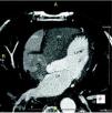
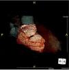
We report the case of a 58-year-old hypertensive man referred for a cardiology consultation due to atypical chest pain. The clinical examination was unremarkable. An electrocardiogram demonstrated sinus rhythm and incomplete right bundle branch block. He had an ambulatory echocardiogram that was described as normal. An exercise test was negative for ischemia and showed intermittent complete right bundle branch block. To exclude coronary disease a contrast computed tomography (CT) scan was performed that revealed a saccular aneurysm of the lateral free wall of the right atrium measuring 45 mm×45 mm×43 mm (Figures 1 and 2). There was no evidence of coronary disease. Given the CT findings, transthoracic echocardiography was repeated and revealed a cystic, pulsatile structure in continuity with the free wall of the right atrium through a narrow neck (Figure 3) permitting low-speed two-way flow, subsequently confirmed by injection of Sonovue® contrast. No cardiac thrombi were visualized. The tricuspid valvular apparatus and right ventricle were morphologically normal, as was biventricular systolic function. Similar findings were visualized on the transesophageal echocardiogram.

Figure 1.

Computed tomography (CT) scan, axial view. LA: left atrium; LV: left ventricle; RA: right atrium; RAA: right atrial aneurysm; RV: right ventricle.

(0.3MB).
Figure 2.

CT scan: three-dimensional reconstruction of the right atrial aneurysm (RAA).

(0.2MB).
Figure 3.

Transthoracic echocardiogram, apical 4-chamber view. LA: left atrium; LV: left ventricle; RA: right atrium; RAA: right atrial aneurysm; RV: right ventricle.

(0.12MB).

Right atrium aneurysm is a rare congenital heart malformation. It occurs in all age groups. Supraventricular arrhythmias and embolic events are the most common reported symptoms. Major complications such as rhythm disturbances, thromboembolic events and aneurysm rupture may occur. Although the best treatment, particularly in asymptomatic patients, has not been established, the patient was started on oral anticoagulation and proposed for surgical aneurysmectomy. He underwent successful resection of the aneurysm (Figure 4) with an uneventful post-operative period.

Figure 4.

Intraoperative photograph (surgeon's view) revealing the right atrial aneurysm (RAA).

(0.15MB).
Ethical disclosuresProtection of human and animal subjects

The authors declare that no experiments were performed on humans or animals for this study.

Confidentiality of data

The authors declare that they have followed the protocols of their work center on the publication of patient data.

Right to privacy and informed consent

The authors have obtained the written informed consent of the patients or subjects mentioned in the article. The corresponding author is in possession of this document.

Conflicts of interest

The authors have no conflicts of interest to declare.

Please cite this article as: da Silva Castro A, Ferreira N, Ferraz R, Bettencourt N, Mota JC, Gama Ribeiro V. Um caso raro de aneurisma congénito da aurícula direita 2014. Rev Port Cardiol. 2014;33:571–572.

Copyright © 2014. Sociedade Portuguesa de Cardiologia
Download PDF
Idiomas
Revista Portuguesa de Cardiologia (English edition)
Article options
Tools
en pt

Are you a health professional able to prescribe or dispense drugs?

Você é um profissional de saúde habilitado a prescrever ou dispensar medicamentos

By checking that you are a health professional, you are stating that you are aware and accept that the Portuguese Journal of Cardiology (RPC) is the Data Controller that processes the personal information of users of its website, with its registered office at Campo Grande, n.º 28, 13.º, 1700-093 Lisbon, telephone 217 970 685 and 217 817 630, fax 217 931 095, and email revista@spc.pt. I declare for all purposes that the information provided herein is accurate and correct.