Sugestões
Partilhar
Informação da revista
Vol. 30. Núm. 10.
Páginas 803 (Outubro 2011)
Vol. 30. Núm. 10.
Páginas 803 (Outubro 2011)
IMAGES IN CARDIOLOGY
Open Access
Prenatal diagnosis of right aortic arch by fetal echocardiography
Diagnóstico pré-natal do arco áortico direito por ecocardiografia fetal
Visitas
7845
Fátima Silva
Autor para correspondência
fatimasilva.15@gmail.com

Corresponding author.
, Cristina Godinho, Ana Carriço
Vila Nova de Gaia Prenatal Diagnosis Centre, Centro Hospitalar Vila Nova de Gaia/Espinho, Vila Nova de Gaia, Portugal
Este item recebeu

Under a Creative Commons license
Informação do artigo
Texto Completo
Baixar PDF
Estatísticas
Figuras (2)
Texto Completo

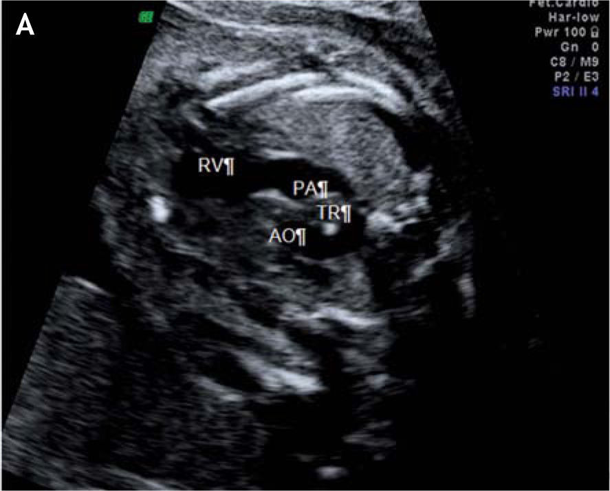
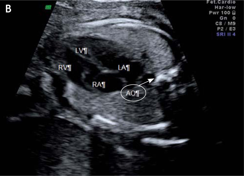
Right aortic arch is a cardiovascular malformation or a variant of normality which can be diagnosed by prenatal ultrasound. This malformation may be associated with DiGeorge syndrome (arising from defects in chromosome 22) and can result in posterior compression of the trachea and esophagus, leading to dysphagia and respiratory impairment after birth.

The authors present images of a case with prenatal diagnosis by echocardiography, in a fetus with right aortic arch as the only structural defect.

A 21-year-old, primigravida, without relevant personal or family history, was referred at 28 weeks of gestation for echocardiographic examination due to fetal arrhythmia, not confirmed. However, in the same exam an isolated right aortic arch was detected (figures A and B). The authors present the images.

Figure A.

Right aortic arch passing posterior to the trachea. AO: aorta; PA: pulmonary artery; RV: right ventricle; SVC: superior vena cava; TR: trachea.

Figure B.

4-chamber view of the fetal heart. AO: aorta; LA: left atrium; LV: left ventricle; RA: right atrium; RV: right ventricle.

Copyright © 2011. Sociedade Portuguesa de Cardiologia
Baixar PDF
Idiomas
Revista Portuguesa de Cardiologia
Opções de artigo
Ferramentas