


Atrial fibrillation (AF) and heart failure (HF) often coexist. AF catheter ablation improves left ventricular ejection fraction (LVEF), but its impact varies between patients. We aimed to identify predictors of LVEF improvement in HF patients with impaired LVEF undergoing AF ablation.
MethodsWe conducted a retrospective single-center study in HF patients with LVEF <50% undergoing AF catheter ablation between May 2016 and May 2022. The primary endpoint was the LVEF recovery rate (‘responders’). Secondary endpoints were one-year safety and effectiveness. We also aimed to validate a prediction model for LVEF recovery.
ResultsThe study included 100 patients (79% male, median age 60 years, 70% with probable tachycardia-induced cardiomyopathy [TIC], mean LVEF 37%, 29% with paroxysmal AF). After a median follow-up of 12 months after catheter ablation, LVEF improved significantly (36±10% vs. 53±10%, p<0.001), with an 82% responder rate. A suspected diagnosis of TIC (OR 4.916 [95% CI 1.166–20.732], p=0.030), shorter QRS duration (OR 0.969 [95% CI 0.945–0.994], p=0.015), and smaller left ventricle (OR 0.893 [95% CI 0.799–0.999], p=0.049) were independently associated with LVEF improvement. Freedom from any documented atrial arrhythmia was 86% (64% under antiarrhythmic drugs), and the rate of adverse events was 2%. The prediction model had a good discriminative performance (AUC 0.814 [95% CI 0.681–0.947]).
ConclusionIn AF patients with HF and impaired LVEF, suspected TIC, shorter QRS duration, and smaller LV diameter were associated with LVEF recovery following AF catheter ablation.
A fibrilhação auricular (FA) e a insuficiência cardíaca (IC) frequentemente coexistem. A ablação de FA melhora a fração de ejeção do ventrículo esquerdo (FEVE), mas o impacto varia entre doentes. O nosso objetivo foi identificar preditores da melhoria da FEVE em doentes com IC e FEVE reduzida submetidos a ablação de FA.
MétodosEstudo retrospetivo unicêntrico de doentes com IC e FEVE < 50% submetidos a ablação de FA entre 05/2016 e 05/2022. O endpoint primário foi a avaliação da taxa de recuperação da FEVE (“Respondedores”). Os endpoints secundários centraram-se na segurança e eficácia a um ano. Também procurámos validar um modelo preditivo de recuperação da FEVE.
ResultadosForam incluídos 100 doentes (79% homens, idade média 60 anos, 70% com provável taquicardiomiopatia, FEVE média 37%, 29% com FA paroxística). Após um seguimento mediano de 12 meses, a FEVE melhorou significativamente (36 ± 10% versus 53 ± 10%, p < 0,001), com 82% de «Respondedores». Suspeita de taquicardiomiopatia (OR 4,916 [95% CI 1,166-20,732], p = 0,030), QRS de menor duração (OR 0,969 [0,945-0,994], p = 0,015) e VE de menor diâmetro (OR 0,893 [0,799-0,999], p = 0,049) associaram-se à melhoria da FEVE. A maioria dos doentes (86%) não apresentou recorrência de arritmia e a taxa de eventos adversos foi de 2%. O modelo preditivo demonstrou bom desempenho (AUC 0,814 [0,681-0,947], 95% CI).
ConclusõesEm doentes com FA e IC com FEVE reduzida, a suspeita de taquicardiomiopatia, QRS de menor duração e menor diâmetro do VE associam-se à recuperação da FEVE após a ablação de FA.
Atrial fibrillation (AF) and heart failure (HF) often coexist, creating a complex cause–effect relationship.1 In HF patients, the development of AF is associated with unfavorable outcomes,2,3 while AF-induced tachycardiomyopathy offers potential reversibility and a better prognosis.4 AF catheter ablation is a well-established therapy to maintain sinus rhythm and alleviate symptoms. The 2020 European guidelines for the diagnosis and management of AF recommend catheter ablation to reverse left ventricular (LV) dysfunction when tachycardiomyopathy is highly probable, regardless of symptoms.5 Catheter ablation has demonstrated the ability to increase LV ejection fraction (LVEF) by restoring sinus rhythm and facilitating reverse cardiac remodeling, although the extent of improvement varies between individuals.6–10 Hence, identifying the subgroup of patients with a higher likelihood of LVEF recovery after catheter ablation is of paramount importance. There are limited national data addressing this issue.10
ObjectivesThis study aims to investigate predictors of LVEF improvement following AF catheter ablation in patients with HF and impaired LVEF.
MethodsStudy design and settingWe conducted a retrospective observational study of consecutive patients with HF and LVEF <50% referred for AF catheter ablation between May 2016 and May 2022 in a tertiary referral center. This study is a continuation of our prior exploratory research which included a small subset of patients.10 All patients provided written informed consent and the study followed the principles of the Declaration of Helsinki.
Patient eligibility criteriaAdult patients referred for AF catheter ablation were eligible for inclusion in the study if they presented the following criteria: LVEF <50% and symptomatic HF. Paroxysmal AF was defined as AF that terminates spontaneously or with intervention (drugs or electrical cardioversion) within seven days. Persistent AF was defined as AF lasting beyond seven days including episodes terminated by intervention, while long-standing persistent AF was defined as continuous AF beyond 12 months with the adoption of a rhythm control strategy. Symptomatic HF was defined as the presence of signs or symptoms typically associated with HF in the presence of LVEF <50%, and the severity of symptoms was described according to the New York Heart Association (NYHA) functional classification.11 Patients with impaired LVEF were categorized into two groups: heart failure with mildly reduced ejection fraction (HFmrEF) with LVEF between 41% and 49%, or heart failure with reduced ejection fraction (HFrEF) with LVEF ≤40%.11 Significant coronary artery disease had to be ruled out by cardiac angiography or stress testing. Patients without severe valvular disease, ischemic cardiomyopathy, hypertrophic cardiomyopathy, neuromuscular disease, congenital heart disease, or other possible etiology, formed the group with suspected tachycardia-induced cardiomyopathy (TIC). To characterize the cohort, relevant data were collected from medical records. This included the echocardiogram performed on the day before ablation and at least six months post-ablation (LVEF, LV end-diastolic diameter [LVEDD], left atrial [LA] volume index) and cardiac magnetic resonance (CMR) measurements performed during the etiologic assessment of HF (LVEF, LV end-diastolic volume, LA area, and the presence of late gadolinium enhancement). Exclusion criteria were redo procedures, contraindication to anticoagulation, and the presence of intracardiac thrombus detected before the ablation procedure.
Ablation procedureDetails of the periprocedural management of AF ablation have been published previously12–14 and are described in detail in the Supplementary Data. The energy source for the procedure (cryoablation or radiofrequency [RF]) was at the discretion of the physician, as was the strategy in patients treated by RF – pulmonary vein (PV) isolation (PVI) or PVI plus ablation of low-voltage areas (these areas were ablated to achieve tissue homogenization and if necessary, lines were performed). All RF procedures were conducted under general anesthesia. In cases of previous history of typical atrial flutter, or if flutter was induced during the procedure, a cavotricuspid isthmus ablation line was also performed. If AF persisted or was inducible at the end of the procedure, electric cardioversion was performed.
Study endpointsThe primary study goal was to determine the number of patients who recovered LVEF after AF catheter ablation and to identify predictors of LVEF improvement. Accordingly, patients were divided into ‘responders’ and ‘non-responders’ in terms of LVEF improvement.15 For patients with HFmrEF, a response was considered positive if LVEF improved to ≥50%, while for patients with HFrEF, a positive response was defined as a ≥10% increase from baseline LVEF, with LVEF reaching >40%. All patients who did not meet these criteria were categorized as non-responders.
Secondary endpoints focused on one-year effectiveness and safety data. Effectiveness was defined as freedom from any documented atrial arrhythmia lasting at least 30 s, irrespective of symptoms, after the three-month blanking period.16 Safety was defined by the occurrence of any of the following events after the index AF ablation: death, major bleeding as defined by the International Society on Thrombosis and Hemostasis,17 any thromboembolic event, atrioesophageal fistula, phrenic nerve palsy, PV stenosis, pericarditis, or vascular access complications.
We also aimed to validate the LVEF recovery prediction model proposed by Bergonti et al.18 This model takes into consideration four variables: established etiology of HF (2 points), QRS >120 ms (2 points), paroxysmal AF (1 point), and LA volume index (1 point).
Follow-upAfter the index procedure, patients were assessed before hospital discharge and at three, six and 12 months. Each appointment included clinical evaluation, electrocardiogram, and 24-hour Holter monitoring. Seven-day Holter monitoring was performed once a year. Additionally, echocardiography was repeated at six or 12 months. No antiarrhythmic drug (AAD) was prescribed after paroxysmal AF ablation, while for persistent AF, AAD prescription was left to the physician's discretion. The first three months post-procedure were considered a blanking period and recurrences in this period were not considered. Anticoagulation therapy was maintained for the first three months and prescribed thereafter according to the CHA2DS2VASc score.19
Statistical analysisContinuous data were described using mean±standard deviation or median and interquartile range, depending on the normality of distribution as assessed by the Kolmogorov–Smirnov test. The Student's t test or the Mann–Whitney U test was used for comparison, depending on normality. Categorical variables were presented as frequency and proportion and compared using Fisher's exact test or the chi-square test. To identify predictors of LVEF improvement, univariate and multivariate logistic regression analyses were conducted. The discriminative performance of the prediction model for LV systolic function recovery in our population was evaluated using the area under the curve (AUC) from receiver operating characteristic (ROC) analysis. Kaplan–Meier curves and the log-rank test were used to assess and compare freedom from AF recurrence during follow-up according to AF type (paroxysmal vs. persistent or long-standing persistent). Statistical significance was set at a two-sided p-value <0.05. Data analysis was performed using IBM SPSS Statistics for Macintosh software, version 20.0 (IBM).
ResultsBaseline characteristicsThe final study sample consisted of 100 patients (79% male, median age 60 [IQR 51–66] years). Baseline characteristics are summarized in Table 1, categorized according to type of HF. Most patients had persistent or long-standing persistent AF (71%). After the exclusion of alternative causes for HF, seventy patients were suspected of having TIC (half of them underwent CMR). Most patients were in NYHA functional class II, while one-third were in class III. Mean LVEF was 37%, with 60% of patients presenting LVEF ≤40% (Table 2). Most patients (71%) were on AADs, of which amiodarone was the most commonly used (Supplementary Table S1).
Baseline characteristics of the study population.
| All patients (n=100) | LVEF ≤40% (n=60) | LVEF 41–49% (n=40) | p | |
|---|---|---|---|---|
| Age, years | 60 (51–66) | 59 (51–67) | 60 (49–65) | 0.381 |
| Male, n (%) | 79 (79.0) | 50 (83.3) | 29 (72.5) | 0.193 |
| Body mass index (kg/m2) | 28.0±3.3 | 27.7±3.1 | 28.5±3.6 | 0.237 |
| CHA2DS2VASc score | 2.1±1.1 | 2.1±1.0 | 2.1±1.3 | 0.815 |
| HAS-BLED score | 1.2±0.9 | 1.3±0.9 | 1.1±1.0 | 0.311 |
| AF pattern, n (%) | ||||
| Persistent | 58 (58.0) | 39 (65.0) | 19 (47.5) | 0.082 |
| Paroxysmal | 29 (29.0) | 13 (21.7) | 16 (40.0) | 0.048 |
| Long-standing persistent | 13 (13.0) | 8 (13.3) | 5 (12.5) | 0.903 |
| Time since AF diagnosis, months | 29 (11–65) | 35 (13–55) | 28 (10–81) | 0.325 |
| Comorbidities, n (%) | ||||
| Type 2 diabetes | 19 (19.0) | 13 (21.7) | 6 (15.0) | 0.405 |
| Hyperlipidemia | 42 (42.0) | 26 (43.3) | 16 (40.0) | 0.741 |
| Hypertension | 61 (61.0) | 37 (61.7) | 24 (60.0) | 0.867 |
| Current smoking | 12 (12.0) | 5 (8.3) | 7 (17.5) | 0.143 |
| Past smoking | 15 (15.0) | 12 (20.0) | 3 (7.5) | 0.086 |
| Excessive alcohol intake | 15 (15.0) | 11 (18.3) | 4 (10.0) | 0.253 |
| Coronary artery disease | 7 (7.0) | 7 (11.7) | 0 | 0.025 |
| Previous stroke | 4 (4.0) | 2 (3.3) | 2 (5.0) | 0.527 |
| Obstructive sleep apnea | 17 (17.0) | 9 (15.0) | 8 (20.0) | 0.514 |
| Obesity (BMI >30 kg/m2) | 21 (21.0) | 9 (14.8) | 12 (30.0) | 0.071 |
| Chronic kidney disease | 10 (10.0) | 5 (8.3) | 5 (12.5) | 0.496 |
| Hyperthyroidism | 8 (8.0) | 4 (6.7) | 4 (10.0) | 0.547 |
| Hypothyroidism | 11 (11.0) | 7 (11.7) | 4 (10.0) | 0.794 |
| HF etiology, n (%) | ||||
| TIC | 70 (70.0) | 43 (71.7) | 27 (67.5) | 0.656 |
| Valvular heart disease | 8 (8.0) | 3 (5.0) | 5 (12.5) | 0.164 |
| Congenital heart disease | 5 (5.0) | 2 (3.3) | 3 (7.5) | 0.314 |
| Ischemic cardiomyopathy | 5 (5.0) | 5 (8.3) | 0 | 0.073 |
| Dilated cardiomyopathy | 5 (5.0) | 5 (8.3) | 0 | 0.073 |
| Hypertrophic cardiomyopathy | 4 (4.0) | 1 (1.7) | 3 (7.5) | 0.174 |
| LVNC cardiomyopathy | 2 (2.0) | 0 | 2 (5.0) | 0.158 |
| Myocarditis | 1 (1.0) | 1 (1.7) | 0 | 0.600 |
| NYHA class, n (%) | ||||
| I | 17 (17.0) | 5 (8.3) | 12 (30.0) | 0.005 |
| II | 50 (50.0) | 29 (48.3) | 21 (52.5) | 0.683 |
| III | 32 (32.0) | 25 (41.7) | 7 (17.5) | 0.011 |
| IV | 1 (1.0) | 1 (1.7) | 0 | 0.600 |
| Device, n (%) | ||||
| CRT | 9 (9.0) | 7 (11.7) | 2 (5.0) | 0.220 |
| ICD | 6 (6.0) | 4 (6.7) | 2 (5.0) | 0.544 |
| Pacemaker | 3 (3.0) | 2 (3.3) | 1 (2.5) | 0.649 |
| Biochemistry parameters | ||||
| Hemoglobin (g/dl) | 14.1±1.6 | 14.3±1.3 | 13.8±1.8 | 0.150 |
| Creatinine (mg/dl) | 0.84 (0.74–1.10) | 0.85 (0.78–1.10) | 0.81 (0.72–0.99) | 0.113 |
| GFR (ml/min) | 99.8±38.4 | 97.3±38.0 | 103.8±39.3 | 0.456 |
| CRP (mg/dl) | 0.23 (0.10–0.41) | 0.18 (0.09–0.36) | 0.32 (0.11–0.49) | 0.100 |
| hs-TnI (ng/l) | 4.4 (2.6–10.0) | 4.4 (2.6–10.8) | 4.4 (2.0–8.6) | 0.559 |
| BNP (pg/ml) | 133.0 (53.5–392.0) | 156.5 (50.5–450.5) | 118.5 (85.2–391.5) | 0.969 |
| TSH (μIU/ml) | 1.75 (1.10–3.10) | 1.70 (1.15–2.85) | 2.10 (1.10–3.50) | 0.585 |
| Thyroxine (T4) (ng/l) | 1.05 (0.90–1.20) | 1.04 (0.95–1.20) | 1.08 (0.90–1.19) | 0.993 |
| QRS duration, ms | 116±26 | 116±26 | 115±26 | 0.832 |
AF: atrial fibrillation; BMI: body mass index; BNP: brain-type natriuretic peptide; CRP: high-sensitivity C-reactive protein; CRT: cardiac resynchronization therapy; GFR: glomerular filtration rate; hs-TnI: high-sensitivity troponin I; ICD: implantable cardioverter-defibrillator; LVNC: left ventricular noncompaction; NYHA: New York Heart Association; TIC: tachycardia-induced cardiomyopathy; TSH: thyroid-stimulating hormone.
Echocardiographic variables.
| All patients (n=100) | LVEF ≤40% (n=60) | LVEF 41–49% (n=40) | p | |
|---|---|---|---|---|
| LVEF, % | 36±9 | 30±7 | 46±2 | <0.001 |
| LVEDD, mm | 56±6 | 58±6 | 53±4 | <0.001 |
| LA volume, ml/m2 | 45±13 | 47±15 | 44±10 | 0.374 |
LA: left atrial; LVEDD: left ventricular end-diastolic diameter; LVEF: left ventricular ejection fraction.
Baseline characteristics were similar between these groups (Table 1). Patients with HFrEF presented a lower prevalence of paroxysmal AF (21.7 vs. 40.0%, p=0.048) and less frequent history of coronary artery disease (11.7 vs. 0%, p=0.025), while having more severe symptoms (NYHA III) (41.7 vs. 17.5, p=0.011). There were no significant differences between groups in brain-type natriuretic peptide levels (156.5 [50.5–450.5] vs. 118.5 [(85.2–391.5] pg/ml, p=0.969). Patients with LVEF ≤40% had greater LVEDD (58±6 vs. 53±4 mm, p<0.001) but similar LA volume (47±15 vs. 44±10 ml/m2, p=0.37) compared to patients with HFmrEF (Table 2).
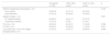
Procedural details and complicationsTable 3 provides an overview of the procedural details. Although 71% of the patients had previously undergone electrical cardioversion (those with persistent or long-standing persistent AF), only 53% were in sinus rhythm at the beginning of the procedure. Most patients (90%) underwent RF-guided ablation and 61% underwent additional ablation besides PVI. At the end of the procedure, 75% of patients were in sinus rhythm, the remainder requiring electrical cardioversion.
Procedural characteristics.
| All patients (n=100) | LVEF ≤40% (n=60) | LVEF 41–49% (n=40) | p | |
|---|---|---|---|---|
| Rhythm at beginning of procedure, n (%) | 0.744 | |||
| Sinus rhythm | 53 (53.0) | 31 (51.7) | 22 (55.0) | |
| Atrial fibrillation | 47 (47.0) | 29 (48.3) | 18 (45.0) | |
| Ablation energy, n (%) | 0.361 | |||
| RF catheter ablation | 90 (90.0) | 55 (91.7) | 37 (87.5) | |
| Cryoballoon ablation | 10 (10.0) | 5 (8.3) | 5 (12.5) | |
| PVI only, n (%) | 39 (39.0) | 22 (36.7) | 17 (42.5) | 0.558 |
| CTI line, n (%) | 50 (50.0) | 33 (55.0) | 17 (42.5) | 0.221 |
| Lines other than CTI or low-voltage homogenization,n (%) | 61 (61.0) | 37 (61.7) | 24 (60.0) | 0.998 |
CTI: cavotricuspid isthmus; LVEF: left ventricular ejection fraction; PVI: pulmonary vein isolation; RF: radiofrequency.
Most patients were discharged under AADs (51%), mainly amiodarone (28%), followed by flecainide (18%), propafenone (4%), and sotalol (1%). Medication for HF is detailed in Supplementary Table S2.
Improvement after catheter ablation of atrial fibrillationAfter a median follow-up of 12 (IQR 7–18) months, there was a significant improvement in LVEF (36±10 vs. 53±10%, p<0.001), with 82% of patients meeting the criteria for responders. The mean absolute increase in LVEF from baseline to the subsequent re-evaluation was 17±13%. A suspected diagnosis of TIC was associated with a substantial increase in LVEF compared to patients with other HF etiologies (21±12% vs. 9±12%, p<0.001). Patients without new episodes of atrial arrhythmia exhibited a significant improvement in LVEF compared to those who experienced recurrences during the follow-up period (22±13 vs. 14±10%, p=0.038).
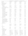
The predictors of LVEF improvement following AF catheter ablation are detailed in Table 4. Multivariate analysis revealed that a suspected diagnosis of TIC rather than another HF etiology (OR 4.916 [95% CI 1.166–20.732], p=0.030), shorter QRS duration (OR 0.969 [95% CI 0.945–0.994], p=0.015) and smaller LVEDD (OR 0.893 [95% CI 0.799–0.999], p=0.049) were associated with LVEF improvement.
Univariate and multivariate predictors of left ventricular ejection fraction improvement after atrial fibrillation ablation.
| Univariate model | Multivariate model | |||
|---|---|---|---|---|
| OR (95% CI) | p | OR (95% CI) | p | |
| Age | 1.026 (0.982–1.072) | 0.248 | ||
| Male gender | 3.630 (0.439–30.004) | 0.232 | ||
| BMI | 1.020 (0.854–1.218) | 0.827 | ||
| CHA2DS2VASc score | 1.346 (0.791–2.291) | 0.274 | ||
| HAS-BLED score | 1.196 (0.632–2.263) | 0.582 | ||
| AF pattern | ||||
| Persistent | 0.461 (0.133–1.592) | 0.221 | ||
| Paroxysmal | 0.990 (0.279–3.507) | 0.988 | ||
| Long-standing persistent | 0.978 (0.854–1.621) | 0.982 | ||
| Time since AF diagnosis | 0.997 (0.986–1.008) | 0.621 | ||
| Comorbidities | ||||
| Type 2 diabetes | 1.538 (0.422–5.612) | 0.514 | ||
| Hyperlipidemia | 0.519 (0.150–1.799) | 0.301 | ||
| Hypertension | 0.664 (0.215–2.052) | 0.477 | ||
| Current smoking | 0.992 (0.191–5.140) | 0.992 | ||
| Past smoking | 0.992 (0.191–5.140) | 0.992 | ||
| Excessive alcohol intake | 1.009 (0.195–5.229) | 0.992 | ||
| Coronary artery disease | 5.417 (0.975–30.090) | 0.053 | ||
| Previous stroke | 0.365 (0.218–1.516) | 0.281 | ||
| Obstructive sleep apnea | 0.651 (0.131–3.246) | 0.600 | ||
| Obesity (BMI >30 kg/m2) | 4.308 (0.525–35.343) | 0.174 | ||
| Chronic kidney disease | 2.583 (0.566–11.782) | 0.220 | ||
| Hyperthyroidism | 0.406 (0.067–2.455) | 0.326 | ||
| Hypothyroidism | 0.271 (0.043–1.727) | 0.167 | ||
| TIC | 7.639 (2.156–27.061) | 0.002 | 4.916 (1.166–20.732) | 0.030 |
| NYHA class | ||||
| I | 0.690 (0.165–2.888) | 0.611 | ||
| II | 1.886 (0.583–6.101) | 0.290 | ||
| III | 0.623 (0.201–1.929) | 0.412 | ||
| IV | 0.511 (0.148–1.322) | 0.718 | ||
| Device | ||||
| CRT | 0.250 (0.050–1.262) | 0.093 | ||
| ICD | 0.406 (0.067–2.455) | 0.326 | ||
| Pacemaker | 0.866 (0.335–3.124) | 0.788 | ||
| Biochemistry parameters | ||||
| Hemoglobin | 1.188 (0.844–1.670) | 0.323 | ||
| Creatinine | 0.784 (0.406–1.513) | 0.468 | ||
| GFR | 1.009 (0.993–1.025) | 0.274 | ||
| CRP | 0.573 (0.284–1.156) | 0.120 | ||
| hs-TnI | 1.000 (0.998–1.003) | 0.807 | ||
| BNP | 1.000 (1.000–1.000) | 0.381 | ||
| TSH | 0.961 (0.828–1.114) | 0.596 | ||
| Thyroxine (T4) | 0.546 (0.040–7.361) | 0.648 | ||
| QRS duration | 0.961 (0.940–0.983) | 0.001 | 0.969 (0.945–0.994) | 0.015 |
| Echocardiographic variables | ||||
| LVEF | 1.009 (0.956–1.065) | 0.755 | ||
| LVEDD | 0.906 (0.829–0.991) | 0.031 | 0.893 (0.799–0.999) | 0.049 |
| LA volume | 0.995 (0.941–1.053) | 0.873 | ||
| Procedural characteristics | ||||
| Rhythm at beginning of procedure | 0.679 (0.321–1.462) | 0.572 | ||
| RF catheter ablation | 1.938 (0.338–11.101) | 0.457 | ||
| Cryoballoon ablation | 0.516 (0.090–2.954) | 0.457 | ||
| PVI only | 0.566 (0.168–1.911) | 0.359 | ||
| CTI line | 2.250 (0.695–7.281) | 0.176 | ||
| Lines (others than CTI) or low-voltage homogenization | 0.429 (0.124–1.483) | 0.181 | ||
AF: atrial fibrillation; BMI: body mass index; BNP: B-type natriuretic peptide; CI: confidence interval; CRP: high-sensitivity C-reactive protein; CRT: cardiac resynchronization therapy; CTI: cavotricuspid isthmus; GFR: glomerular filtration rate; hs-TnI: high-sensitivity troponin I; ICD: implantable cardioverter-defibrillator; LA: left atrial; LVEDD: left ventricular end-diastolic diameter; LVEF: left ventricular ejection fraction; NYHA: New York Heart Association; OR: odds ratio; PVI: pulmonary vein isolation; RF: radiofrequency; TIC: tachycardia-induced cardiomyopathy; TSH: thyroid-stimulating hormone.
The mean score derived from Bergonti et al.’s prediction model18 was 1.98±1.78, with a higher score being significantly associated with a lack of LVEF improvement (3.75±1.39 in non-responders vs. 1.79±1.90 in responders (p=0.004)). Among subjects with a score ≤1, the majority (82%) were responders. In our study population, the scoring system demonstrated good ability to discriminate between responders and non-responders, as evidenced by an area under the curve (AUC) of 0.814 (0.681–0.947, 95% CI) (Figure 1).
![Receiver operating characteristic analysis demonstrating good discriminative power between responders and non-responders with Bergonti et al.’s scoring system18 (area under the curve [AUC] 0.814, 95% confidence interval 0.681–0.947).](https://static.elsevier.es/multimedia/08702551/0000004300000011/v1_202410292055/S087025512400115X/v1_202410292055/en/main.assets/gr1.jpeg?xkr=ue/ImdikoIMrsJoerZ+w94UphxYc+GPca8Z7OggvdfJQF4SIqTc4zp8SrbcUWBiK2zRV+4/JKcouWkO4IeVBdTd/wm35wdmHMa8XooXbH7OXiv4uAJd0RWEx9yB+2jATg+XvpP0GcjqtlSdJ8ImLOhCSCtBsHantaRoQ0ZTL2FfUnqoQ4ct4n1mSQ7blqAoJCX+UkQm8RwcFNl5GVE5fGWXLl1BHkYv8zimCr/+0P0qpIMcPazPnNTehg4fYlUnKpBkAm40+I0eA8aJ9G7AbBUJ4MlZnCFzs1Mpp0DmmT6M= "Receiver operating characteristic analysis demonstrating good discriminative power between responders and non-responders with Bergonti et al.’s scoring system18 (area under the curve [AUC] 0.814, 95% confidence interval 0.681–0.947).")
Receiver operating characteristic analysis demonstrating good discriminative power between responders and non-responders with Bergonti et al.’s scoring system18 (area under the curve [AUC] 0.814, 95% confidence interval 0.681–0.947).
After a median follow-up of 12 (IQR 7–18) months (Figure 2), freedom from arrhythmia recurrence was observed in 86% of the patients (64% under AADs). The median time to recurrence was 7.5 (6.0–9.0) months. There was no statistically significant difference in atrial arrhythmia recurrence rate between patients with paroxysmal vs. persistent or long-standing persistent AF (11.3 vs. 20.7%, p=0.293). Among those in whom atrial arrhythmia recurred, nine patients had AF (five persistent and four paroxysmal), while the remainder presented atrial tachycardia. The most frequent strategy involved consisted of initiating or increasing the AAD dose (64.4%), while 21.4% of them underwent another catheter ablation procedure. No predictors of recurrence were identified (Supplementary Table S2).
 Recurrence was observed in 14% of the overall cohort at 12 months of follow-up; (B) there was no significant difference in recurrence rate between patients with paroxysmal vs. persistent or long-standing persistent AF (11.3 vs. 20.7%, p=0.293). AF: atrial fibrillation.")
Freedom from atrial fibrillation over time. (A) Recurrence was observed in 14% of the overall cohort at 12 months of follow-up; (B) there was no significant difference in recurrence rate between patients with paroxysmal vs. persistent or long-standing persistent AF (11.3 vs. 20.7%, p=0.293). AF: atrial fibrillation.
There were only two reported complications (vascular fistula and hematoma), both managed conservatively.
DiscussionTo our knowledge, this is the first national study to analyze predictors for LVEF improvement after AF ablation in patients with HF and impaired LVEF. Our findings suggest that (1) most patients recovered LV function, and a suspected diagnosis of TIC, shorter QRS duration, and smaller LVEDD were independently associated with LVEF improvement; (2) the newly proposed scoring system18 aiming to predict LVEF recovery after AF ablation demonstrated good discrimination in our population; (3) the effectiveness of AF ablation in this subset of patients persisted over one year of follow-up, with a low rate of adverse events.
Our study highlights the beneficial impact of AF catheter ablation on LVEF improvement in patients with HF. Although in the AMICA trial no difference was found in outcomes between catheter ablation and medical treatment,8 several other clinical trials supported the benefit of catheter ablation in comparison to medical therapy in patients with HF.6,7,9,20 Although our study did not set out to compare these two strategies, it was notable that 82% of the patients who underwent catheter ablation were responders and mean LVEF improved by 17% at one year of follow-up, despite guideline-directed medical therapy. This improvement may be attributed, in part, to the higher proportion of patients with a suspected diagnosis of TIC, leading to an increased likelihood of substantial LVEF recovery.11 On the other hand, it may also be explained by the low recurrence rate observed in our cohort, which may have contributed to the high rate of responders. In the CASTE-AF trial, patients undergoing AF ablation spent longer in sinus rhythm and therefore had a better outcome than those under medical therapy.6 In animal studies, inducing persistent tachycardia through continuous rapid atrial or ventricular pacing resulted in LV systolic dysfunction, while interrupting pacing promoted LVEF recovery. Within these models, impairment of excitation-contraction coupling and diastolic function can be attributed to factors such as subclinical ischemia, disturbances in energy metabolism and mitochondrial function, oxidative stress, and abnormal calcium homeostasis.21,22 Additional research is needed to elucidate the mechanisms underlying both impairment and restoration of systolic function.
Identifying patients with AF and HF who may benefit from catheter ablation is of paramount importance, since catheter ablation is an invasive procedure and the degree of LVEF improvement varies between patients. Recently, Bergonti et al. introduced a novel model18 with several parameters (HF etiology, QRS duration, AF type, and LA volume index) for predicting LV systolic function recovery following AF catheter ablation in patients with HF. This score presented a good discriminative value in our population, with some of the variables included in the model being independent predictors of LVEF improvement, including suspected TIC or shorter QRS duration. As previously mentioned, TIC was associated with an increased likelihood of LVEF improvement,11,21,22 probably due to a lesser degree of fibrosis,23 which has previously been demonstrated to be associated with LVEF recovery.24–26 QRS duration was also associated with LVEF improvement in our study. It is well known that a wide QRS complex is associated with the extent of myocardial scar in both ischemic and nonischemic cardiac disease,27 promoting mechanical dyssynchrony in patients with HF.28 Hence, a wider QRS complex might indicate more profound and enduring structural alterations that are less amenable to remodeling after AF catheter ablation.18 However, in our study LA volume index and persistent AF were not predictors of LVEF improvement, while LV diameter was. In Bergonti et al.’s study, LV dilatation was associated with LVEF recovery, but only in univariate analysis.18 Similarly to our findings, Ukita et al. found that a smaller LVEDD predicted LVEF improvement after AF catheter ablation.30 Compared to Bergonti et al.’ sample,18 our study had fewer patients with severe LA dilatation (29% vs. 72%) and fewer with persistent AF (71% vs. 82%), which may explain the lack of predictive power of these variables in our study. However, it should be borne in mind that both studies included a limited sample of patients, and therefore more data are required from larger multicenter randomized studies.
Our study presented good freedom from arrhythmia recurrence at one-year follow-up, despite 71% of the patients presenting persistent or long-standing persistent AF, which is usually associated with greater arrhythmia recurrence.29,30 Several factors may explain our results: (1) a significant percentage of patients had probable TIC, which is associated with better outcomes, including reverse atrial and ventricular remodeling4,10,22; (2) a tailored approach has been associated with high medium-term effectiveness in patients with persistent AF12,31,32; (3) a significant proportion of responders, who normally have reverse atrial and ventricular remodeling and therefore present a better outcome33,34; and (4) nearly two-thirds of patients were still on AADs at one-year follow-up. The latter probably derives from the concern that potential AF recurrence could lead once again to LVEF deterioration and HF symptoms. Importantly, even in this population with low ejection fraction, catheter ablation was a safe procedure with a low rate of adverse events, in line with data from other studies.35
Study limitationsWe acknowledge several limitations inherent to our work. First, this was a single-center study and therefore our results cannot be generalized to other centers. Although the sample size of our study was relatively modest, it was similar to that of other studies analyzing predictors of LVEF improvement.18,36 Larger clinical trials are required to verify our results. Second, we acknowledge that not all patients underwent CMR and therefore we might have erroneously categorized some subjects as having a suspected diagnosis of TIC. Third, we recognize that allowing patients to undergo electrical cardioversion before ablation could have affected our results. The goal was to reduce time in AF, which has an impact on AF ablation outcomes.37 Nevertheless, since echocardiographic assessment was performed on the day before ablation, the impact of this measure was limited. Fourth, we acknowledge that arrhythmia recurrence was underestimated in our study, since our assessment relied on 24-hour and seven-day Holter monitoring. However, as demonstrated in CASTLE-AF,6 perhaps more important than the arrhythmia recurrence rate is reduction in arrhythmia burden. The assessment of arrhythmia burden in our study was limited, since only 17% of patients had an implantable device, precluding a comprehensive analysis. Finally, there will always be a degree of intra- and interobserver variability in LVEF measurement. Nonetheless, LVEF continues to be the primary parameter employed to assess LV systolic function in clinical settings, providing crucial therapeutic guidance and prognostic insights across a range of cardiac conditions, including AF.38
ConclusionsIn patients with impaired LVEF and HF, a suspected diagnosis of TIC, shorter QRS duration, and smaller LV diameter were associated with LVEF improvement following AF catheter ablation.
Conflicts of interestP.A.S. has received consulting fees from Abbott, Biosense Webster, Boston Scientific, and Medtronic. Natália António has received consulting fees from Microport and Medtronic. All other authors have reported that they have no relationships relevant to the contents of this paper to disclose.
The following are the supplementary data to this article:

