


A 61-year-old woman was referred to our hospital for assessment of a cardiac mass depicted by transthoracic echocardiography (TTE). She had a medical history of stroke 10 years prior to admission and reported new-onset fatigue, with no other cardiovascular symptoms.
TTE revealed a highly mobile round 1.4 cm×1.4 cm isoechogenic mass at the mid left ventricular (LV) level with no obvious insertion base (Figure 1A-C). LV ejection fraction was normal and the valves had no pathological changes. For better characterization of the anatomy and mass we performed three-dimensional echocardiography (Figure 1D) which showed the mass attached to the base of the anterolateral papillary muscle. The myocardium underlying the mass appeared normal, with no signs of infiltration. Given the high predicted embolic risk, the patient underwent surgical excision of the mass. The pathological diagnosis was papillary fibroelastoma (Figure 1E and F). The patient's postoperative course was uneventful and two months after surgery she was asymptomatic with no residual mass on echocardiographic reassessment.
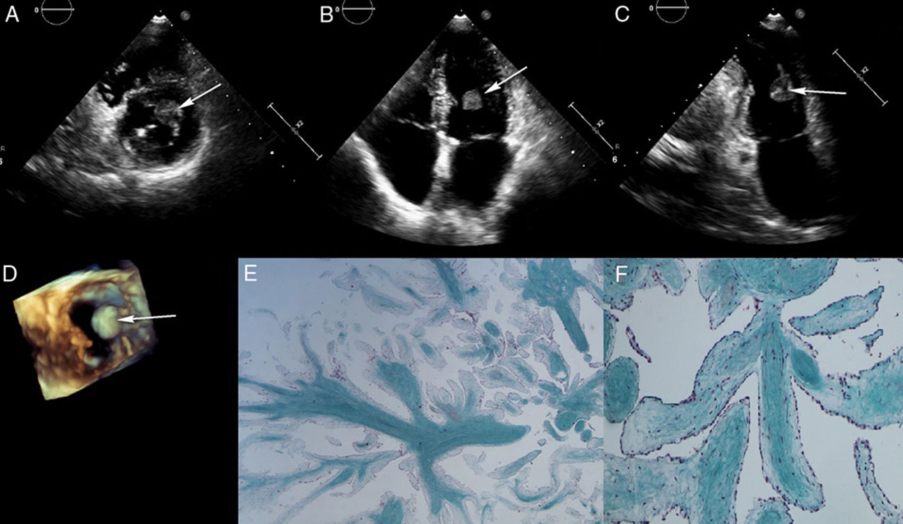
(A-C) Short-axis, apical 4- and 2-chamber transthoracic echocardiographic images, respectively, showing a round mass inside the left ventricle, attached to the papillary muscle (arrow); (D) three-dimensional echocardiographic short-axis view using the 2-Click Crop function from the apex; (E and F) histopathology; (E) neoform of papillary architecture (Masson's trichrome, 25×); (F) papillary axes consisting of collagen, stained green, and lax stroma with endothelial/endocardial coating (Masson's trichrome, 100×).
Papillary fibroelastoma is a rare benign tumor that accounts for fewer than 10% of primary cardiac tumors. These tumors can arise anywhere in the heart, but usually involve the surface of the aortic and mitral valves, and rarely the atria or ventricles. We described a rare case of a papillary fibroelastoma originating from the anterolateral papillary muscle. Surgical resection is generally curative.
Conflicts of interestThe authors have no conflicts of interest to declare.
