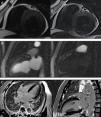
A 63‐year‐old man was admitted for non‐ST‐elevation myocardial infarction. Echocardiography showed preserved biventricular systolic function with apical hypokinesia. Apical 4‐chamber color Doppler view revealed abnormal diastolic flow, apparently intramyocardial (Figure 1A and Video 1). Coronary angiography revealed moderate (60%) coronary stenosis in the mid right coronary artery (Figure 1B), with a fractional flow reserve of 0.91, compatible with a functionally non‐significant lesion. Multiple small fistulae originating from both the right and left coronary arteries were observed, draining into the left ventricle (Figure 1B and C and Videos 2 and 3). Cardiac magnetic resonance imaging (MRI) provided further characterization of these fistulae, which were visible in T2‐weighted sequences in short axis view in the anterior and posterior interventricular grooves (Figure 2A). First‐pass myocardial rest perfusion imaging in two‐chamber view revealed epicardial hyperenhancement of the anterior wall simultaneous with aortic opacification (Figure 2B and Video 4), as well as earlier perfusion of the mid and apical anterior segments. There was subendocardial late gadolinium enhancement (LGE) in the apical inferior segment, compatible with non‐transmural myocardial infarction (Figure 2C). Dobutamine stress echocardiography under optimized therapy showed no inducible ischemia and the patient was discharged, and has remained stable ever since, now at two‐year follow‐up.
(A) Two‐dimensional echocardiography in 4‐chamber color Doppler view showing abnormal intramyocardial diastolic flow in the apical lateral segment (arrow); (B) selective right coronary angiography showing 60% stenosis in the mid right coronary artery and multiple coronary fistulae arising from its distal segment and draining into the left ventricle (arrow); (C) selective left coronary angiography showing multiple coronary fistulae arising from the distal segments of the left coronary artery, also draining into the left ventricular cavity (arrow).
(A) Short‐axis T2‐weighted images with multiple unexpected small vascular structures in the anterior and posterior interventricular grooves; (B) first‐pass myocardial rest perfusion imaging in 2‐chamber view, showing epicardial hyperenhancement of the anterior wall simultaneous with aortic opacification (left) and earlier perfusion of the mid and apical anterior segments (right); (C) subendocardial late gadolinium enhancement in the apical inferior myocardial segment, in 4‐chamber and 2‐chamber views.
Multiple coronary fistulae constitute a very rare entity and probably correspond to the persistence of embryonic myocardial sinusoids. They are an uncommon cause of subendocardial ischemia, due to a coronary steal phenomenon.
This case highlights the role of multimodality imaging in the presence of such coronary anomalies: echocardiography raised the suspicion, coronary angiography made the diagnosis and cardiac MRI provided further characterization of anatomy, physiology (rest perfusion) and myocardial structure (LGE).
To our knowledge this is the first report describing the anatomical appearance of multiple coronary fistulae in T2‐weighted images as well as their peculiar pattern on first‐pass myocardial rest perfusion imaging.
Ethical disclosuresProtection of human and animal subjectsThe authors declare that no experiments were performed on humans or animals for this study.
Confidentiality of dataThe authors declare that they have followed the protocols of their work center on the publication of patient data and that all the patients included in the study received sufficient information and gave their written informed consent to participate in the study.
Right to privacy and informed consentThe authors have obtained the written informed consent of the patients or subjects mentioned in the article. The corresponding author is in possession of this document.
Conflicts of interestThe authors have no conflicts of interest to declare.
The following are the supplementary data to this article:


