


A 65-year-old man had undergone percutaneous revascularization of the left anterior descending artery four years earlier, with two non-overlapping stents being implanted in the same procedure: a first-generation drug-eluting stent (DES) in the proximal segment (2.75 mm×16 mm TAXUS Element paclitaxel-eluting stent) and a second-generation DES in the mid segment (2.5 mm×24 mm PROMUS Element everolimus-eluting stent). The patient underwent new coronary angiography for effort angina, which demonstrated diffuse restenosis of the proximal stent, with a 71% obstructive lesion in the mid portion (Figure 1A).
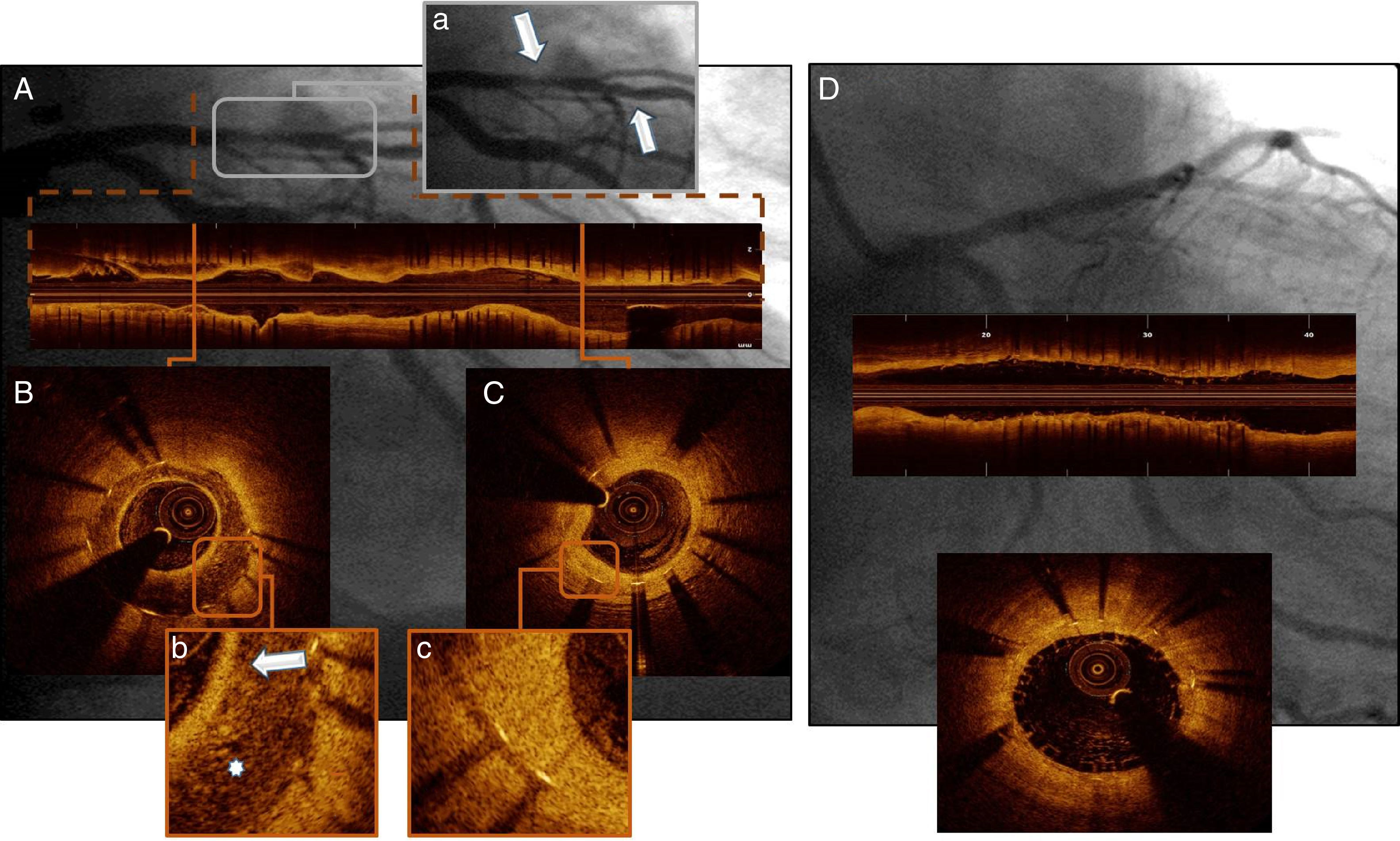
Initial coronary angiography (A, a) showing diffuse restenosis of the proximal stent and a good late result of the distal stent (arrows); optical coherence tomography (OCT) demonstrating a heterogeneous hypointensive neointima with a bright superficial layer (arrow) and presence of macrophage infiltration and microvessels (asterisk) into the neointima in the first-generation DES (B and b), and a homogeneous hyperintensive neointima in the second-generation DES (C and c); OCT post-stent (D) showing good expansion and apposition of the bioresorbable scaffold.
Optical coherence tomography demonstrated two different patterns of in-stent coverage in the two stents: mild proliferation of a homogeneous hyperintense neointima highly suggestive of classic (‘healthy’) neointimal thickening in the distal stent, with minimal luminal area (MLA) of 3.8 mm2 (Figure 1C and c); and a heterogeneous hypointense neointima with low attenuation suggesting an active inflammatory process causing severe restenosis of the TAXUS stent (MLA 2.1 mm2) (Figure 1B and b).
Following predilatation, an Absorb everolimus-eluting bioresorbable scaffold (BRS) (Abbott Vascular, CA) was implanted into the proximal stent, with good results (Figure 1D).
This case reveals a process of restenosis in a first-generation DES and normal neointimal coverage in a second-generation DES implanted in a single patient and artery in the same procedure.
Conflicts of interestThe authors have no conflicts of interest to declare.
