


Post-COVID-19 condition or “Long COVID” is a complex heterogeneous entity characterized by incomplete recovery after acute COVID-19 infection. Prevalence of chest pain varies from 21% to 53% and the potential pathophysiological mechanisms include myocardial infarction, myocarditis and pericarditis.1 Preliminary reports suggest that microvascular dysfunction may be a cause of chest pain in these patients.2,3 However, the extent of microvascular dysfunction contribution in relation to other factors causing chest pain in patients with long COVID has not been thoroughly characterized.
This prospective study included consecutive patients with long COVID reporting typical chest pain, from February 2021 to May 2023. The study was approved by the institution's human research committee (PI-21-065). Epicardial coronary artery disease was ruled out via coronary computed tomography angiography. Adenosine stress cardiac magnetic resonance (CMR) was performed in a 1.5 Tesla scanner. Medis software was used for CMR data analysis. Microvascular dysfunction was considered positive if first-pass stress perfusion showed a significant circumferential subendocardial perfusion defect. Statistical analysis was conducted using SPSS v.28, with significance set at p<0.05.
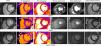
Of the 548 individuals with long COVID-19 symptoms at our center, 59 patients reported typical chest pain and underwent stress CMR. Median time between COVID infection and CMR was 13 months. Mean age was 46.9 years, and the majority were female (71%), and a low prevalence of cardiovascular risk factors was observed (Table 1). Most patients had experienced a previous mild form of COVID-19 infection. Twenty-three patients (39%) were positive for microvascular dysfunction (Case A from Figure 1). No significant differences were observed in clinical or risk factors between patients with and without microvascular dysfunction. There were no disparities in cardiac chamber dimensions, myocardial T1 mapping or extracellular volume between the two groups. While all patients exhibited T2 mapping values within the normal range, those without microvascular dysfunction presented a slightly higher T2 mapping (47.6 ms compared to 45.2 ms, p=0.022). LGE was present in five patients (8.5%). The most common pattern of LGE was epicardial (three patients – see Case B from Figure 1). In five patients (8.5%), CMR data suggested the presence of prior myocarditis. Neither myocardial infarction nor pericardial disease were detected in any case.
Clinical and cardiac magnetic resonance findings.
| Cohort(n=59) | No microvascular dysfunction(n=36) | Microvascular dysfunction(n=23) | p-Value | |
|---|---|---|---|---|
| Age (years) | 46.9 (8.9) | 45.9 (9.6) | 48.4 (7.6) | 0.310 |
| Sex (male) | 17.0 (28.8) | 13.0 (36.1) | 4.0 (17.4) | 0.150 |
| BMI (kg/m2) | 27.2 (5.6) | 27.4 (6.1) | 27.0 (4.9) | 0.825 |
| Family history of coronary artery disease | 10.0 (16.9) | 5.0 (13.9) | 5.0 (21.7) | 0.490 |
| Smoking | 20.0 (23.9) | 11.0 (30.6) | 9.0 (39.1) | 0.551 |
| Hypertension | 11.0 (18.6) | 5.0 (13.9) | 6.0 (26.1) | 0.310 |
| Dyslipidemia | 10.0 (16.9) | 7.0 (19.4) | 3.0 (13.0) | 0.725 |
| Type 2 diabetes | 5.0 (8.5) | 4.0 (11.1) | 1.0 (4.3) | 0.639 |
| COVID-related inappropriate tachycardia | 15.0 (25.4) | 9.0 (25.0) | 6.0 (26.1) | 0.925 |
| Asthma | 1.0 (1.7) | 1.0 (2.8) | 0.0 (0.0) | 0.610 |
| CMR findings | ||||
| LVDVi (ml/m2) | 74.0 (17.4) | 71.6 (18.3) | 77.8 (15.6) | 0.188 |
| LVSVi (ml/m2) | 28.3 (9.2) | 28.1 (9.8) | 28.8 (8.2) | 0.779 |
| LVEF (%) | 62.5 (5.4) | 63.0 (8.0) | 63.0 (6.0) | 0.629 |
| LVMi (g/m2) | 55.3 (24.1) | 55.2 (21.1) | 64.9 (26.4) | 0.351 |
| RVDVi (ml/m2) | 72.4 (19.1) | 68.8 (19.0) | 78.2 (18.1) | 0.065 |
| RVSVi (ml/m2) | 29.0 (14.5) | 27.9 (10.0) | 30.8 (9.3) | 0.280 |
| RVEF (%) | 60.3 (8.0) | 59.9 (5.9) | 61.0 (6.1) | 0.497 |
| T1 mapping (ms) | 989.0 (49.0) | 984.0 (66.0) | 991.0 (38.0) | 0.731 |
| T2 mapping (ms) | 46.7 (4.2) | 47.6 (4.7) | 45.2 (2.8) | 0.022 |
| ECV (%) | 25.0 (3.7) | 24.8 (3.2) | 25.2 (4.4) | 0.702 |
| LGE | 5.0 (8.5) | 3.0 (8.3) | 2.0 (8.7) | 0.654 |
| Epicardic | 3.0 (5.1) | 2.0 (66.7) | 1.0 (50.0) | |
| Mid-myocardial | 2.0 (3.4) | 1.0 (33.3) | 1.0 (50.0) | |
BMI: body mass index; CMR: cardiovascular magnetic resonance; COVID: coronavirus disease; ECV: extracellular volume; LGE: late gadolinium enhancement; LVDVi: indexed left ventricular end-diastolic volume; LVEF: left ventricular ejection fraction; LVMi: left ventricular mass index; LVSVi: indexed left ventricular end-systolic volume; RVDVi: indexed right ventricular end-diastolic volume; RVEF: right ventricular ejection fraction; RVSVi: indexed right ventricular end-systolic volume.

Among patients with long COVID-19 and typical chest pain, the prevalence of microvascular dysfunction, as assessed by stress CMR, is 39%. Furthermore, 8.5% patients exhibited sequelae of previous myocarditis, while none displayed myocardial infarction or pericardial disease. Given the absence of cardiac structural or functional differences between patients with and without microvascular dysfunction, it is plausible that an immune-mediated inflammatory mechanism may underlie these findings.4 In patients without COVID-19 infection, a recent meta-analysis showed a pooled prevalence of 43% microvascular dysfunction in a general cohort of patients without obstructive coronary artery disease.5 Therefore, further research is needed to ascertain causality rather than just the coexistence of microvascular dysfunction and long COVID symptoms.
Ethical approvalGermans Trias I Pujol Hospital's human research committee (PI-21-065).
Conflicts of interestAntoni Bayes-Genis reports personal fees and/or advisory board from AstraZeneca, Novartis, Boehringer Ingelheim, Abbott, Roche Diagnostics, and Vifor Pharma.
Victoria Delgado has received speaker fees from Abbott Vascular, Medtronic, Edwards Lifesciences, MSD, and GE Healthcare.
The remaining authors have nothing to disclose.
